The cerebral cortex is formed from gray matter, covering the deep areas of the brain, formed from white myelinated nerve fibers. The cerebral cortex has a gray tint - it is given by neurons and capillaries of the blood flow system. The thickness of the bark layer in some places reaches 4.5 mm. The minimum thickness is 1.3 mm. The functions of the cortex are associated with the regulation of mental activity, which is a reflection of the reflex reaction of the brain to external stimuli.
Psyche is a function of the brain determined by the interaction between the body and the outside world. The physiology of the psyche is based on the formation of neural connections (conditioned reflexes), which are temporary and controlled by centers located in the cerebral cortex. Conditioned reflexes are formed on the basis of unconditioned ones under the control of the higher parts of the brain, which include the cerebral cortex, pituitary gland, hypothalamus, and thalamus.
Structure of the cerebral cortex
Environmental conditions are constantly changing. The faster and more plastic the reaction of brain structures to external changes, the easier a person adapts to reality, the faster he achieves personal growth and success. The parts of the cerebral cortex are responsible for the formation of a system of conditioned reflex connections, which is a consequence and reflection of life experience. The system is called the motor stereotype.
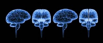
Based on the motor stereotype, individual habits and skills are formed - gait, manner of speaking, plastic movements, gestures, posture, handwriting. Having once learned to ride a bicycle, a person subsequently does not think about the sequence of movements, performing them automatically. Externally, the structure of the cortex resembles a walnut, because the surface of the cerebrum is dotted with curved grooves - convolutions.
The main feature that characterizes the cortex is tortuosity, due to which the human brain can accommodate many billions of neurons, regardless of how much space the organ itself occupies. Thanks to the deepening of the grooves, the total area of the cortical surface expands. The morphological structure of the cortex is determined by the cells that make up this area of the brain.
Gray matter is composed of neurons, glial cells (protoplasmic astrocytes), processes of neurons - dendrites and axons, processes of glial cells. Interaction between neurons occurs using processes. The processes of motor neurons reach a length of more than 1 meter. One neuron can contact 10 thousand other neurons, ensuring interaction in the functioning of organs and systems. Neurons of the cerebral cortex work synchronously, performing the following functions:
- Perception of information from the outside world.
- Processing and analysis of incoming data.
- Generating new information based on the results obtained.
- Consciousness, self-awareness, personality development.
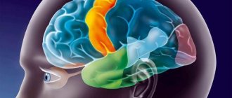
The cortex is the least ancient part of the brain, appearing later than all other departments. The cortex, like other areas of the cerebrum, is characterized by a high rate of metabolic and oxidative processes. The share of the cortex covering the cerebral hemispheres in the structure of the total body weight is 2%, but this zone, located in the brain, consumes the largest amount of oxygen entering the body - 18% (3-5 ml/min). To get an idea of the structure of the cortex, you need to consider that it consists of layers and divides the cerebral hemispheres into lobes.
Despite the clear distinction between the functions of the shares, they work in a coordinated and interconnected manner. Heteromodal areas receive information from several sensory or association areas. Heteromodal regions integrate sensory signals, conditioned motor patterns, and other impulses into instinctive behavior patterns and learned skills.
Frontal lobe
The largest area of the cortex is the frontal lobes, located in the frontal part of the cerebral hemispheres. To identify all the functions of the frontal lobe, you need to remember what parts it consists of: prefrontal (medial, dorsolateral, orbitofrontal zones) and mediobasal. The anterior lobe of the cortex, covering the brain, is responsible for planning, cognitive abilities, voluntary movements, and determines goal-directed behavior. Regulates speech function, controls the center of working memory - information recently received.
Parietal lobe
The parietal lobe consists of sections: somatosensory, posterolateral, midparietal, subdominant. Visual-spatial perception (understanding the trajectory of movement), features of the position and movement of an object relative to a landmark, the relationships of objects within three-dimensional space are controlled by the parietal cortex, located on top of the deep layers of the human brain.
Occipital lobe
The functions and tasks of the occipital lobe include the perception of visual, visual information. Controls the organs of vision - interconnected eye movements, accommodation, changes in pupil diameter. Damage to this part of the brain leads to visual agnosia, a condition in which a person cannot distinguish between familiar objects based on visual images.
Temporal lobe
The temporal lobe controls auditory function, perception of speech information, memory based on verbal and visual sensations, emotions, while simultaneously coordinating the received data with other parts of the cortex covering the cerebral hemispheres. Regulates the activity of statokinetic and taste analyzers.
Insula
Receives, adapts and reacts to vegetative and sensory impulses that come from vital systems and internal organs. Involved in the control of speech function, interacts with receptors responsible for pain and temperature sensations.
Cortical zones
Another type of classification of areas of the cerebral cortex is also noted. It marks three zones of the cortex, differing in purpose and structure.
Among them:
- Primary zone. Consists of highly differentiated cells and receives data from receptors.
- Secondary zone. Responsible for processing the received information and consists of departments of analyzer cores.
- Associative. Forms conditioned reflexes and helps to understand the world around us.
This determines not only the individual structure of the zones, but also the individual functions for each of them.
The human cerebral cortex has a complex structure, distributed into lobes and layers. Each section is responsible for its own functions, regulating various life processes. In total there are 5 lobes and 6 layers, which together make up the cortex.
Functions of the cortex covering the brain
To understand the significance of the cortex, you need to understand what it is, where it is located in the brain and what it is responsible for. With the participation of cortical brain structures, new movements are mastered and habitual physical skills are improved, as well as any meaningful and unconscious activity. The main function of the cortex located in the brain is to maintain the process of homeostasis.
Homeostasis is the body’s ability to self-regulate, the ability to maintain a constant internal state and overcome negative influences directed from the external environment. Sections of the cortex, covering the deep layers of the brain, coordinate all physiological processes occurring in the body. Thanks to its multi-layered, finely organized structure, the cortex, located in the brain, performs the following functions:
- Maintains balance of internal state when interacting with the external environment.
- Reacts to the slightest impulses, signaling changes within the body due to the penetration of toxic, foreign substances.
- Regulates all physiological processes, including the functioning of the circulatory and respiratory systems.
Control of organs, systems and processes occurs through excitation and inhibition of neurons. At the same time, a balance of states is maintained. If excitation occurs in one of the functional areas of the cortex, inhibition occurs in another area of the brain.
The interaction of the cortex with the subcortical and deep centers located in the brain is also carried out according to the principle of balanced inhibition and excitation. The higher parts of the central nervous system are interconnected with all reflex reactions. Signals entering the brain centers along afferent pathways are perceived in a complex manner, which allows you to accurately and objectively perceive the surrounding reality.
Pulse processing area
Perception of information occurs through sensory systems. The impulse processing zones are located mainly in the posterior parts of the cortical structures of the hemispheres. As we move towards the cortical sections, information is processed at least at three levels - receptor-effector (receptors, muscles), segmental (spinal cord, stem complexes), subcortical (brain sections).
The sequence reflects the process of movement of an impulse to the cortical sections and the order of making a chosen decision with the subsequent commission of a purposeful action. Data enters the cortical zones in a compressed form - as it moves from the receptors to the brain, unimportant, unimportant details are eliminated.
Sensory zone
The sensory zones constantly receive auditory, visual, olfactory, gustatory, and somatosensory signals from peripheral receptors. Processing of the received data occurs in associative zones, where information about models and images of information coming from outside is stored. During the analysis, processing, comparison of existing and new information, the images are adjusted - updated, specified, detailed.
Association zone
Information from the outside enters the brain, in particular to the centers of the cortex, along afferent pathways. The pathways of conscious sensitivity continue to cortical structures. The pathways of unconscious sensitivity end in the subcortical layers. During the perception of information, it is compared with the data in memory and signals sent by other receptors. Afferent pathways of general sensitivity carry impulses coming from pain, temperature, and tactile receptors.
The structural organization of the cortex includes association zones, which are also called functional zones. The comparative analysis takes place in the associative zones of the cortex covering the cerebral hemispheres, which is of greatest importance in the development of intellectual (cognitive) abilities. Sensory signals entering the associative zones are interpreted, differentiated, and comprehended. Based on the results of the analysis, an adequate response is selected, and the corresponding information is sent to the motor zone.
The work of associative zones is interconnected with the processes of memorizing data, learning, and mental activity, and therefore play a decisive role in increasing intelligence. In the occipital region there is an associative zone that interacts with the organs of vision, which works in concert with the sensory zone and is responsible for the interpretation of visual sensations. Among the main associative zones:
- Sound. Sound analysis.
- Speech. Perception and comprehension of words, phrases, expressions.
- Motor. Planning and reproduction of complex motor activity.
The division of zones in the cortical region is carried out according to the somatotopic principle. Information coming from the face area is projected into the central posterior gyrus, into its lower sections, hands - into the middle part of the same gyrus, legs - into the upper part. The more complex the functional tasks of body parts, the larger the area of projection of impulses in the cortex.
Diseases associated with disorders of its activity
There are many diseases that affect the human brain. Some of them affect its cortex, disrupting its processes and reducing performance. However, not much is known about them.
A common disease of the cortex is atrophy or Pick's disease. This disease develops in older people and is characterized by the death of neurons. The external condition of the brain is similar to Alzheimer's disease and resembles a dried walnut. The disease is not curable; individual symptoms are eliminated.
There are also diseases that indirectly affect the cortex. With hypertension, foci of excitation appear in the cortex, creating powerful vasoconstrictor impulses. This leads to increased blood pressure.
In addition, diseases can occur against the background of external infections. The same meningitis that occurs due to infections of pneumococcus, meningococcus and the like. The development of the disease is characterized by pain in the head, fever, pain in the eyes and many other symptoms such as weakness, nausea and drowsiness.
Many diseases that develop in the brain and its cortex have not yet been studied. Therefore, their treatment is complicated by a lack of information. So it is recommended to consult a doctor at the first non-standard symptoms, which will prevent the disease by diagnosing it at an early stage.
There are many diseases of the cerebral cortex. Among them are infectious diseases, illnesses associated with other diseases of the body, as well as diseases with an unclear cause. But most of them can be cured with medicine. Therefore, it is recommended not to delay if you feel unwell and undergo a bark examination, which is carried out in many clinics.
Diseases
Damage to tissue in the centers of the cortex covering the cerebral hemispheres leads to disruptions in the functioning of the entire organism. Damage to various cortical lobes is accompanied by deterioration of visual, auditory, motor, and mental functions. The main types of diseases are atrophy, the appearance of foci of ischemia, necrosis, inflammatory processes, the formation of a cyst or malignant tumor.
The main causes of disease are genetic predisposition, intoxication, infection and injury to the brain. All types of disorders lead to deterioration of memory, cognitive abilities, and gross and fine motor functions. The result of long-term pathological processes is dementia, disability, and the need for constant medical monitoring and care.
Pathologies and signs of damage to departments
Extensive damage to the medial areas of the frontal lobe provokes the development of abulia, which is manifested by slow reactions, indifference, and indifference to what is happening. If an area of the prefrontal orbital cortex is damaged, the patient experiences a lack of critical assessment of his own behavior and emotional lability.
Bilateral injury in the frontal region is accompanied by signs: agitation, restless behavior, obsessiveness, verbosity. Abnormal behavior is a sign of dementia, which develops against the background of degenerative processes affecting the frontal lobes. Damage to the motor cortex medulla causes hemiparesis, or muscle weakness.
Disorders develop on the side opposite to the location of the pathological focus in the brain. Damage to the visual area in one hemisphere leads to the development of bilateral blindness in half the visual field. A lesion in area 19 is associated with visual agnosia, a disturbance in visual perception. The patient sees an object, but cannot recognize it.
The information that comes through the visual analyzer is not processed or processed incorrectly, which leads to the inability to distinguish between familiar objects and people's faces. In such patients, color perception is impaired - they cannot distinguish shades.
Damage to field 22 leads to the development of musical deafness (impaired perception of musical works), the appearance of auditory hallucinations, and impaired reactions focused on auditory stimuli. Damage to field 41 is accompanied by the development of cortical deafness (inability to perceive sound signals).
Damage to field 34 is accompanied by impaired perception of smells, including olfactory hallucinations. Pathological structural changes in the nerve tissue of area 39 lead to the inability to read and write. When the field tissue is damaged, 37 people do not remember the names of objects.
Areas of the brain are divided into sensory and motor, as well as associative - and all areas interact with each other. Each department is endowed with certain functions, which together determine higher mental and complex motor activity.
Diagnostic methods
To identify disorders and their causes, blood and cerebrospinal fluid tests are prescribed. Hardware diagnostic methods:
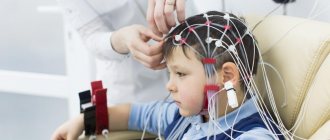
- Electroencephalography. Registration of bioelectrical brain activity. Shows diffuse slowing of signaling rates.
- Magnetoencephalography. Measuring the strength of the magnetic field generated by brain activity. It is used to identify the localization of foci of epileptic activity. The method is widely used in neurology for the diagnosis of multiple sclerosis, Alzheimer's disease, trigeminal neuralgia and other facial nerves.
- Positron emission tomography. Assessment of the state of the nigrostriatal pathways (control of motor activity), identification of foci that cause epileptic activity, areas of tissue damage that provoke dementia.
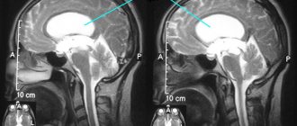
- Magnetic resonance introscopy. Visual, layer-by-layer visualization of the internal structure of the brain.
Modern instrumental methods make it possible to detect neurological disorders at an early stage. Degenerative changes during the study are observed in the preclinical stage.
Cortical structures of the brain are the most important elements of the central nervous system, which control the functioning of the body, ensure the relationship between a person and the environment, and regulate motor and mental functions. Timely diagnosis and therapy will help to avoid serious consequences associated with degenerative processes in cortical tissues.
072
White matter
It is presented in the form of numerous fibers. They are divided into three groups:
- Projection. This category is represented by bundles of efferent and afferent fibers. Through them, there are connections between the projection centers and the basal, stem and spinal nuclei.
- Associative. These fibers provide connection to the cortical areas within the boundaries of one hemisphere. They are divided into short and long.
- Commissural. These elements connect the cortical zones of the opposite hemispheres. Commissural formations are considered: the corpus callosum, posterior and anterior commissure and commissure of the fornix.
General information about GM bark
It is a superficial layer up to 0.2 cm thick that covers the hemispheres. It provides vertically oriented nerve endings. This organ contains centripetal and centrifugal nerve processes, neuroglia. Each share of this department is responsible for certain functions:
- temporal – auditory function and sense of smell;
- occipital – visual perception;
- parietal – touch and taste buds;
- frontal – speech, motor activity, complex thought processes.
In fact, the cortex predetermines the conscious activity of the individual, participates in the control of thinking, and interacts with the outside world.
Differences from old bark
The old cortex (archicortex) is an earlier emerging part of the cerebral cortex than the neocortex. But in the process of evolution, the new cortex became more developed and extensive. In this regard, the archicortex ceased to play a dominant role and became one of the constituent parts of the limbic system.
If we compare the old one in terms of the functions performed, then the first is assigned the role of fulfilling innate reflexes and motivation, and the second - managing emotions and actions at a higher level.
In addition, the neocortex is significantly larger in size than the old cortex. So the first occupies about 96% of the total surface of the hemispheres, and the size of the second is no more than 3%. This ratio shows that the archicortex cannot perform higher nervous functions.
The role of the neocortex in emotions and stereogynesis
Emotions in humans initially appear in the limbic system of the brain. But in this case they are represented by primitive concepts, which, once in the new cortex, are processed using the associative function. As a result, a person can operate with emotions at a higher level, which makes it possible to introduce concepts such as joy, sadness, love, anger, etc.
The neocortex also has the ability to dampen strong outbursts of emotion in the limbic system by sending calming signals to areas of high neuronal excitability. This leads to the fact that in a person the dominant role in behavior is played by the mind, and not by instinctive reflexes.
Anatomy
The functions performed by the cortex are often determined by its anatomical structure. The structure has its own characteristic features, expressed in a different number of layers, dimensions, and anatomy of the nerve endings that form the organ. Experts identify the following types of layers that interact with each other and help the system as a whole function:
- Molecular layer. Helps create chaotically connected dendritic formations with a small number of spindle-shaped cells that determine associative activity.
- Outer layer. Expressed by neurons having different outlines. After them, the external contours of structures having a pyramidal shape are localized.
- The outer layer is pyramidal. Assumes the presence of neurons of different sizes. These cells are similar in shape to a cone. The largest dendrite emerges from the top. Neurons are connected by dividing into small units.
- Granular layer. Provides nerve endings of small size, localized separately.
- Pyramidal layer. It assumes the presence of neural circuits of different sizes. The upper processes of neurons are able to reach the initial layer.
- A covering containing neural connections resembling a spindle. Some of them, located at the lowest point, can reach the level of white matter.
- Frontal lobe
- Plays a key role for conscious activity. Participates in memory, attention, motivation and other tasks.
Provides for the presence of 2 paired lobes and occupies 2/3 of the entire brain. The hemispheres control opposite sides of the body. So, the left lobe regulates the work of the muscles on the right side and vice versa.
The frontal parts are important in subsequent planning, including management and decision making. In addition, they perform the following functions:
- Speech. Helps express thought processes in words. Damage to this area can affect perception.
- Motor skills. Allows you to influence physical activity.
- Comparative processes. Contributes to the classification of objects.
- Memorization. Each area of the brain is important in memory processes. The frontal part forms long-term memory.
- Personal formation. It makes it possible to interact with impulses, memory and other tasks that form the main characteristics of an individual. Damage to the frontal lobe radically changes personality.
- Motivation. Most of the sensory nerve processes are located in the frontal region. Dopamine helps maintain the motivational component.
- Attention control. If the frontal parts are not able to control attention, then attention deficit syndrome is formed.
Parietal lobe
Covers the upper and lateral parts of the hemisphere, and is also separated by the central sulcus. The functions that this area performs differ for the dominant and non-dominant sides:
- Dominant (mostly left). Responsible for the ability to understand the structure of the whole through the relationship of its components and for the synthesis of information. In addition, it makes it possible to carry out interrelated movements that are required to obtain a specific result.
- Non-dominant (predominantly right-wing). A center that processes data coming from the back of the head and provides a 3-dimensional perception of what is happening. Damage to this area leads to the inability to recognize objects, faces, and landscapes. Since visual images are processed in the brain separately from data coming from other senses. In addition, the side takes part in the orientation of a person in space.
Both parietal parts are involved in the perception of temperature changes.
Temporal
It implements a complex mental function - speech.
It is located on both hemispheres in the lateral lower part, closely interacting with nearby sections. This part of the cortex has the most pronounced contours. The temporal areas process auditory impulses, converting them into a sound image. They are important in providing verbal communication skills. Directly in this department, the recognition of heard information and the selection of linguistic units for semantic expression occur.
To date, it has been confirmed that the occurrence of difficulties with the sense of smell in an elderly patient signals the development of Alzheimer's disease.
A small area inside the temporal lobe (hippocampus) controls long-term memory. The immediate temporal part accumulates memories. The dominant department interacts with verbal memory, the non-dominant one promotes visual memorization of images.
Simultaneous damage to two lobes leads to a serene state, loss of the ability to identify external images and increased sexuality.
Island
The insula (closed lobule) is located deep in the lateral sulcus. The insula is separated from adjacent sections by a circular groove. The upper section of the closed lobule is divided into 2 parts. The taste analyzer is projected here.
Forming the bottom of the lateral sulcus, the closed lobule is a projection, the upper part of which is directed outward. The insula is separated by a circular groove from nearby lobes that form the operculum.
The upper section of the closed lobule is divided into 2 parts. The precentral sulcus is localized in the first, and the anterior central gyrus is located in the middle of them.
Furrows and convolutions
They are depressions and folds located in the middle of them, which are localized on the surface of the cerebral hemispheres. The grooves contribute to the enlargement of the cerebral cortex without increasing the volume of the cranium.
The significance of these areas lies in the fact that two-thirds of the entire cortex is located deep in the grooves. There is an opinion that the hemispheres develop unequally in different departments, as a result of which the tension will also be uneven in specific areas. This can lead to the formation of folds or wrinkles. Other scientists believe that the initial development of the furrows is of great importance.