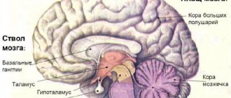
The cerebral cortex is represented by a uniform layer of gray matter 1.3-4.5 mm thick, consisting of more than 14 billion nerve cells. Due to the folding of the bark, its surface reaches large sizes - about 2200 cm2.
The thickness of the cortex consists of six layers of cells, which are distinguished by special staining and examination under a microscope. The cells of the layers vary in shape and size. From them, processes extend deep into the brain.
Structure of the cerebral cortex
It was found that different areas - fields of the cerebral cortex differ in structure and function. There are from 50 to 200 such fields (also called zones, or centers). There are no strict boundaries between the zones of the cerebral cortex. They constitute an apparatus that provides reception, processing of incoming signals and response to incoming signals.
Areas of the cerebral cortex
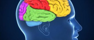
In the posterior central gyrus, behind the central sulcus, there is an area of cutaneous and joint-muscular sensitivity . Here the signals that arise when touching our body, when it is exposed to cold or heat, or when it is painful are perceived and analyzed.
Areas of the cerebral cortex
In contrast to this zone, in the anterior central gyrus, in front of the central sulcus, the motor zone . It identifies areas that provide movement of the lower extremities, muscles of the trunk, arms, and head. When this area is irritated by electric current, contractions of the corresponding muscle groups occur. Injuries or other damage to the motor cortex lead to paralysis of the body muscles.
The temporal lobe contains the auditory area . The impulses arising in the receptors of the cochlea of the inner ear are received here and analyzed. Irritation of areas of the auditory zone causes sensations of sounds, and when they are affected by the disease, hearing is lost.
The visual zone is located in the cortex of the occipital lobes of the hemispheres. When it is irritated by electric current during brain surgery, a person experiences sensations of flashes of light and darkness. When it is affected by any disease, vision deteriorates and is lost.
Near the lateral sulcus there is a gustatory zone , where taste sensations are analyzed and formed based on signals arising in the receptors of the tongue. The olfactory zone is located in the so-called olfactory brain, at the base of the hemispheres. When these areas are irritated during surgery or during inflammation, people smell or taste something.
purely speech zone .
It is represented in the cortex of the temporal lobe, the inferior frontal gyrus on the left, and parts of the parietal lobe. Their diseases are accompanied by speech disorders.
The work of the frontal lobes of the brain. Frontal lobe: functions, structure and damage
The frontal lobes occupy about 28% of the total area of cortical structures.
Their mass is approximately half the weight of the entire brain - about 450 g. The frontal lobes are structures located in the frontal plane of the brain that are responsible for human mental activity, which determines their most important role in the formation and use of the mind.
The functions of the frontal lobes, located in the brain, include the ability to reflect, analyze, abstract, and generalize.
Parietal
In order to understand the functions of the parietal lobes, it is important to understand that the dominant and non-dominant side will do different jobs. The dominant parietal lobe of the brain helps to understand the structure of the whole through its parts, their structure, order
Thanks to her, we know how to put individual parts into a whole. The ability to read is very indicative of this. To read a word, you need to put the letters together, and you need to create a phrase from the words. Manipulations with numbers are also carried out
The dominant parietal lobe of the brain helps to understand the structure of the whole through its parts, their structure, order. Thanks to her, we know how to put individual parts into a whole. The ability to read is very indicative of this. To read a word, you need to put the letters together, and you need to create a phrase from the words. Manipulations with numbers are also carried out.
The parietal lobe helps to link individual movements into a complete action. When this function is disrupted, apraxia is observed. Patients cannot perform basic actions, for example, they are not able to get dressed. This happens with Alzheimer's disease. A person simply forgets how to make the necessary movements.
The non-dominant side (in right-handed people it is the right side) combines information that comes from the occipital lobes and allows you to perceive the world around you in three-dimensional mode. If the non-dominant parietal lobe is disrupted, visual agnosia may occur, in which a person is unable to recognize objects, landscapes, or even faces.
The parietal lobes are involved in the perception of pain, cold, and heat. Their functioning also ensures orientation in space.
brain
The human brain is an organ weighing 1.3-1.4 kg, located inside the cranium. The human brain is made up of more than one hundred billion neuron cells that form the gray matter, or cortex, the vast outer layer of the brain.
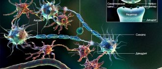
Neuronal processes (kind of like wires) are the axons that make up the white matter of the brain. Axons connect neurons to each other through dendrites.
The adult brain consumes about 20% of all the energy that the body needs, while the child's brain consumes about 50%.
- How does the human brain process information?
- Functions of the right and left hemispheres of the brain
- Emotions
- The structure of the human brain, the trinity of the brain
- white and gray matter
- prefrontal cortex
- hippocampus
- islet of Reil
- Broca's area
Brain reward system Brain differences between men and women Aging of the human brain Sources
How does the human brain process information?
Today it is considered proven that the human brain can simultaneously process on average about 7 bits of information. These can be individual sounds or visual signals, shades of emotions or thoughts distinguished by consciousness.
The minimum time required to distinguish one signal from another is 1/18 of a second. Thus, the perceptual limit is 126 bits per second. Conventionally, we can calculate that over the course of a 70-year life, a person processes 185 billion bits of information, including every thought, memory, and action.
Information is recorded in the brain through the formation of neural networks (a kind of routes).
First and second signaling systems
The role of the cerebral cortex in improving the first signaling system and developing the second is invaluable. These concepts were developed by I.P. Pavlov. The signaling system as a whole is understood as the entire set of processes of the nervous system that carry out perception, processing of information and the response of the body. It connects the body with the outside world.
First signaling system
The first signaling system determines the perception of sensory-specific images through the senses. It is the basis for the formation of conditioned reflexes. This system exists in both animals and humans.
In the higher nervous activity of man, a superstructure has developed in the form of a second signaling system. It is peculiar only to humans and is manifested by verbal communication, speech, and concepts. With the advent of this signaling system, abstract thinking and generalization of countless signals from the first signaling system became possible. According to I.P. Pavlov, words turned into “signals of signals.”
Functions
In addition to the physiological division of the brain into lobes, the need arose to divide it into areas that are responsible for certain functions.
Frontal lobes
This is the so-called command center. What is the frontal lobe responsible for? It is a point of independence, self-awareness, and initiative. The defeat of these areas or the presence of pathologies in their functioning will affect a person’s attitude towards the world - almost everything will become indifferent to him, motivation will disappear, interest in current events will disappear, and laziness will appear.
The main functions of the frontal lobes are to control human behavior. It generates responses to social phenomena. When zones are violated, the limiter is deactivated, establishing a ban on certain actions, called uncultural.
The frontal lobes also allow you to analyze, plan, and learn new skills. Repeated repetition of the same sequences of movements over time becomes automatic and does not require effort or thought to perform them. Damage will force you to repeat monotonous movements every time as if for the first time, putting a lot of effort into it.
Perseveration is another consequence of deviations in the functioning of the frontal lobes. This is looping or repetition, such as repeating one phrase or word during a conversation
On the left side (for right-handed people) there are centers that are responsible for speech and attention.
These areas of the brain are also involved in coordinating and maintaining the body in an upright position when sitting and walking.
Temporal
They are located on the sides in the upper part of the brain, in the temple area. Thanks to them, the sound perceived by sound receptors turns into images, a person understands what he heard, certain sound vibrations are associated with images and are assigned to them. With the help of this part of the brain, people understand each other, their sound vibrations are filled with meaning, they choose the necessary words to describe certain phenomena.
Usually the left, non-dominant lobe is involved in determining the intonation of speech and reads emotions from facial expressions alone. Thanks to a small formation, the hippocampus, access to long-term memory is provided. Where it is located, on what medium and in what way our memories are recorded, this should be found out. The non-dominant part is used in visual memory, but the dominant part is used in verbal memory.
With problems with the temporal lobes, deviations in the functioning of the speech apparatus appear, in particular aphasia.
Parietal
MRI has shown that for left-handers and right-handers these lobes perform different functions, in fact directly opposite.
The left one gives us the ability to create a whole from fragments, that is, it helps to form a holistic picture of the world from small, at first glance, unrelated pieces.
They allow you not only to put together a mosaic from fragments, but also from letters into words, from a sequence of actions into a dance or technique, etc.
The non-dominant part allows you to perceive the world in three dimensions by processing information coming from the occipital lobes. Due to impairments, a person loses the ability to recognize faces, outline objects, and determine the distance to them and between them. These areas are also involved in the perception of pain and cold.
Occipital
Visual information processing center. They interpret the photons reflected from obstacles arriving at the biological light sensor - the retina - and form the resulting image, rotating it 180 degrees. Data about the size, color, shape, and material of an object are processed in separate centers and then recombined to form a single three-dimensional picture.
From the editor: Types, causes and treatment of hemorrhage in the eye
cerebral cortex,
a layer of gray matter 1-5
mm
covering the cerebral hemispheres of mammals and humans.
This part of the brain,
which developed in the later stages of the evolution of the animal world, plays an extremely important role in the implementation of mental, or
higher nervous activity,
although this activity is the result of the work of the brain as a whole.
Thanks to bilateral connections with the underlying parts of the nervous system, the cortex can participate in the regulation and coordination of all body functions. In humans, the cortex makes up on average 44% of the volume of the entire hemisphere as a whole. Its surface reaches 1468–1670 cm2.
The structure of the cortex.
A characteristic feature of the structure of the cortex is the oriented, horizontal-vertical distribution of its constituent nerve cells across layers and columns;
Thus, the cortical structure is distinguished by a spatially ordered arrangement of functioning units and connections between them (
Fig. 1 )
. The space between the bodies and processes of cortical nerve cells is filled with neuroglia
and a vascular network (capillaries).
neurons
are divided into 3 main types: pyramidal (80-90% of all cortical cells), stellate and fusiform.
The main functional element of the cortex is the afferent-efferent (i.e., perceiving centripetal and sending centrifugal stimuli) long-axon pyramidal neuron (
Fig. 2 )
. Stellate cells are distinguished by weak development of dendrites
and powerful development
of axons,
which do not extend beyond the diameter of the cortex and cover groups of pyramidal cells with their branches.
Stellate cells play the role of perceiving and synchronizing elements capable of coordinating (simultaneously inhibiting or exciting) spatially close groups of pyramidal neurons. The cortical neuron is characterized by a complex submicroscopic structure (see Cell
)
.
Areas of the cortex that are different in topography differ in the density of cells, their size and other characteristics of the layer-by-layer and columnar structure.
All these indicators determine the architecture of the cortex, or its cytoarchitectonics (
see Fig. 1 and 3 )
.
The largest divisions of the cortex are the ancient (paleocortex), old (archicortex), new (neocortex) and interstitial cortex. The surface of the new cortex in humans occupies 95.6%, old 2.2%, ancient 0.6%, interstitial 1.6%.
If we imagine the cerebral cortex as a single cover (cloak) covering the surface of the hemispheres, then the main central part of it will be the new cortex, while the ancient, old and intermediate will take place on the periphery, i.e., along the edges of this cloak. The ancient cortex in humans and higher mammals consists of a single cell layer, indistinctly separated from the underlying subcortical nuclei; the old bark is completely separated from the latter and is represented by 2-3 layers; the new cortex consists, as a rule, of 6-7 layers of cells; interstitial formations - transitional structures between the fields of the old and new cortex, as well as the ancient and new cortex - from 4-5 layers of cells. The neocortex is divided into the following areas: precentral, postcentral, temporal, inferior parietal, superior parietal, temporo-parietal-occipital, occipital, insular and limbic. In turn, areas are divided into subareas and fields. The main type of direct and feedback connections of the new cortex are vertical bundles of fibers that bring information from subcortical structures to the cortex and send it from the cortex to these same subcortical formations. Along with vertical connections, there are intracortical - horizontal - bundles of associative fibers passing at various levels of the cortex and in the white matter under the cortex. Horizontal beams are most characteristic of layers I and III of the cortex, and in some fields for layer V. Horizontal bundles ensure the exchange of information both between fields located on adjacent gyri and between distant areas of the cortex (for example, frontal and occipital).
Functional features of the cortex
are determined by the above-mentioned distribution of nerve cells and their connections across layers and columns.
Convergence (convergence) of impulses from various sensory organs is possible on cortical neurons. According to modern concepts, such a convergence of heterogeneous excitations is a neurophysiological mechanism of integrative activity of the brain, that is, analysis and synthesis of the body’s response activity. It is also significant that the neurons are combined into complexes, apparently realizing the results of the convergence of excitations on individual neurons. One of the main morpho-functional units of the cortex is a complex called a column of cells, which passes through all cortical layers and consists of cells located at the same perpendicular to the surface of the cortex. The cells in the column are closely connected to each other and receive a common afferent branch from the subcortex. Each column of cells is responsible for the perception of predominantly one type of sensitivity. For example, if at the cortical end of the skin analyzer
one of the columns reacts to touching the skin, then the other reacts to the movement of the limb in the joint.
In the visual analyzer,
the functions of perceiving visual images are also distributed across columns. For example, one of the columns perceives the movement of an object in the horizontal plane, the adjacent one in the vertical plane, etc.
The second complex of cells in the neocortex, the layer, is oriented in the horizontal plane. It is believed that small cell layers II and IV consist mainly of perceptive elements and are “entrances” to the cortex. Layer magnocellular V —
exits from the cortex to the subcortex, and the middle cellular layer III is associative, connecting various cortical zones
(
see Fig. 1 )
.
The localization of functions in the cortex is characterized by dynamism due to the fact that, on the one hand, there are strictly localized and spatially delimited zones of the cortex associated with the perception of information from a specific sensory organ, and on the other hand, the cortex is a single apparatus in which individual structures are closely connected and if necessary, they can be interchanged (the so-called plasticity of cortical functions). In addition, at any given moment, cortical structures (neurons, fields, areas) can form coordinated complexes, the composition of which varies depending on specific and nonspecific stimuli that determine the distribution of inhibition
and
excitations
in the cortex.
Finally, there is a close interdependence between the functional state of cortical zones and the activity of subcortical structures. Cortical territories differ sharply in their functions. Most of the ancient cortex is included in the olfactory analyzer system. The old and interstitial cortex, being closely related to the ancient cortex both by systems of connections and evolutionarily, are not directly related to smell. They are part of the system responsible for the regulation of vegetative reactions and emotional states of the body (see Reticular formation, Limbic system
)
.
The new cortex is a set of final links of various perceptive (sensory) systems (cortical ends
of analyzers
)
.
It is customary to distinguish projection, or primary, and secondary fields, as well as tertiary fields, or associative zones, in the zone of a particular analyzer. Primary fields receive information mediated through the smallest number of switches in the subcortex (in the thalamus, or thalamus, of the diencephalon). It is as if the surface of peripheral receptors is projected onto these fields ( Fig. 4
)
.
In the light of current data, projection zones cannot be considered as devices that perceive point-to-point stimuli. In these zones, the perception of certain parameters of objects occurs, i.e., images are created (integrated), since these areas of the brain respond to certain changes in objects, their shape, orientation, speed of movement, etc. In addition, the localization of functions in the primary zones duplicated many times using a mechanism reminiscent of holography,
when each smallest section of a storage device contains information about the entire object.
Therefore, the preservation of a small area of the primary sensory field is sufficient for the ability to perceive to be almost completely preserved. Secondary fields receive projections from the senses through additional switching in the subcortex, which allows for a more complex analysis of a particular image. Finally, tertiary fields, or associative zones, receive information from nonspecific subcortical nuclei, which summarize information from several sense organs, which makes it possible to analyze and integrate a particular object in an even more abstracted and generalized form. These areas are also called analyzer overlap areas. Primary and partly secondary fields are a possible substrate of the first signaling system,
and tertiary zones (associative) are a
substrate of the second signaling system,
specific to humans (I. P. Pavlov).
These inter-analyzer structures determine complex forms of brain activity, including professional skills (inferior parietal region), thinking, planning and purposefulness of actions (frontal region), and written and oral speech (inferior frontal subregion, temporal, temporo-parietal-occipital and inferior parietal regions ). the organ of Corti
is projected in the precentral region - field 4, where the projection of proprioceptors is carried out in accordance with the location of the muscles, in the postcentral region - fields 3 and 1 , where exteroceptors are projected in accordance with their distribution in the skin.
Secondary zones are represented by fields 8 and 6 (motor analyzer)
, 5 and 7 (skin analyzer), 18 and 19 (visual analyzer), 22
(auditory analyzer).
Tertiary zones are represented by large areas of the frontal region (fields 9, 10, 45, 44 and 46), inferior parietal (fields 40 and 39), temporo-parietal-occipital (field 37).
Cortical structures play a primary role in learning in animals and humans. However, the formation of some simple conditioned reflexes,
mainly from internal organs, can be provided by subcortical mechanisms.
These reflexes can also form at lower levels of development, when there is no cortex yet. Complex conditioned reflexes that underlie integral acts of behavior
require the preservation of cortical structures and the participation of not only the primary zones of the cortical ends of the analyzers, but also the associative - tertiary zones.
Cortical structures are also directly related to memory mechanisms.
Electrical stimulation of certain areas of the cortex (for example, the temporal cortex) evokes complex patterns of memories in people.
A characteristic feature of the activity of the cortex is its spontaneous electrical activity, recorded in the form of an electroencephalogram (EEG). In general, the cortex and its neurons have rhythmic activity, which reflects the biochemical and biophysical processes occurring in them. This activity has a varied amplitude and frequency (from 1 to 60 Hz
) and changes under the influence of various factors.
The rhythmic activity of the cortex is irregular, but several different types can be distinguished by the frequency of potentials (alpha, beta, delta and theta rhythms). The EEG undergoes characteristic changes in many physiological and pathological conditions (various phases of sleep,
for tumors, seizures, etc.).
The rhythm, i.e. frequency, and amplitude of the bioelectric potentials
of the cortex are set by subcortical structures that synchronize the work of groups of cortical neurons, which creates the conditions for their coordinated discharges. This rhythm is associated with the apical (apical) dendrites of pyramidal cells. The rhythmic activity of the cortex is influenced by influences coming from the senses. Thus, a flash of light, a click or a touch on the skin causes the so-called. a primary response consisting of a series of positive waves (downward deflection of the electron beam on the oscilloscope screen) and a negative wave (upward deflection of the beam). These waves reflect the activity of the structures of a given area of the cortex and change in its different layers.
Phylogeny and ontogeny of the cortex.
The cortex is the product of a long evolutionary development, during which the ancient cortex first appears, arising in connection with the development of the olfactory analyzer in fish. With the emergence of animals from water onto land, the so-called. a mantle-shaped part of the cortex, completely separate from the subcortex, which consists of old and new cortex. The formation of these structures in the process of adaptation to the complex and diverse conditions of terrestrial existence is associated with the improvement and interaction of various perceptive and motor systems. In amphibians, the cortex is represented by an ancient and rudiment of the old cortex; in reptiles, the ancient and old cortex is well developed and the rudiment of a new cortex appears. The greatest development the new cortex reaches mammals, and among them primates (monkeys and humans), proboscis (elephants) and cetaceans (dolphins, whales). Due to the uneven growth of individual structures of the new cortex, its surface becomes folded, covered with grooves and convolutions. Improvement of the cortex The telencephalon in mammals is inextricably linked with the evolution of all parts of the central nervous system. This process is accompanied by an intensive growth of direct and feedback connections connecting cortical and subcortical structures. Thus, at higher stages of evolution, the functions of subcortical formations begin to be controlled by cortical structures. This phenomenon is called corticolization of functions. As a result of corticolization, the brain stem forms a single complex with the cortical structures, and damage to the cortex at higher stages of evolution leads to disruption of the vital functions of the body. The association zones undergo the greatest changes and increase during the evolution of the neocortex, while the primary sensory fields decrease in relative size. The growth of the new cortex leads to the displacement of the old and ancient cortex onto the lower and middle surfaces of the brain.
The cortical plate appears relatively early in the process of intrauterine development of a person - at the 2nd month. The lower layers of the crust (VI-VII) are distinguished first, then the higher ones (V, IV, III and II; see Fig. 1
).
By 6 months, the embryo already has all the cytoarchitectonic fields of the cortex characteristic of an adult. After birth, three turning points can be distinguished in the growth of the cortex: at 2-3 months of life, at 2.5-3 years and at 7 years. By the last period, the cytoarchitecture of the cortex is fully formed, although the cell bodies of neurons continue to increase until 18 years of age. The cortical zones of the analyzers complete their development earlier, and the degree of their increase is less than that of the secondary and tertiary zones. There is great diversity in the timing of maturation of cortical structures in different individuals, which coincides with the diversity of timing of maturation of the functional characteristics of the cortex. Thus, individual (ontogeny)
and historical
(phylogeny)
development of the cortex is characterized by similar patterns.
Lit.:
Orbeli L. A., Questions of higher nervous activity, M.-L., 1949; Cytoarchitecture of the human cerebral cortex. Sat. Art., M., 1949; Filimonov I.N., Comparative anatomy of the cerebral cortex of mammals, M., 1949; Pavlov I.P., Twenty years of experience in the objective study of higher nervous activity of animals, Complete. collection op., 2nd ed., vol. 3, book. 1-2, M., 1951; Brazier M., Electrical activity of the nervous system, trans. from English, M., 1955; Sepp E.K., History of the development of the nervous system of vertebrates, 2nd ed., M., 1959; Luria A. R., Higher human cortical functions and their disorders in local brain lesions, M., 1962; Voronin L.G., Course of lectures on the physiology of higher nervous activity, M., 1965; Polyakov G.I., On the principles of neural organization of the brain, M., 1965; Cortical regulation of the activity of subcortical formations of the brain. Sat. Art., Tb., 1968; Anokhin P.K., Biology and neurophysiology of the conditioned reflex, M., 1968; Beritov I. S., Structure and functions of the cerebral cortex, M., 1969.
L. G. Voronin.
Contents TSB

