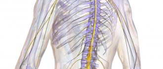
What makes it possible to detect NSG of the brain of newborns?
A cyst is a pathology that is a choroid plexus resembling a bubble containing fluid inside.
in newborns they can form during the passage of the birth canal. In this case, they usually resolve on their own and do not require any treatment. If the reason for their formation is different, then this requires additional examination and appropriate treatment. Increased levels can be detected using NSG. This study allows you to detect various abnormalities of brain development, which are caused by circulatory disorders or birth injuries.
A serious pathology that manifests itself in the displacement of one of the hemispheres. The cause may be a tumor, hemorrhage or a large cyst. This pathology requires prompt attention to a specialist.
Intraventricular or parenchymal hemorrhages in newborns can also be detected using an NSG study. Intraventricular hemorrhages are more common in children with hypoxia or premature babies. Parenchymal ones most often develop in the fetus in utero. With this pathology, treatment begins immediately from the moment of birth.
Hydrocephalus is an enlargement of one or more ventricles of the brain. This pathology requires urgent attention to a neurologist and intensive care.
Serious disorders of the nervous system can be detected almost from the first days of life using NSG of the brain of newborns. Reviews from parents whose children, thanks to this study, were completely cured and did not become disabled from early childhood, indicate the advisability of carrying out the procedure in case of any suspicion. Experts are of the same opinion.
The transcript of this study is read exclusively by a doctor. In this case, all the nuances of labor are taken into account:
- How the birth went - with or without complications.
- How long did they last?
- Did the fetus have hypoxia?
- Did the newborn have any birth injuries?
- Child's weight, etc.
Taking into account all this data, the doctor makes a conclusion. In a study such as NSG of the brain of newborns, deciphering the data may turn out to be the norm for some babies, but not for others (taking into account complications during childbirth). The study evaluates the following data:
- Symmetry or asymmetry of brain structures. Normally there should be complete symmetry.
- Clarity of grooves and convolutions in the cerebral cortex.
- Symmetry and homogeneity of the ventricles of the brain, anechoicity. The presence of so-called flakes (seals) indicates hemorrhages.
- Hyperechogenicity and homogeneity of vascular accumulations.
- No leukomalacia (excessive softness of the brain matter structure).
- No cysts.
Difference between the mantle and other structures of the telencephalon, 3 stages of development of the cortex
The cloak covers all other parts of the telencephalon. Consists of gray and white matter.
Gray matter - the cortex, is represented by clusters of neuron bodies lying on the surface (above the white matter).
White matter - pathways.
Three stages of cortex development:
- The ancient cortex - the cells above the white matter - are not organized and are located chaotically. Located on the basal surface, closer to the amygdala.
- Old cortex - the appearance of elements of ordered nerve cells by similarity and function. Layers are formed (no more than three). Located on the medial surface, next to the central part of the olfactory brain.
- New cortex - cells are strictly ordered, differ in shape and function. Form layers (6 layers). Cells in different areas are different: Cytoarchitectonic fields (about 130),
- Myelarchitectonic fields (100) - differences in the structure of processes,
- Neuroarchitectonic fields (80) - along neuroglia,
- Angioarchitectonic fields - according to the vascular pattern.
Ependymocytes
The cells of this epithelium have many organelles and a large nucleus. Their outer surface is covered with a large number of microvilli; they help promote the cerebrospinal fluid, as well as its absorption. Outside the ependyma are Kolmer cells, which are considered a special type of macrophages capable of moving throughout the body.
Through multiple small gaps in the basement membrane of epindemocytes, blood plasma sweats into the ventricular cavity. Proteins produced directly by the cells of the internal epithelium of the brain cavities are added to it, and this is how cerebrospinal fluid is obtained.
What is included in the basal ganglia? Their localization
The basal ganglia is a collection of gray matter within the telencephalon, surrounded by white matter.
1) The striatum - consists of alternating areas of gray and white matter.
- caudate nucleus,
- Lenticular nucleus: shell (lateral part),
- globus pallidus (lateral and medial nuclei (lamellae)).
2) The fence is the shape of a thin plate (lateral to the lenticular nucleus).
3) Amygdala - basal nuclei, located in the temporal lobe of the hemispheres (closer to the lower surface).
Possible consequences and complications
The consequences of cerebral edema in a newborn can have varying degrees of severity. This depends on how timely therapeutic measures were taken and at what stage the pathology was identified. Possible consequences:
- intracranial hypertension (increased pressure inside the skull, which is characterized by periodic bursting headaches);
- impairment of a child’s intellectual ability at an older age, which occurs against the background of cell death in the cortical structure;
- frequent consequences of edema - impaired extensor function of the limbs, inability to hold the head, impaired grasping and sucking reflexes;
- complications include the development of cerebral palsy and epilepsy;
- formation of a bone marrow tumor with expansion of the 4th ventricle.
The most severe consequence is death, which occurs when therapy is not started in a timely manner or with primary extensive organ damage. In other cases, with early detection of pathology, the prognosis is relatively favorable.
Not in all cases, cerebral ventricular edema in a newborn is treated. If, in accordance with the results of the diagnostic measures carried out, it was possible to detect a physiological increase in the structural element (for example, with a large child, this is the norm), specific therapy is not prescribed.
We recommend further reading: Causes of bags under the eyes in a one-year-old child and methods of treating red eyes
An increase in cerebrospinal fluid pressure requires the use of therapeutic measures, which include the use of medications. Self-medication and traditional medicine are not effective in such cases.
Drug therapy
If pronounced clinical manifestations are present, drugs from the following groups are used:
- diuretics are prescribed to reduce swelling of the ventricle and normalize the process of urination (Furosemide, etc.);
- products containing potassium replenish its insufficient content in the body, which occurs against the background of accelerated functioning of the urinary tract organs (Asparkam, etc.);
- vitamin preparations have a preventive effect on the development of rickets and accelerate regeneration processes (B6, D3, etc.);
- nootropics normalize blood circulation in the brain, strengthen vascular walls (Encephabol, etc.);
- drugs with a sedative effect reduce symptoms of nervous system disorders (Glycine, etc.).
In most cases, drug therapy helps to get rid of the pathology. Treatment of brain cysts in a child, tumors and other neoplasms that are accompanied by swelling is carried out surgically.
Surgical
Surgery is prescribed for existing tumors in the ventricular cavity. This is excised with surgical instruments. In the postoperative period, massage courses and a physical therapy complex are prescribed, which helps normalize muscle tone and prevent atrophy.
What 3 morphological substrates provide analytical and synthetic function?
Cloak functions:
1) Analytical - decomposition of objects and phenomena into individual characteristics.
For this purpose, there is a cortical center - a section of the cortex that contributes to the perception of one sign.
The bark itself consists of:
- Cortical nucleus (contains cells specialized in one trait);
- The scattered part (contains cells that perform the sensation of one sign, but can be rearranged to another sign).
2) Synthetic function - an idea of an object or phenomenon is made up of individual features. Synthetic functions perform:
- Associative fields are the zone of intersection of the peripheral (scattered) parts of several cortical centers. Here are cells that are initially capable of performing two functions.
- Association nerve fibers.
Treatment of the disease
Treatment can only be prescribed by a neurosurgeon or neurologist. Drug therapy is usually used. Not all episodes require treatment, but it is used in cases of pronounced neuropathological abnormalities. The main medications are:
- Diuretics are used to reduce cerebral edema, normalize and accelerate fluid excretion.
- Potassium-containing preparations compensate for the deficiency of the required amount of potassium while accelerating the process of urination.
- Vitamin complexes are used to replenish lost vitamins, as well as to restore the patient’s body.
- Nootropics improve blood supply to the brain, circulation in microtissues and vascular elasticity.
- Sedatives have a calming effect and reduce neurological signs such as tearfulness, moodiness, and irritability.
If the cause of deviations in the size of the brain cavities is mechanical damage to the head, then surgical intervention is required.
This article will be relevant for parents whose children have been diagnosed with ventricular enlargement
The human brain contains structures that contain cerebrospinal fluid (CSF). These structures are the largest in the ventricular system.
They can be divided into the following types:
- Lateral;
- Third;
- Fourth.
The lateral ventricles are designed to store cerebrospinal fluid. Compared to the third and fourth, they are the largest among them. On the left side there is a ventricle, which can be called the first, on the right side - the second. Both ventricles work with the third ventricle.
The ventricle, called the fourth, is one of the most important formations. The fourth ventricle contains the spinal canal. It appears to be diamond-shaped.
- Decreased appetite of the child; it often happens that the child refuses breastfeeding.
- Muscle tone is reduced.
- Tremors of the upper and lower extremities appear.
- A distinct manifestation of veins on the forehead, the cause is from the cranial cavity.
- The child's swallowing and grasping abilities are reduced.
- High likelihood of developing strabismus.
- Disproportionality of the head.
- Frequent regurgitation due to increased cerebrospinal fluid pressure.
The child often complains of the inability to raise his eyes and lower his head, dizziness and weakness appear, and the skin begins to turn pale.
From the editor: Features, diagnosis and treatment of cerebral hematomas
Functional impairment
Age-related changes such as cerebral atherosclerosis; vascular lesions caused by toxic causes or diseases such as diabetes mellitus, dysfunction of the thyroid gland, can lead to the death of a large number of capillaries of the choroid and their replacement by growing connective tissue. Such growths are scars, which are always larger than the original area before the lesion. As a result, large areas of the brain will suffer from deterioration in blood supply and nutrition.
The surface area of affected vessels is always less than that of normally functioning vessels. In this regard, the speed and quality of metabolic processes between blood and cerebrospinal fluid decrease. Because of this, the properties of the cerebrospinal fluid change, its chemical composition and viscosity change. It becomes thicker, disrupting the activity of nerve pathways, and even puts pressure on the areas of the brain bordering the 4th ventricle. One type of such condition is hydrocephalus, or dropsy. It spreads to all areas of the liquor supply, thereby affecting the cortex, expanding the gap between the grooves, exerting a pressing effect on them. At the same time, the volume of gray matter is significantly reduced, and a person’s thinking abilities are impaired. Dropsy, affecting the structures of the midbrain, cerebellum and medulla oblongata, can affect vital centers of the nervous system, such as the respiratory, vascular and other zones of regulation of biological processes in the body, which causes an immediate threat to life.
First of all, disorders manifest themselves at the local level, as indicated by the symptoms of damage to those same pairs of cranial nerves from the fifth to the twelfth. Which, accordingly, is manifested by local neurological symptoms: changes in facial expressions, impaired peripheral vision, hearing impairment, impaired coordination of movements, speech defects, taste abnormalities, problems with spoken language, secretion and swallowing of saliva. There may be disturbances in the activity of the muscles of the upper shoulder girdle.
The causes of dropsy may lie not only at the cellular level. There are tumor diseases (primary from nervous or vascular tissue, secondary - metastasis). If the tumor occurs near the boundaries of the 4th ventricle, then the result of an increase in size will be a change in its shape, which again will lead to hydrocephalus.
Intermediate brain
It consists of 2 parts:
- ventral (hypothalamus);
- dorsal (metathalamus, thalamus, epithalamus).
It is the thalamus that receives stimuli and sends them to the hemispheres. This is a reliable and always busy intermediary. Its second name is the visual thalamus. The thalamus ensures successful adaptation to an ever-changing environment. The limbic system reliably connects it to the cerebellum.
The hypothalamus is a subcortical center that regulates all autonomic functions. It affects through the nervous system and glands
The hypothalamus ensures the normal functioning of individual endocrine glands and participates in metabolism, which is so important for the body. The hypothalamus is responsible for the processes of sleep and wakefulness, eating and drinking
Below it is the pituitary gland. It is the pituitary gland that provides thermoregulation, the functioning of the cardiovascular and digestive systems.
Ventricular diseases
The brain, like all other internal human organs, is prone to various diseases.
Pathological processes affecting parts of the central nervous system and the ventricles, including, require immediate medical intervention. In pathological conditions developing in the organ cavities, the patient’s condition rapidly deteriorates because the brain does not receive the required amount of oxygen and nutrients. In most cases, the cause of ventricular diseases is inflammatory processes resulting from infections, injuries or neoplasms.
Hydrocephalus
Hydrocephalus is a disease characterized by excessive accumulation of fluid in the ventricular system of the brain. The phenomenon in which difficulties arise in its movement from the site of secretion to the subarachnoid space is called occlusive hydrocephalus.
If the accumulation of fluid occurs due to a violation of the absorption of cerebrospinal fluid into the circulatory system, then this pathology is called aresorptive hydrocephalus.
Hydrocele of the brain can be congenital or acquired. The congenital form of the disease is usually detected in childhood. The causes of the acquired form of hydrocephalus are often infectious processes (for example, meningitis, encephalitis, ventriculitis), neoplasms, vascular pathologies, injuries and developmental defects.
Dropsy can occur at any age. This condition is dangerous to health and requires immediate treatment.
Hydroencephalopathy
Another common pathological condition due to which the ventricles in the brain may suffer is hydroencephalopathy.
In this pathological condition, two diseases are combined at once - hydrocephalus and encephalopathy. As a result of impaired circulation of cerebrospinal fluid, its volume in the ventricles increases, intracranial pressure increases, and because of this, brain function is disrupted. This process is quite serious and without proper control and treatment leads to disability.
Ventriculomegaly
When the right or left ventricles of the brain become enlarged, a disease called ventriculomegaly is diagnosed. It leads to disturbances in the functioning of the central nervous system, neurological abnormalities and can provoke the development of cerebral palsy. This pathology is most often detected during pregnancy at a period of 17 to 33 weeks (the optimal period for detecting pathology is 24-26 weeks).
A similar pathology often occurs in adults, but ventriculomegaly does not pose any danger to a mature organism.
Ventricular asymmetry
Changes in the size of the ventricles can occur under the influence of excessive production of cerebrospinal fluid. This pathology never occurs on its own. Most often, the appearance of asymmetry is accompanied by more serious diseases, for example, neuroinfection, traumatic brain injury, or a tumor in the brain.
Hypotensive syndrome
A rare phenomenon, usually a complication after therapeutic or diagnostic procedures. Most often it develops after a puncture and leakage of cerebrospinal fluid through the hole from the needle.
Other causes of this pathology may be the formation of cerebrospinal fluid fistulas, disruption of the water-salt balance in the body, and hypotension.
Clinical manifestations of low intracranial pressure: the appearance of migraine, apathy, tachycardia, general loss of strength. With a further decrease in the volume of cerebrospinal fluid, pallor of the skin, cyanosis of the nasolabial triangle, and breathing problems appear.
How and when is diagnosis carried out?
Regular ultrasound examination of the brain is prescribed in the first month of the baby’s life, if there are alarming symptoms, for example, weak reflexes or causeless restlessness of the child.
If pathology is present, the examination in children under one year of age is repeated every three months.
Deviation from the norm at this age does not always require treatment. A wait-and-see approach and regular examinations are required to determine the dynamics of changes in the condition of brain tissue. Often, enlarged ventricles are temporary and quickly return to normal without any treatment.
In case of complicated childbirth, ultrasound is performed in the first hours of life. In all other cases, a neurologist may refer you for examination if the child exhibits the following symptoms:
- head too big;
- weakening of reflexes;
- anxiety;
- fontanelle injuries;
- strabismus;
- elevated body temperature.
Also, diagnosis of the state of the brain is carried out in cases of suspected cerebral palsy, rickets and a number of other congenital disorders.
Reasons for the increase
Enlargement of the ventricles of the brain is formed due to the influence of such factors:
- Skull trauma at birth. This happens if the mother's birth canal does not match the size of the fetus's head. For example, if the mother has a narrow pelvis and the child has a large head circumference.
- Congenital anatomical features. Some have long fingers, some have big ears, some have wide ventricles in the brain.
- Violation of the outflow of cerebrospinal fluid, resulting in an excess of fluid in the cavities. This is observed in diseases accompanied by mechanical compression of the cerebrospinal fluid ducts. For example, a tumor in the cerebral hemispheres or a herniation of the spinal cord.
- In adults, ventriculomegaly develops as a result of hemorrhagic stroke - an acute circulatory disorder in which blood enters the brain and can enter the ventricles.
Treatment
If the diagnostician’s suspicions of enlarged or asymmetrical cerebral ventricles are confirmed, the child receives a referral to a neurosurgeon or neurologist, who will develop an individual treatment regimen for his little patient. Usually, drug therapy methods are used to normalize the patient’s condition. For children with obvious neuropathological abnormalities, doctors recommend:
- Diuretics. Accelerated removal of fluids from the body helps eliminate cerebral edema.
- Vitamin and mineral complexes with high potassium content. Due to frequent urination, the body loses many useful substances, the deficiency of which must be compensated in a timely manner. In addition, regular intake of vitamins helps the patient recover faster.
- Nootropic medications. Improved blood supply due to increased elasticity of blood vessels facilitates the normal functioning of brain structures.
- Sedatives. Sedatives reduce the manifestation of neurological symptoms of the disease (tearfulness, irritability, etc.).
From the editor: What can cause a spot in the eye, how to get rid of it
For the first time, a doctor can pay attention to deviations in the size of brain structures from the norm during an intrauterine ultrasound examination of the fetus. If the head size does not return to normal, a repeat ultrasound is performed after the baby is born.
Enlargement of the ventricles of the brain in newborns is diagnosed after neurosonography - ultrasound performed through the skin of an undeveloped fontanel. This study can be carried out until the child’s skull bones have completely fused.
If the disease develops chronically, the doctor may pay attention to the fact that the ventricles of the brain are larger than normal when examining the child with an ultrasound scan at three months of age. To clarify the diagnosis, it is recommended to undergo additional examination:
- Ophthalmological examination - helps to identify swelling of the eye discs, indicating increased intracranial pressure, hydrocephalus.
- Magnetic resonance imaging can be used to monitor the growth of the cerebral ventricles after the bones of the child’s skull have fused. MRI is a long procedure, the time spent under the machine is 20-40 minutes. In order for the child to lie motionless for such a long time, he is immersed in medicated sleep.
- When undergoing a CT scan, you do not need to remain motionless for a long time. Therefore, this type of study is suitable for children for whom anesthesia is contraindicated. Using CT and MRI, you can obtain accurate images of the brain, determine how much the size of the ventricular system deviates from the norm, and whether there are neoplasms or hemorrhages in the medulla.
It is recommended to undergo an ultrasound of the brain for children in the first month of life if pregnancy or childbirth was accompanied by complications. If the ventricles are enlarged but there are no neurological symptoms, it is recommended to be re-examined after three months.
Enlargement or asymmetry of ventricular structures does not always require treatment. If the child develops correctly, eats and sleeps well, it is considered that enlargement of the ventricular horns is an acceptable deviation from the norm.
If pronounced neurological symptoms occur, the baby is prescribed special medications:
- Diuretics (Diacarb, Furosemide) - to reduce cerebral edema, speed up urination, and normalize fluid excretion from the body.
- Potassium preparations (Panangin, Asparkam) - to replenish potassium deficiency, which occurs during accelerated work of the urinary tract.
- Vitamins (Multitabs, B6, D3, Magne B6) - to prevent rickets and accelerate regeneration processes in the body of a newborn.
- Nootropic drugs (Cavinton, Vinpocetine, Noofen, Ecephabol, Cerebrolysin) - to normalize cerebral circulation, strengthen blood vessels, improve microcirculation in brain tissue.
- Sedative medications (Glycine) – help reduce nervous symptoms: tearfulness, moodiness, irritability; stabilize the process of falling asleep, normalize sleep.
When enlarged ventricles of the brain are diagnosed in a child, treatment takes a long period of time. Newborns need to undergo massage courses and constantly perform physical therapy exercises to restore muscle tone and prevent atrophy.
Diseases
The ventricles are susceptible to a number of diseases. The most common among them is the above-mentioned hydrocephalus. With this disease, the cerebral ventricles can grow to pathologically large sizes. In this case, the head hurts, a feeling of pressure appears, coordination may be impaired, nausea and vomiting appear. In severe cases, it is difficult for a person to even move. This can lead to disability and even death.
The appearance of the mentioned signs may indicate congenital or acquired hydrocephalus. Its consequences are detrimental to the brain and the body as a whole. Blood circulation may be impaired due to constant compression of soft tissues, and there is a risk of hemorrhage.
The symptoms of this pathology are associated with the fact that more cerebrospinal fluid is produced than necessary. This substance quickly accumulates in the cavities, and since there is a decrease in outflow, the cerebrospinal fluid does not drain away as it should normally. The accumulated cerebrospinal fluid can be in the ventricles and stretch them, it compresses the vascular walls, impairing blood circulation. Neurons do not receive nutrition and quickly die. It is impossible to restore them later.
Hydrocephalus often affects newborns, but it can appear at almost any age, although it is much less common in adults. Proper circulation of cerebrospinal fluid can be established with proper treatment. The only exception is severe congenital cases. During pregnancy, ultrasound can reveal possible hydrocephalus in the baby.
To diagnose pathologies in the functioning of the ventricles, MRI and CT are used. These methods help to identify abnormal processes at a very early stage. With adequate treatment, the patient's condition can improve. Even a complete recovery is possible.
The ventricles may be subject to other pathological conditions. For example, their asymmetry has a negative impact. Tomography can reveal it. Asymmetry is caused by vascular dysfunction or degenerative processes.
If there is an increased volume of cerebrospinal fluid, this can happen not only due to its excessive production, but also due to the fact that there is no normal outflow of fluid. This may be the result of the appearance of neoplasms, hematomas, or blood clots.
With diseases of the ventricles, the patient is concerned about serious health problems. The brain suffers from a deficiency of nutrients, oxygen, and hormones. In this case, the protective function of the cerebrospinal fluid is disrupted, poisoning of the body begins, and intracranial pressure increases.
Types of ventricles
Each part of the brain's central nervous system requires its own self-care, and therefore has its own storage facilities for spinal cerebrospinal fluid. Thus, the lateral stomachs (which include the first and second), third and fourth are distinguished. The entire ventricular organization has its own message system. Some (the fifth) are pathological formations.
From the editor: How to easily learn the text
Lateral ventricles - 1 and 2
The anatomy of the ventricle of the brain involves the structure of the anterior, inferior, posterior horn and the central part (body). These are the largest in the human brain and contain cerebrospinal fluid. The lateral ventricles are divided into the left - the first, and the right - the second. Thanks to the foramina of Monroe, the lateral cavities connect to the third ventricle of the brain.
The lateral ventricle of the brain and the nasal bulb as functional elements are closely interconnected, despite their relative anatomical distance. Their connection lies in the fact that between them there is, according to scientists, a short path along which pools of stem cells pass. Thus, the lateral stomach is a supplier of progenitor cells for other structures of the nervous system.
Speaking about this type of ventricles, it can be argued that the normal size of the ventricles of the brain in adults depends on their age, skull shape and somatotype.
In medicine, every cavity has its normal values. Lateral cavities are no exception. In newborns, the lateral ventricles of the brain normally have their own dimensions: the anterior horn is up to 2 mm, the central cavity is 4 mm. These dimensions are of great diagnostic importance when studying pathologies of the infant’s brain (hydrocephalus, a disease discussed below). One of the most effective methods for studying any cavity, including the cavities of the brain, is ultrasound. It can be used to determine both the pathological and normal size of the ventricles of the brain in children under one year of age.
3rd ventricle of the brain
The third cavity is located below the first two, and is located at the level of the intermediate section of the central nervous system between the visual tuberosities. The 3rd ventricle communicates with the first and second through the foramina of Monroe, and with the cavity below (4th ventricle) through the aqueduct.
Normally, the size of the third ventricle of the brain changes with the growth of the fetus: in a newborn – up to 3 mm; 3 months – 3.3mm; in a one-year-old child – up to 6 mm. In addition, an indicator of the normal development of cavities is their symmetry. This stomach is also filled with cerebrospinal fluid, but its structure differs from the lateral ones: the cavity has 6 walls. The third ventricle is in close contact with the thalamus.
4th ventricle of the brain
This structure, like the previous two, contains cerebrospinal fluid. It is located between the Sylvian water supply and the valve. The fluid in this cavity enters the subarachnoid space through several channels - two foramina of Luschko and one foramen of Magendie. The rhomboid fossa forms the bottom and is represented by the surfaces of the brain stem structures: the medulla oblongata and the pons. Also, the fourth ventricle of the brain provides the foundation for the 12th, 11th, 10th, 9th, 8th, 7th, and 5th pairs of cranial nerves. These branches innervate the tongue, some internal organs, the pharynx, facial muscles and facial skin.
5th ventricle of the brain
In medical practice, the name “fifth ventricle of the brain” is used, but this term is not correct. By definition, the stomachs of the brain are a set of cavities interconnected by a system of messages (channels) filled with spinal cerebrospinal fluid. In this case: the structure called the 5th ventricle does not communicate with the ventricular system, and the correct name would be “cavity of the septum pellucida.” From this follows the answer to the question of how many ventricles there are in the brain: four (2 lateral, third and fourth).
This hollow structure is located between layers of transparent partition. It, however, also contains cerebrospinal fluid, which enters the “stomach” through pores. In most cases, the size of this structure does not correlate with the frequency of pathology, however, there is evidence that in patients with schizophrenia, stress disorders and people who have suffered a traumatic brain injury, this part of the nervous system is enlarged.
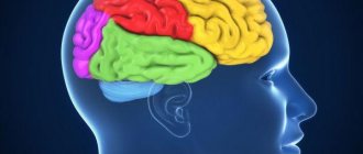
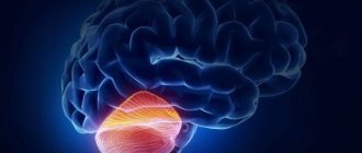
What are the left and right hemispheres responsible for?
In general, the left hemisphere of the brain is responsible for language and speech and is called the “dominant” hemisphere. The right hemisphere plays a large role in interpreting visual information and spatial processing.
For about a third of people who are left-handed, speech function may be located in the right hemisphere of the brain. Left-handers may need special testing to determine whether their speech center is on the left or right side before any surgery in that area.
Aphasia is a speech disorder that affects speech production, comprehension, reading, or writing. Occurs due to brain injury, most often from a stroke or injury. The type of aphasia depends on the area of the brain that is damaged.
Broca's area: lies in the left frontal lobe (Fig. 3). If this area is damaged, the person may have difficulty moving the tongue or facial muscles to produce speech sounds. The person can still read and understand spoken language, but has difficulty speaking and writing (i.e., forming letters and words, not writing in lines)—called Broca's aphasia.
Wernicke's area: lies in the left temporal lobe (Fig. 3). Damage to this area causes Wernicke's aphasia. A person may speak in long sentences that do not make sense, add unnecessary words, and even create new words. They can make speech sounds, but they have difficulty understanding speech and are therefore unaware of their mistakes.
Treatment of pathology
When treating a pathology, the doctor sets two goals: finding and eliminating the cause of abnormal organ enlargement and neutralizing its consequences for the newborn. In case of a mild isolated form, which is not caused by chromosomal abnormalities, the expectant mother is prescribed drug therapy: taking diuretics, vitamins, injections of drugs that prevent hypoxia and placental insufficiency.
In the postpartum period, infants are given several courses of massage aimed at relieving muscle tone, strengthening, and eliminating neurological symptoms. Monitoring by a neurologist is required in the first weeks, months and years of life. Severe forms of pathology require surgical treatment after the birth of the baby.
The prognosis for a child with a severe form and chromosomal abnormalities is unfavorable.
There are a number of anatomical features of the brain of each person. Sometimes such specificity is considered physiological; in other situations, deviations from the norm can give rise to manifestations of a pathological process.
One such condition is asymmetry of the lateral ventricles of the brain. On the one hand, such cerebral specificity is not considered a separate nosological entity, and its clinical symptoms may be absent.
However, often ventricular asymmetry may indicate the presence of a number of diseases.
The ventricles of the brain are a cerebral system of cavities communicating with each other, the subarachnoid space and the spinal canal. Their inner surface is lined with ependyma. Under this layer are the choroid plexuses, which produce cerebrospinal fluid.
The lateral (or lateral) ventricles are the most voluminous. They are localized on both sides of the midline and have pairs of anterior, posterior and lower horns that are symmetrical relative to each other.
The lateral ventricular cavities communicate with each other with the third ventricle, located between the thalami, through the foramen of Monroe. Between the cerebellum and the brain stem is the fourth ventricle.
From it, the cerebrospinal fluid enters the subarachnoid space through the foramina of Luschka (paired) and Magendie (unpaired).
An increase in the size of the lateral ventricles is formed as a result of impaired circulation of the cerebrospinal fluid. This picture emerges when:
- overproduction of cerebrospinal fluid;
- impaired adsorption of cerebrospinal fluid;
- difficulty in the outflow of cerebrospinal fluid.
- Changes in liquor dynamics due to overproduction and slower reabsorption of cerebrospinal fluid occur as a result of irritation of the ventricular choroidal plexuses and the arachnoid membrane of the brain by a pathological process (most often as a result of neuroinfection).
- Difficulty in the outflow of cerebrospinal fluid is caused by blockage of the cerebrospinal fluid pathways by neoplasms and cysts.
- The main reasons for the expansion of the lateral ventricles are:
- neuroinfections (meningitis, meningoencephalitis);
- skull injuries;
- brain tumors;
- idiopathic hydrocephalus;
- formed hematomas;
- hemorrhagic stroke;
- thrombosis of cerebral vessels;
- atypical embryonic anlage of the ventricular system.
Manifestations of asymmetry of the lateral ventricles in an adult are often absent. In this case, such a structure of cerebral structures is not considered a pathology; it is revealed as a finding during neuroimaging. Along with this, a pronounced disturbance of liquor dynamics leads to the following clinical symptoms:
- headaches;
- feeling of heaviness and fullness in the head;
- dizziness;
- nausea;
- vomiting that does not bring relief;
- anxiety-phobic syndrome;
- apathy.
Diagnostics
Enlargement of the lateral ventricles is diagnosed exclusively using instrumental methods. The required scope of procedures includes:
- neuroimaging (CT, MRI);
- echoencephaloscopy;
- electroencephalography;
- fundus examination.
In this case, only neuroimaging allows one to accurately assess the width and size of the ventricular cavities from the anterior to posterior horns, as well as analyze the state of the ventricular system as a whole. Other diagnostic methods are auxiliary and are used as additional procedures.
Embryology
Rice. 1. Schematic representation of the development of the ventricles of the brain (according to Patten): a - the stage of three brain vesicles in the 4th week of development: 1 - optic vesicle, 2 - mesocele; b - stage of five brain vesicles at the 5th week of development: 1 - vesiculae laterales telencephali, 2 - telocele, 3 - diocele, 4 - mesocele, 5 - metacele, 6 - myelocele, 7 - optic vesicle, 8 - interventricular foramen ; c - formation of the lateral vesicles of the telencephalon at the 6-10th week of development: 1 - lateral ventricle, 2 - telocele, 3 - diocele, 4 - mesocele, 5 - metacele, 6 - myelocele; d - definitive (final) structure of the ventricles: 1-3 - right lateral ventricle (1 - anterior horn, 2 - lower horn, 3 - posterior horn), 4 - fourth ventricle, 5 - cerebral aqueduct, 6 - third ventricle.
The ventricles of the brain, as well as the cavities of the spinal cord, are formed as a result of transformations of the primary cavity of the neural tube - the neural canal. The nerve canal along the spinal cord gradually narrows and turns into the central canal and the terminal ventricle. The anterior end of the neural tube expands and then dismembers, forming at the 4th week. development of three brain vesicles (Fig. 1): anterior, middle and rhomboid. At 5-6 weeks. development through the differentiation of three brain vesicles, five vesicles are formed, giving rise to five main parts of the brain: telencephalon, diencephalon, midbrain (mesencephalon), hindbrain (metencephalon), medulla oblongata (myelencephalon).
The telencephalon rapidly grows to the sides, forming two lateral bubbles - the rudiments of the cerebral hemispheres. The primary cavity of the telencephalon (telocele) gives rise to the cavities of the lateral vesicles, which represent the anlage of the lateral ventricles. At 6-7 weeks. development, the growth of the lateral bubbles occurs in the lateral and anterior directions, which leads to the formation of the anterior horn of the lateral ventricles; at 8-10 weeks. growth of the lateral vesicles in the opposite direction is observed, as a result of which the posterior and lower horns of the ventricles appear. Due to the increased growth of the temporal lobes of the brain, the inferior horns of the ventricles move laterally, downward and forward. The part of the cavity of the telencephalon, located in connection with the cavities of the lateral vesicles, turns into interventricular foramina (foramina interventricularia), which communicate the lateral ventricles with the anterior part of the third ventricle. The primary cavity of the diencephalon (diocele) narrows, maintaining a connection with the middle part of the telencephalon cavity, and gives rise to the third ventricle. The cavity of the midbrain (mesocele), which passes in front into the third ventricle, narrows very strongly even at the 7th week. turns into a narrow channel - the cerebral aqueduct (aqueductus cerebri), connecting the third ventricle with the fourth. At the same time, the cavity of the rhombencephalon, which gives rise to the hindbrain and medulla oblongata, expanding laterally, forms the fourth ventricle with its lateral recesses (recessus lat.). The vascular base of the fourth ventricle (tela chorioidea ventriculi quarti) initially almost completely closes its cavity (with the exception of the opening of the cerebral aqueduct). By the 10th week. During development, openings are formed in it and in the wall of the ventricle: one median (apertura mediana) at the lower corner of the rhomboid fossa and two paired lateral ones (aperturae lat.) at the tops of the lateral recesses. Through these openings, the fourth ventricle communicates with the subarachnoid space of the brain. The cavity of the fourth ventricle passes below into the central canal of the spinal cord.
Is it important Fenzitate or phenazepam?