Veins of the brain
The cerebral veins (vv. cerebri) are divided into superficial, forming in the cortex of the cerebral hemispheres, and deep, starting from the central parts of the cerebral hemispheres.
The following belong to the superficial veins of the brain (Fig. 416).
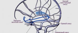
416. Veins of the brain. 1 - sinus signoideus; 2 - sinus transversus; 3 - confluence sinuum; 4 - sinus rectus; 5 - sinus sagittalis superior; 6 - vv. cerebri superiores
1. The superior veins of the cerebrum (vv. cerebri superiores) collect blood from the cortex of the dorsolateral surface of the cerebral hemispheres, forming a network of veins in the choroid. Large venous vessels are located mainly in the cortical grooves. Vv. cerebri superiores pierce the arachnoid membrane and flow into the sinus sagittalis superior.
2. The superficial middle vein of the cerebrum (v. cerebri media superficialisis) is a paired large vein that runs in the central sulcus and connects the sinus sagittalis superior and sinus cavernosus.
3. The anterior cerebral vein (v. cerebri anterior) originates on the medial surface of the cerebral hemisphere, extends to the base of the brain and connects the great cerebral vein with the inferior sagittal sinus.
4. The inferior veins of the cerebrum (vv. cerebri inferiores) originate in the basal cortex of the brain, flow into the sinus caroticus, intersigmoideus et. sphenoparietalis.
5. The basal vein (v. basalis) is formed in the area of the substantia perforata anterior, then accompanies the optic nerve tract. Having gone around the cerebral peduncles, it flows over the pineal gland into v. cerebri magna.
6. The superior veins of the cerebellum (vv. serebelli superiores) begin on the upper surface of the cerebellar hemispheres, flow into the sinus rectus and v. cerebri magna.
7. The inferior veins of the cerebellum (vv. serebelli inferiores) are located on the lower surface of the cerebellum and anastomose with the previous ones. They flow into sinus transversus and sinus petrosus inferior.
The internal veins of the cerebral hemispheres begin in the basal ganglia and white matter. They are represented by the following trunks.
1. The internal veins of the cerebrum (vv. cerebri internae) collect blood from the white matter of the cerebral hemispheres, the walls of the ventricles, the optic thalamus and the basal ganglia. In the transverse fissure of the brain near the quadrigeminal, all branches of the veins merge into the great vein of the brain (v. cerebri magna).
2. The vein of the choroid plexus (v. choroidea) is formed from the veins of the choroid plexus of the lateral ventricle, penetrating through the for. interventriculare into the central part of the lateral ventricle. It flows into v. cerebri magna.
3. The veins of the transparent septum (vv. septi pellucidi) begin in the substance of the brain, forming the anterior horn of the lateral ventricle. They flow into v. choroidea.
Great vein of the brain
The great vein of the brain (v. cerebri magna) is single, represents a short trunk 0.5-1 cm long. It is formed by the fusion of the above branches of the deep veins of the cerebral hemispheres. In the transverse sulcus of the brain above the superior colliculus, the midbrain flows into the sinus rectus.
Veins of the spongy substance of the bones of the cranial vault
Diploic veins (vv. diploicae) are located in the spongy substance of the bones of the cranial vault. They are oriented towards the large openings of the outer plate of the skull bones, through which the so-called exhaust veins (vv. emissariae) pass. The graduates anastomose with the saphenous veins of the skull, and the diploic veins with the venous sinuses. Due to the absence of valves in the veins of the spongy substance, blood flow through them is possible in two directions. The following diploic veins are distinguished.
1. The frontal diploic vein (v.diploica frontalis) is located in the scales of the frontal bone. Connects the supraorbital vein with the superior sagittal venous sinus.
2. The anterior temporal diploic vein (v.diploica temporalis anterior) is located in the parietal bone and the squama of the temporal bone. Connects the deep temporal veins and sinus sphenoparietalis, anastomoses with the frontal diploic vein.
3. The posterior temporal diploic vein (v.diploica temporalis posterior) connects the parietal outlet with the mastoid outlet and flows into the posterior auricular vein.
4. The occipital diploic vein (v. diploica occipitalis) begins in the squama of the occipital bone and flows into the occipital outlet.
Emissary veins of the skull
Emissary veins (vv. emissariae) are located in various parts of the skull.
1. The parietal emissary vein (v. emissaria parietalis) is a pair, connects the superficial temporal vein with the superior sagittal sinus.
2. The mastoid emissary vein (v. emissaria mastoidea) establishes an anastomosis between the sinus sigmoideus occipital and posterior temporal veins.
3. The condylar emissary vein (v. emissaria condylaris) connects the sinus sigmoideus with the venous plexuses of the spinal column and the deep vein of the neck.
4. The occipital emissary vein (v. emissaria occipitalis) is located on the external occipital eminence, reports vv. occipitales with the transverse sinus or the confluence of the sinuses.
Venous plexuses in the skull
The venous plexuses (plexus venosus) surround the contents of the foramen ovale, carotid and sublingual canals.
1. The venous plexus of the foramen ovale (plexus venosus foraminis ovalis) is located in the foramen ovale and connects the cavernous sinus with the pterygoid venous plexus.
2. The venous plexus of the carotid canal (plexus venosus canalis carotid) surrounds the internal carotid artery in the cranial canal of the same name, collects blood from the mucous membrane of the tympanic cavity and establishes a connection between the cavernous sinus and the pterygoid plexus.
3. The venous plexus of the hypoglossal canal (plexus venosus canalis hypoglossi) connects the occipital sinus with the sinus petrosus inferior and the internal vertebral plexus.
Veins of the orbit
From the contents of the orbit, the frontal region, and partly the upper jaw, small veins originate, which are the sources of the upper and lower ophthalmic veins, which flow into the cavernous sinus and veins of the head.
1. The nasofrontal vein (v. nasofrontalis) originates in the forehead and external nose. In the medial corner of the orbit it connects with v. angularis, representing the beginning of the facial vein.
2. Ethmoid veins (vv. ethmoidales) collect blood from the mucous membrane of the cells of the ethmoid bone, exit through the openings of the same name into the orbit.
3. The lacrimal vein (v. lacrimalis) originates in the lacrimal gland.
4. Veins of the eyelids (vv. palpebrales) and veins of the connective tissue membrane (vv. conjunctivales), vorticose veins (vv. vorticosae), ciliary veins (vv. ciliares), central retinal vein (v. centralis retinae), suprascleral veins (vv. episclerales) are formed in formations of the same name.
All of the listed orbital veins gather on the upper surface of the eyeball and merge into the superior ophthalmic vein (v. ophthalmic superior).
The superior ophthalmic vein has no valves and is characterized by a well-developed muscle layer. Initially, the vein is located in the medial corner between the superior and medial walls of the orbit, then it goes to the outer wall of the orbit, crossing the optic nerve under the superior rectus muscle of the eye. It leaves the orbit through the superior orbital fissure, flowing into the cavernous sinus.
The inferior ophthalmic vein (v. ophthalmica inferior) is formed from small veins of the lacrimal sac, medial, inferior rectus and oblique muscles of the eye. From the medial corner of the orbit, the vein passes to its lower wall and accompanies m. rectus inferior. In the upper part of the orbit, the vein divides into two branches: one of them flows into the sinus cavernosus or the superior ophthalmic vein, the other, passing through the inferior orbital fissure, connects with the deep facial vein. Anastomoses with the venous pterygoid plexus and the infraorbital vein. There are no valves in the system of these veins, so blood can pass both from the veins of the face to the cavernous sinus and back. This creates conditions for inflammation, when it is possible for infection to spread from the upper jaw, orbit and nasal cavity into the venous sinuses of the dura mater.
Veins of the meninges
Veins of the dura mater (vv. meningeae) originate in the dura mater and accompany the arteries. Veins from the shell of the calvarium flow into the sinus sagittalis superior, from the shell of the base of the skull into the venous sinuses of the base of the skull. Among the veins of the hard shell of the base of the skull, the middle vein (v. meningea media) is distinguished, accompanying a. meningea media. The vein flows into the sinus sphenoidalis and anastomoses in the area of the foramen ovale with the plexus of the same name and the pterygoid plexus.
Description of the emissary
The emissary veins are located in the openings of the skull, connecting the capillaries of the cavity and the outer integument of the brain center.
Types of emissary branches:
- parietal, passing through the parietal foramen in the cerebellar region;
- occipital, surrounding the external occipital protuberance;
- condylar, located in the canaliculi of the occipital bone;
- mastoid, passing through the mastoid outlet of the temporal bone.
The emissary group includes not only tubes, but also plexuses - the foramen ovale, carotid and sublingual reflex.
Diagnostics
Accurate and objective diagnostic information plays an important role in choosing a treatment method, especially surgical, the place and time of its implementation. The therapeutic and diagnostic capabilities of modern neurosurgery are increasing rapidly thanks to new diagnostic equipment. At the same time, traditional diagnostic methods also remain relevant.
Diagnostic lumbar puncture is an informative method for determining subarachnoid hemorrhage, which often occurs when an intracranial aneurysm ruptures. Using echoencephalography (EchoEG), when an aneurysm ruptures with the formation of a hematoma, the side of its location is specified based on the pronounced (more than 4-6 mm) displacement of the M-echo.
Doppler ultrasound allows you to non-invasively determine the linear speed and direction of blood flow, the degree and level of circulatory disorders in the main arteries. Duplex (double) ultrasound scanning makes it possible to simultaneously determine changes in blood flow and obtain an image of the vessel itself, identify carotid artery stenosis (less than 50%), as well as the location and structure of the atherosclerotic plaque.
Changes in cerebral blood flow and cerebral vascular tone can be judged from the data of rheoencephalography, impedance rheoplethysmography and telethermography.
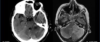
The study of regional cerebral blood flow using the clearance of a radioactive isotope (rXe) allows us to determine the degree of reduction in cerebral circulation. Computed tomography (CT) allows one to differentiate focal cerebral ischemia from hemorrhage. The size and location of the intracranial hematoma and the focus of cerebral infarction, as well as the state of the surrounding brain space, are clearly determined. Magnetic resonance imaging (MRI) makes it possible, even without the use of a contrast agent, to evaluate not only anatomical structures, but also the level of energy, enzymatic and metabolic processes in the brain. Non-invasive nuclear magnetic resonance angiography has even greater diagnostic capabilities, which allows you to obtain angiograms in any projection and identify not only aneurysms, but also atherosclerotic plaques in the arteries.
Recently, nuclear magnetic resonance spectroscopy has been used, which makes it possible to draw conclusions about the dynamics of focal brain lesions both in areas of irreversible changes and in the “ischemic penumbra” zone.
Treatment in each case is selected individually, depending on the cause and location of the damage.
How to do an MRI of cerebral veins
The classic MR venography protocol is a cycle of several sequential actions:
- the patient is placed on a horizontal platform in the “supine” position, a special RF coil is installed in the head area, which amplifies the tomograph signal;
- the moving part with the person is directed into a round magnet;
- Doctors go into the next room to monitor the operation of the devices on the computer.
You need to be prepared that during operation, medical equipment may make sounds in the form of pops, clicks or knocks. To make the process more comfortable, medical patients are offered to wear headphones and undergo the screening accompanied by pleasant musical accompaniment.
Etiology of malformation
The reasons for the development of the disease are poorly understood. But factors include:
- congenital pathologies established during the development of the embryo;
- antenatal and birth injuries;
- sclerotic and atherosclerotic lesions of blood vessels.
Contributing factors include intrauterine infections, exposure to radiation, and bad habits of the mother. Sexual and genetic predisposition was also established.
Main services of Dr. Zavalishin’s clinic:
- consultation with a neurosurgeon
- treatment of spinal hernia
- brain surgery
- spine surgery
How does vascular malformation manifest?
According to the clinical course, all malformations are divided into hemorrhagic and torpid. Hemorrhagic course is the most common pathology (approximately 70% of all cases). It is characterized by sharply increased pressure and the formation of a vascular tangle of small diameter.
Torpid flow is characterized by the formation of a pathological node of medium and large sizes. The favorite location is the structural units of the cerebral cortex. The formation receives its blood supply thanks to the branches of the cerebral arteries, and the clinical symptoms resemble the course of the disease in patients with organic brain damage.
In the initial stages, the disease practically does not manifest itself and is discovered completely by accident - during the examination of the patient for another pathology. Only 12% of all patients experience characteristic neurological symptoms, which may be a reason to consult a doctor. It manifests itself in different ways - depending on the location of the pathological plexus. The main symptoms include:
- weakness in the muscles and throughout the body;
- dizziness;
- dysarthria, as well as impaired coordination and decreased vision;
- decreased ability to remember information and impaired consciousness.
A number of patients experience convulsive syndrome, which can be either partial or total. It is accompanied by various forms of impairment of consciousness. Headaches also occur, varying in intensity and frequency. Very often the localization of the pain point does not correspond to the localization of the vascular tangle.
According to statistics, approximately 2-4% of all cases of vascular malformation are accompanied by cerebral hemorrhage. In terms of wall rupture, small blood vessels are much more dangerous. The deeper the vascular tangle is located, the more pronounced the neurological symptoms will be.
Characteristic for children is head size that is disproportionate to the body, as well as the development of congestive cardiac and acute respiratory pathologies against the background of vascular pathology.
Why are malformations dangerous?
Most often, cerebral vascular malformations are the cause of subarachnoid hemorrhage.
It is characterized by:
- sudden, severe headache;
- feeling of pulsation in the occipital region;
- vomiting that does not bring relief;
- disturbance of consciousness;
- positive meningeal symptoms;
- convulsions.
Without timely medical care, the patient's condition can deteriorate significantly, even leading to death.
Cerebral infarction and paralysis may also occur.


