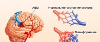
The spread of head tumors among the population is growing alarmingly every year. This worries doctors in all countries. These pathologies include angioma or benign brain tumor. It consists mainly of cells of blood and lymphatic vessels, abnormally intertwined with each other.
An angioma looks like a slightly elevated neoplasm of a dark cherry or bluish color. Despite the fact that the pathology is benign in origin, it is considered very dangerous and can damage the nervous system. Among the victims, the most common are children, mainly newborn girls.
What is cerebral venous angioma
The disease is not aggressive, but its presence should not be ignored. The interweaving of blood vessels are fused balls that expand over time, provoking the development of inflammatory processes in the brain.
The more the brain is affected, the greater the health risk caused by the presence of the formation. This pathology is dangerous for the body due to the rapid growth of angioma, which, affecting the tissues of nearby organs, leads to disruptions in the functioning of the entire body.
It is the brain that is responsible for the quality functioning of all systems. Pathological changes occur and, if not treated in a timely manner, destruction occurs. A patient with a tumor is at high risk of cerebral hemorrhage, which can be fatal.
At the first symptoms of the disease, it is necessary to begin treatment immediately. Therapy in the early stages of tumor development has a favorable outcome. At an advanced stage of the disease, drug and surgical interventions will not give positive results.
Skin angioma
Almost every person has a certain number of new growths on their skin - moles. They differ in color, shape and size. Red moles are angiomas. Most often they are congenital, but can also appear during life.
Causes of red moles
There are several reasons why skin angioma develops. Red moles in newborns appear as a result of maternal illnesses during pregnancy: ARVI, influenza, pyelonephritis.
The provoking factors of acquired skin angioma are:
- hormonal disorders;
- prolonged exposure to UV rays;
- skin injuries.
In old age, skin angioma develops due to the expansion and proliferation of blood vessels. People with fair skin and hair are most susceptible to the disease.
Symptoms of skin angioma
Skin angioma appears as reddish or purplish-blue spots; they can be located separately from each other or form clusters of several moles. New growths have clearly defined edges, usually rise above the surface of the skin, and are painless. When you press on the angioma, it turns pale as it is filled with blood. When the pressure is removed, it returns to its normal color. Small angiomas do not cause any discomfort, but require constant monitoring as they can develop into malignant tumors. This is especially true for polymorphic angiomas.
Diagnosis of skin angioma
A visual examination allows a diagnosis of skin angioma to be made. Characteristic features of the neoplasm are a typical color and disappearance when pressed. If a skin angioma is detected, it is recommended to undergo a full diagnosis of the body to exclude vascular neoplasms in other organs: magnetic resonance and computed tomography, radiography and angiography.
Treatment of vascular skin tumors
The need and tactics of treatment are selected depending on the location of vascular moles, their size and the dynamics of the disease. Single small angiomas that do not cause discomfort and are not prone to proliferation do not require treatment.
Doctor intervention is required in the following situations:
- the mole is located on the neck, head or in places where there is a high risk of damage;
- the neoplasm increases in size and is often injured;
- there is a risk of the tumor developing into a malignant one.
Treatment is selected depending on the location of the tumor. Surgery is performed when the angioma affects the deep layers of the skin and subcutaneous tissue. Superficial tumors are removed using a laser. Sometimes infrared coagulation, cauterization with carbon dioxide and cryodestruction (exposure to low temperatures) are used to remove angioma. These methods are only suitable for small flat tumors located on the surface of the skin.
Prevention of skin angioma
It is impossible to prevent the appearance of congenital angiomas. To detect angioma at an early stage, conduct periodic examinations of the entire body. If you discover new moles, be sure to consult a dermatologist.
In recent years, there has been an increase in skin diseases, including angiomas. In pursuit of a beautiful tan, girls often forget about basic safety rules. Prolonged exposure to the sun and frequent visits to the solarium are dangerous to your health!
Kinds
Venous angioma is an enlarged venous column of vessels collected in a tangle. Depending on the location of the formations, the disease is divided into several types.
Frontal lobe angioma
She is responsible for initiative, correct decision-making, responsibility and showing interest. Therefore, when education is localized in this area, the character of a person changes greatly.
He becomes apathetic, a change in self-esteem occurs, speech is impaired, and inadequacy appears in his behavior and actions.
It is difficult for a person to think logically; he often commits unconscious actions. The patient cannot take responsibility and is unable to independently solve various problems.
Performance decreases, the emotional state is unstable, and over time the person suffers from depression. The patient does not feel cold or heat when touched.
Cerebellar angioma
If there is a tumor in this area of the brain, functional disturbances occur in the body. The patient's muscle movements are not coordinated, he cannot perform smooth movements, so all actions are abrupt. A person’s motor ability is greatly slowed down, all actions are performed in the form of jerks.
There are disturbances in speech, there are serious problems when writing words and sentences. Breathing is impaired, resulting in problems with the respiratory system and skeletal muscles.
It is impossible to cure the disease using medications. With this form of angioma, surgical intervention is indicated.
Angioma of the left hemisphere
There is inconsistency in the work of the muscles of the arms and legs. The patient has speech impairment and changes in gait.
Cerebellar venous angioma
Brain angioma is a benign neoplasm consisting of a plexus of small blood vessels or lymphatic spaces.
Taste preferences change completely. Vision decreases, nystagmus of the extraocular muscles is observed.
Due to impaired blood flow, a person is susceptible to epileptic seizures and convulsions. At an advanced stage of the disease, partial paralysis may occur.
Angioma of the right hemisphere
With this type of disease, the patient experiences a movement disorder. It becomes slow and sharp. Speech becomes slow, writing style changes. A person is bothered by tremors of the arms and legs.
Angioma of the parietal lobe
The patient's coordination of movements is impaired, tactile sensations and pain threshold change, and speech abilities may deteriorate.
Cavernous angiomas on MRI
Cavernous angiomas represent about 1% of all intracranial vascular lesions and 15% of cerebrovascular malformations. With the development and introduction of MRI, cavernous angiomas have become the most commonly detected vascular malformations of the brain. In early studies on autopsy material, the frequency of their occurrence was 0.02-0.53%. Using MRI, the incidence of formations similar to cavernous hemangiomas was 0.39-0.9%, and the detection of previously unidentified asymptomatic formations using MRI increased their incidence to 0.45-0.9%.
Get an MRI of the brain in St. Petersburg
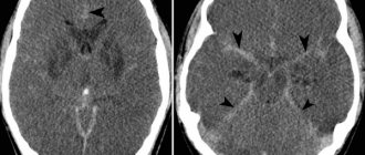
On MRI, parenchymal cavernous angiomas are represented by a characteristic “popcorn”-type formation, clearly defined, with a smooth border. The internal part is represented by multiple foci of signal of varying intensity, which correspond to hemorrhages at different stages of resolution.
MRI signs of cavernous angioma. Large cavernous angiomas of the right frontal lobe and left occipital lobe on T1-weighted axial section. These two heterogeneous space-occupying lesions have a central mesh structure with alternating areas of high and low signal intensity, surrounded by a hypointense rim of hemosiderin.
A fresh hematoma containing deoxyhemoglobin is isointense on T1-weighted images and significantly hypointense on T2-weighted images. A subacute hematoma containing extracellular methemoglobin is hyperintense on both T1- and T2-weighted images due to the paramagnetic effect exerted by methemoglobin.
Intermediate fibrous elements are characterized by a weakly hypointense signal on T1- and T2-weighted images, since they contain calcifications and hemosiderin. The heterogeneous interior of the mass is surrounded by a hemosiderin rim, which has low intensity on T1-weighted images. The hypointensity of this rim becomes more pronounced, resembling a halo, on T2-weighted and gradient-echo images due to the higher sensitivity of these sequences to changes in the magnetic field.
Axial gradient-echo MR images provide better visualization of large cavernous angiomas in the right frontal and left occipital lobes. The hemosiderin rim appears as a halo due to the increased magnetic susceptibility of hemosiderin.
Smaller cavernomas appear as low-intensity nodules on T1- and T2-weighted images.
Small lesions are better visualized on gradient echo images due to the increased sensitivity to changes in the magnetic field that is inherent in such pulse sequences. It has also been shown that in sequential gradient echo images, small punctate formations are better visualized with longer echo times; these data suggest that such formations contain paramagnetic substances.
Gradient-echo MR imaging shows multiple bilateral small, punctate and round, low-intensity lesions in the periventricular and subcortical white matter. The largest lesion is visualized in the periventricular white matter of the frontal lobe anterior to the anterior (frontal) horn of the left lateral ventricle near the genu of the corpus callosum. Multiple smaller lesions are visible anterior and posterior to it.
On time-of-flight angiography images, methemoglobin in the center of a cavernous malformation may resemble moving blood. However, on a subsequent phase-contrast MR angiogram obtained with a low blood flow speed setting during encoding (10-20 cm/s), blood flow or pathological vascularization is not visualized, which makes it possible to exclude vascular lesions.
Typically, cavernous angiomas do not have a bulking effect on adjacent tissue or cause edema, and they do not have a feeding artery or draining vein unless they are associated with other similar vascular malformations. Cavernous angiomas are often associated with venous malformations, which are characterized by the presence of a draining vein. In such mixed cases, standard angiography may be useful.
T2-weighted image of a cavernoma of the pons.
Cavernous malformations detected on MRI include other occult vascular malformations (AVM/aneurysm thrombosis, capillary telangiectasia), hemorrhage in a primary or secondary tumor (metastasis of melanoma, choriocarcinoma, thyroid or kidney cancer), amyloid angiopathy, treated or primary infection (toxoplasmosis or cysticercosis), multiple hemorrhages associated with damage to the blood system (disseminated intravascular coagulation, leukemia), as well as the consequences of diffuse axonal damage.
Symptoms
In addition to the differences between different types of angiomas, there are common symptoms that are characteristic of all types of cerebral venous angioma:
- frequent fainting;
- dizziness;
- paralysis;
- noise in the head;
- tremor of hands and feet;
- disorders ;
- headache ;
- imbalance and coordination of movements ;
- disturbances in the functioning of the cardiovascular system;
- epilepsy attacks
- problems ;
- convulsions;
- disorders ;
- blurred vision;
- decreased mental activity;
- noise in the head;
- change in taste preferences;
- impairment ;
- increased fatigue;
- nausea and vomiting;
- loss of ability to work.
Diagnostics
To perform an accurate analysis, a specialist carries out diagnostic measures.
First of all, laboratory tests are prescribed. The patient must undergo a general and biochemical blood test, as well as a general and biochemical urine test.
If the norm deviates, the patient is then prescribed an ultrasound examination to confirm the diagnosis. Magnetic resonance imaging will determine the presence of formations, identify the location and size of the angioma.
Why can our articles be trusted?
We make health information clear, accessible and relevant.
- All articles are checked by practicing doctors.
- We take scientific literature and the latest research as a basis.
- We publish detailed articles that answer all questions.
It is also possible to perform a computed tomography, angiography or x-ray. Only after all diagnostic procedures have been completed can treatment be prescribed.
Kidney angioma
Kidney angioma develops from the blood vessels that supply the organ, is benign, and has a low tendency to grow. However, renal angiomas require observation, since there is still a minimal risk of the tumor degenerating into a malignant one.
Causes of kidney angioma
As is the case with angiomas of other organs, the factors causing the appearance of vascular neoplasms of the kidneys are not fully understood.
Among the main causes of kidney angioma, researchers identify:
- genetic predisposition;
- congenital developmental anomalies;
- hormonal disorders;
- injuries;
- infections of the genitourinary system.
Many scientists are inclined to believe that most vascular tumors appear during fetal development or in early childhood.
Symptoms of vascular kidney tumors
When small in size, renal angiomas do not reveal themselves in any way. Symptoms of the disease occur only with large tumors.
Most often, patients with renal angioma complain of:
- pain in the lumbar region;
- urinary disorders;
- increased blood pressure;
- weakness and fatigue.
Similar symptoms are typical for other kidney diseases, so additional testing is required to make a diagnosis.
Diagnosis of kidney angioma
The following research methods allow diagnosing renal angioma:
- magnetic resonance imaging (MRI);
- computed tomography (CT);
- ultrasound examination (ultrasound);
- angiography;
- intravenous urography.
All of these methods are informative and help to accurately determine the location and size of the angioma. Usually one or two tests are enough to make a diagnosis.
Treatment of kidney angioma
The choice of treatment method for renal angioma is determined depending on the prevalence of the tumor process and the growth rate of the tumor. Small isolated angiomas require only observation. In most cases, they do not grow or increase significantly in size and therefore do not require treatment. In rare cases, when an angioma grows rapidly and the patient’s condition worsens, medical intervention cannot be avoided.
Currently, modern methods are used to treat renal angioma: embolization and cryoblation. In the first case, doctors block the vessels feeding the tumor; in the second, they act on the tumor cells with low temperatures. Both methods are highly effective and can reduce or completely destroy the tumor. Surgery to remove a kidney angioma is performed as a last resort, only for very large tumors with the threat of rupture and internal bleeding.
Note! In the vast majority of cases, the prognosis for renal angioma is favorable. However, even after removal of the tumor, relapses are possible, so patients with this disease are recommended to undergo kidney examinations at least once a year.
Treatment
Treatment of education includes taking medications, using traditional medicine, and in advanced cases, surgery.
Self-medication is dangerous with complications!
Attention
Despite the fact that our articles are based on trusted sources and have been tested by practicing doctors, the same symptoms can be signs of different diseases, and the disease may not proceed according to the textbook.
Pros of seeing a doctor:
- Only a specialist will prescribe suitable medications.
- Recovery will be easier and faster.
- The doctor will monitor the course of the disease and help avoid complications.
find a doctor
Do not try to treat yourself - consult a specialist.
Drug treatment
It is impossible to cure venous angioma by taking medications. This type of therapy is used only to alleviate the patient’s condition.
Drug therapy is prescribed if there is no threat of hemorrhage. Vascular medications are indicated to improve blood circulation.
Sedatives occupy an important place.
All drugs will only alleviate the patient’s condition, but will not contribute to recovery. A benign formation can only be removed surgically.
Surgical intervention
There are several methods of surgical treatment of the disease.
Treatment of angioma
Angioma is a group of benign neoplasms in the formation of which blood and lymphatic vessels take part.
In severe cases of the disease, surgical removal of the accumulation of blood vessels is performed. A plastic spiral is inserted into the patient's lumen.
If the sclerotherapy method is chosen, a special substance is injected using a special catheter, which clogs the lumens of the vascular bundle and promotes scarring of the tumor. The operation is painful and lengthy.
It is possible to apply a superficial radiation beam to the tumor using a gamma knife. With its help, blood vessels are blocked. Education stops growing and developing, no longer presenting a danger to human life and health.
Liquid embolism can be injected into the collection of vessels, which easily penetrates into the smallest cavities and prevents the growth of angioma. Over time, the area where the tumor grew is replaced by connective tissue.
Diet
Diet plays a big role in treating the disease. The patient should exclude fatty and fried foods from his diet. Minimize the consumption of salt and sugar, coffee, chocolate and baked goods.
Causes of angiomas
Angiomas are minor skin neoplasms consisting of epithelial tissue and blood vessels.
Avoid eating butter, pork, full-fat milk, liver and kidneys. It is necessary to eat vegetables and fruits, fish and seafood, cereals, dried fruits, and herbs.
Brain angioma is a serious disease. Therefore, it is strictly forbidden to self-medicate.
Tumor formation and its types
With angioma, the cells of the vascular body divide, increasing the growth of venous tissue. A benign process occurs in vascular and lymphoid tissues:
- On the inside;
- On the outer surface;
- Simultaneously in both types of epithelium.
The neoplasm is divided by type into two classifications:
- In appearance;
- By source of education.
Division of neoplasms by appearance
- Flattened - a spot of varying sizes with a reddish tint. The main place of distribution is in the neck and face.
- Lumpy - an irregularly shaped speck of dark color. Affects the epidermis and mucous membranes. Inherent in childhood, resolves after eight years.
- Cavernous, or deep, type - growth is directed deep into the layers of the skin. In superficial places, the color varies from reddish to flesh-colored. The preferred location is internal organs.
Venous angioma
Division by origin
- Normal – the tissue of the vascular channels for blood flow grows.
- Capillary, vascular - formed by the plexus of capillary vessels rising above the dermis layer.
- Pustular or inflamed - mainly formed on the mucous membrane of the inner side of the cheek, tongue or gum.
- Lymphangiomas.
- Glomangiomas - formed from arterioles, usually under the nails of the upper and lower extremities.
- Telangiectasia - small blood vessels on the skin become tortuous, tangled, and red. They are clearly visible through the skin.
- Cobweb-like - looks like a cobweb. The color depends on the pressure in the circulatory system.
- Bacterial – bacterial infection caused by microorganisms of a certain type.
- The cavernous is the main place of formation of a blood vessel; it expands and does not narrow, slowing down the flow of blood. The appearance of new cells increases the risk of bleeding due to abnormal connection with the rest. The main localization is internal organs.
Can it develop into cancer?
Venous angioma of the brain is a benign formation. Therefore, it does not degenerate into a cancerous tumor.
Over time, it can lead to negative consequences and threaten human life. Therefore, it is important to consult a doctor in a timely manner for diagnosis and adequate treatment.
If the tumor does not manifest itself in any way, the person does not have behavioral disturbances or deterioration in health, you can live with this type of disease until old age. But in most cases, when diagnosing a tumor, surgical intervention is indicated.
Forecast
An intact angioma may not be detected throughout life and may not cause serious trouble to the patient.
But if a hemorrhagic stroke or vasospasm occurs, the person may become disabled for life or die. Often, rupture of the vascular bundle leads to irreversible processes in brain activity.
Depending on the patient’s age, his general health, and the presence of vascular diseases, a prognosis for the course of the disease is made. A properly prescribed treatment regimen and timely surgical intervention will allow the patient to lead a healthy and fulfilling life for a long time.
Prevention
There are no preventive measures to prevent the development of cerebral venous angioma. Once diagnosed, a person should monitor their health carefully and regularly.
It is important to constantly monitor your blood pressure, not self-medicate and take medications only under the supervision of a doctor. Taking medications without consultation with a specialist can cause hemorrhage.
Women should take oral contraceptives with caution. If you have education, you should limit physical activity. It is important to follow a diet, get proper rest, give up bad habits and monitor your weight.
Compliance with all doctor’s recommendations, a healthy lifestyle, and timely removal of the angioma will allow a person to feel healthy and lead a full life.