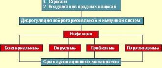
Diencephalic syndrome (hypothalamic) is a complex of endocrine, metabolic, and autonomic disorders.
During these disorders, damage to the hypothalamic region of the diencephalon is observed. In most cases, these disorders occur due to dysfunction of the hypothalamus.
Diencephalic syndrome is characterized by:
- change in body weight, in most cases it increases;
- the appearance of headaches;
- mood problems, frequent changes;
- the appearance of hypertension;
- problems with the menstrual cycle;
- increased appetite;
- a feeling of increased thirst;
- Sometimes sex drive increases and decreases.
This disorder often occurs in endocrinological, gynecological, and neuralgic practice, and during diagnosis difficulties arise that are associated with the variety of symptoms and varieties of the syndrome.
Diencephalic syndrome in most cases occurs in adolescents aged 13 to 15 years, as well as during reproductive age from 31 to 40 years. During reproductive age, it mainly predominates in women - from 12.5% to 17.5%.
The disease leads to serious reproductive health problems for many women. The development of endocrine infertility, the appearance of polycystic ovary syndrome, as well as various obstetric and perinatal pathologies are observed.
Causes and provoking factors
Disturbances in the functioning of the hypothalamus can occur due to a number of reasons and factors:
- the presence of tumors in the brain , which lead to compression of the hypothalamic region;
- traumatic brain injuries , during which direct damage to the hypothalamus occurs;
- state of neurointoxication - substance abuse, drug addiction, alcoholism, harm from industrial activities, the influence of environmental harmful components;
- various disorders of a vascular nature - stroke, osteochondrosis;
- neuroinfections of viral and bacterial types - influenza, malaria, chronic tonsillitis;
- factors with a psychogenic nature - the manifestation of stressful and shock situations, excessive mental stress;
- the period of pregnancy and hormonal changes that are associated with this period;
- chronic diseases that manifest themselves together with elements of the vegetative type - bronchial asthma, hypertension, ulcerative lesions of the stomach and duodenum, obesity.
Dysfunction of midline brain structures according to EEG
The concept of “midline brain structures” in electroencephalography combines the formations of the medulla oblongata, pons, midbrain, thalamus and hypothalamus, as well as in some cases some sections of the mediobasal formations included in the so-called limbic system (hippocampus, amygdala, orbital cortex, anterior parts of the cingulate gyrus ), and the transverse commissures of the brain, uniting these formations, located in the two hemispheres. There are a number of practical and theoretical justifications for including the listed entities into a unified system. As numerous experimental and clinical studies show, the activity of these formations is characterized by a high degree of interaction between homologous nuclei and formations of the two hemispheres, as well as systems located vertically.
Thus, there is numerous evidence that the modulating influence of limbic formations on the level of functional activity of the brain, the function of attention and the regulation of affective tone is carried out through the systems of the ascending reticular formation of the midbrain. The somnogenic mechanisms of the forebrain also realize their action indirectly through the caudal deactivating and thalamic synchronizing mechanism of the reticular formation1. Experimental studies and EEG recordings in patients through long-term implanted electrodes have also shown bilateral involvement in the pathological activity of the mediobasal limbic systems when the dominant pathological source of impulse is localized in one of the hemispheres, and most often this secondary bilateral synchronization and generalization of pathological activity is realized through the reticular stem and thalamic mechanisms 23. All this information is of great importance for understanding the origin of changes in the EEG with damage to the midline structures of the brain.
These brain structures can be involved in the pathological process in certain types of encephalitis that selectively affect the brainstem, in circulatory disorders in the system of vertebrobasilar arteries that supply the brainstem, in tumors of both the brainstem itself and adjacent formations (cerebellum, base of the skull, pineal gland) , pituitary gland)4.
Rice. 1. EEG of a 34-year-old patient with a brainstem tumor
slowing down the main rhythm to 7-8 Hz. Bilaterally synchronous bursts of 4 Hz θ waves in all leads.
Due to the diffuse and symmetrical projections of the midline brain structures onto the cortex, the changes in electrical activity that occur when they are involved in the pathological process are diffuse and bilaterally synchronous. The most typical sign of damage to the midline structures of the brain is generalized bilateral synchronous θ- and δ-waves. Depending on the severity of the lesion, they can be more or less constant, or occur periodically or in outbreaks (Fig. 1). In this case, the pathological activity is more regular and symmetrical, the lower in the trunk the pathological focus is localized.
Obviously, this is due to less differentiation of the influences of the lower brainstem (rhombencephalic and mesencephalic) parts of the reticular formation on brain activity than the influences of higher lying thalamic and limbic mechanisms56.
Rice. 2. EEG of a 66-year-old patient with a tumor of the posterior cranial fossa
Bilaterally synchronous generalized bursts of a-waves with an amplitude of up to 200 µV in the occipital regions.
In some cases, damage to the midline structures of the brain can manifest itself as bilaterally synchronous generalized bursts of high-amplitude (above 100-120 μV) α-waves (Fig. 2) or β-oscillations with an amplitude of more than 30 μV (Fig. 3).
Rice. 3. EEG in case of cerebrovascular accident in the basilar artery basin
Bilaterally synchronous bursts of p-like oscillations with an amplitude of up to 80 μV.
With damage to the lower brain stem of traumatic, desontogenetic and vascular origin, apparently, due to dysfunction of reticular synchronizing mechanisms, changes in the type of desynchronization can be observed on the EEG78. In the absence of an α rhythm, low-amplitude (<20 μV), polymorphic, predominantly high-frequency activity is recorded in all leads. Low-amplitude EEGs are a normal variant. In this regard, they can only be interpreted as a manifestation of brainstem damage if the clinic is taken into account. The EEG conclusion in this case should be conditional; categorical formulations are unacceptable (Fig. 4).
Rice. 4. EEG for circulatory failure syndrome in the vertebrobasilar system
There is no a-rhythm on the EEG. All leads record activity with a frequency of 13-25 Hz and an amplitude not exceeding 25 μV. Low-amplitude EEG is a normal variant. In the context of clinical data, a decrease in EEG amplitude with a predominance of fast rhythms may indicate dysfunction of brainstem structures.
As a rule, the EEG does not allow us to speak with certainty about the level of damage, since the nature of its changes depends on the specific involvement of synchronizing or desynchronizing mechanisms, localized at all levels, from the bulbar to the frontal limbic cortex, on the nature of this involvement (exciting or destructive), from neurodynamic changes in the interaction of multiple brain nonspecific systems in pathology and, finally, most importantly, from the massiveness of damage to brain structures. However, the experience of many years of diagnostic use of EEG in many laboratories around the world, in some cases verified by special model experimental studies on animals, allows us to outline some general patterns of relationships between certain types of lesions and certain levels of brainstem-median structures.
Brainstem lesion
In this context, by brainstem we mean the international definition, which includes sections ranging from the medulla oblongata to the midbrain inclusive. Considering the limited volume of this formation, most pathological processes always involve, to one degree or another, several of its levels or influence them, therefore it is practically impossible to narrowly isolate changes characteristic of limited sections. It seems reasonable to at least analyze in more detail the data based on cases where the level of the lesion was accurately verified by imaging methods (CT, NMRI) and on autopsy. First, we will analyze the data for ischemic disorders, since they do not cause dislocation and liquorodynamic effects at a distance, which makes it possible to associate the observed EEG changes with the affected structure.
Thrombosis of the basilar artery causes infarctions mainly of the medulla oblongata and the pons; the midbrain and higher levels are affected to a lesser extent, since compensation there occurs through the circle of Willis. Thus, these disorders can be considered as a model of predominant damage to the lower brainstem level (medulla oblongata and pons). They are usually not accompanied by the appearance of slow oscillations in the EEG. In half of the cases, the a-rhythm is preserved with normal reactions to afferent stimulation; in some cases, low-amplitude EEG is observed. A number of observations have described chronic insomnia, which is adequate to the idea of the role of the bulbo-pontine synchronizing reticular formation in the organization of slow-wave sleep. Due to damage at the pontine level to structures associated with the REM stage of sleep, disturbances in the form of its absence, reduction in duration, or dissociation of its components may be observed. Slow oscillations are observed with the addition of ischemic softening at a higher mesencephalic and (or) diencephalic level910111213.
When predominantly affected at the mesencephalic level, generalized bilateral synchronous slow waves or a beta coma pattern with a sleep pattern are observed. With limited mesencephalic destruction, slow-wave activity alternates with periods of normal alpha rhythm, and changes in stages with the presence of REM sleep are noted in the sleep pattern. This is obviously due to the preservation of the pontine mechanism of REM sleep and thalamic nonspecific systems for generating a-activity14.
With massive destructive bilateral lesions of the brainstem including the midbrain, a picture of α-coma occurs. It is assumed that in these cases, the preservation of α-activity is due to the intactness of the thalamo-cortical systems of its generation, and the total destruction of lower-brain synchronizing and mesencephalic desynchronizing mechanisms with the simultaneous switching off of specific and nonspecific systems of ascending activation determines the autonomy and unresponsiveness of the EEG15.
It should be noted that the data presented above relate to irreversible destructive, often vital, disorders. In cases where the disorders manifest themselves mainly as clinical symptoms without gross morphological disturbances of the stem structures, EEG changes can be milder and have a transient nature, according to the phase of the pathological process.
Here it is appropriate to discuss the opinion, widespread in Russian literature and practice, that lesions of the caudal sections of the trunk lead to the appearance of bilaterally synchronous slow or hypersynchronous waves in the posterior sections, and lesions of the rostral sections of the trunk - in the anterior sections1617. Analyzing the quoted messages, the following conclusions can be drawn:
- observations concern the children's age group;
- in the above cases, occlusive hydrocephalus of the third ventricle of the brain occurs;
- In not a single case are there pathohistologically verified indications of damage to specific stem structures, and in a number of the reported observations there is no adequate noso-syndromic diagnosis;
- analysis of the reported EEGs suggests that slow activity in the posterior leads fully corresponds to the characteristics of the “slow α-variant” of the EEG.
Thus, it is obvious that the discussed EEG changes cannot be interpreted specifically as a manifestation of the pathology of the lower brainstem. In cases of occlusive lesions at the level of the posterior cranial fossa, they are a symptom at a distance - a consequence of dysfunction of the thalamic nuclei surrounding the third ventricle and associated with the generation of the α rhythm, as a result of mechanical hydrocephalic and hyperhydration effects18. In childhood and adolescence, the slow αa variant is much more common than in adults and reflects a significant variation in the rate of maturation of normal midline structures19. In conditions of hypertensive hydrocephalus of the third ventricle, which occurs in the publications cited above, a delay in the normal increase in the frequency of the dominant rhythm is a natural consequence of dysfunction of the diencephalic, i.e. rostral midline brain structures, and not a direct result of unproven damage to the lower brainstem levels. Thus, the idea of a predominant predominance of bilateral synchronous oscillations in the anterior sections with rostral lesions, and in the posterior sections with caudal lesions of the trunk, seems unfounded, and diagnostic conclusions based on this criterion are arbitrary and inadequate.
To summarize, we can say that EEG changes with damage to brain stem structures are symmetrical in nature and, with the exception of disturbances such as desynchronization and, accordingly, a decrease in amplitude, are manifested by high-amplitude bilaterally synchronous a-waves, bilaterally synchronous slow waves or bursts of high-amplitude oscillations. In this case, the higher the lesion is localized, the greater the probability of the appearance of slow waves, the amplitude and degree of frequency slowdown:
- lower brainstem lesions are most often manifested by low-amplitude EEG or high-amplitude α-oscillations,
- lesions of the middle level of the trunk - θ-waves,
- upper trunk (midbrain and mesodiencephalic junction) - δ waves. The lower the level of damage, the higher the regularity and rhythm of activity.
Damage to the medial structures of the hemispheres
rice.
1. EEG of a 39-year-old patient with a craniopharyngioma compressing the hypothalamic regions. Bilaterally synchronous generalized δ waves.
Bilateral or medially localized damage to the hypothalamic structures leads to the appearance of bilaterally synchronous bursts or constant bilaterally synchronous slow oscillations in the EEG. Most often these are δ-band waves (Fig. 1). Similar changes are observed with bilateral lesions of the thalamus (Szirmai I. et al., 1977).
Rice. 2. EEG of a 38-year-old patient with a tumor of the mediobasal regions of the left hemisphere of the brain involving the thalamic nuclei
Sopor. Generalized δ-waves (frequency 1-3 Hz, amplitude up to 200 μV), occasionally predominant in amplitude in the left hemisphere.
Lateralized lesions of the thalamus typically produce bilaterally synchronous widespread δ waves with amplitude predominance on the affected side (see Fig. 2). Sometimes gross destructive lateralized thalamic lesions give a picture of symmetrical bilaterally synchronous or even slow oscillations predominant on the intact side. This is due to the fact that due to powerful transverse adhesions that connect both thalami essentially into a single rhythmogenic structure, a single pathological pattern of activity is formed that unites both thalami. However, due to damage to one of the visual thalamus by a destructive process and disruption of its connection with the cortex of the corresponding hemisphere, pathological activity on the affected side turns out to be less regular and pronounced20. In such cases, a test with light rhythmic stimulation can provide significant assistance in determining the localization of the pathological focus. As already indicated, with this test, assimilation of the EEG rhythms of light flickers is observed. This effect occurs due to the functioning of thalamocortical relays. In case of disruption of thalamocortical connections and damage to the nuclei of the thalamus, the transmission of rhythmic impulses from the periphery to the cortex is disrupted, and on the affected side the rhythm assimilation reaction is either absent or poorly expressed, while on the side of the morphologically intact thalamus the disappearance of slow pathological activity and its replacement by rhythmic activity are observed. activity corresponding to the frequency of light flickers.
Rice. 3. EEG of a 43-year-old patient with glioma of the rostral part of the corpus callosum
Against the background of diffuse changes, there are flashes of asymmetrical δ-oscillations of irregular shape in the homotopic sections of both hemispheres, predominant in the anterior sections, respectively, the projections of the affected fibers.
In the absence of the possibility of presenting rhythmic light stimulation or in a severe comatose state, in which the reaction of rhythm assimilation may be absent, it is possible to use any other activating stimuli: sound, tactile, nociceptive. In this case, on the side of the morphologically intact hemisphere, an activation reaction occurs in the form of a restructuring of the rhythm (usually towards an increase in frequency and a decrease in amplitude), while on the affected side monotonous background activity remains without noticeable changes in response to stimulation. The described tests are effective for lateralized lesions of any midline structures, not just the thalamus, when the background EEG does not provide clarity regarding the side of the lesion. A severe destructive lesion of the corpus callosum causes the appearance of widespread slow oscillations in the symmetrical parts of the hemispheres, mainly in the areas of projection of the fibers of the corpus callosum corresponding to the lesion. Their peculiarity is that, arising simultaneously in homotopic sections of both hemispheres, they, however, as a rule, are asymmetrical and not strictly bilaterally synchronous. Violations of bilateral synchrony and symmetry are due to the fact that the corpus callosum is one of the most important mechanisms for bilateral synchronization of the activity of the hemispheres and, accordingly, the destruction of this structure by the pathological process causes a violation of the coherence of their activity2122 (Fig. 3).
Rice. 4. EEG of a 66-year-old patient with a tumor of the posterior cranial fossa
Bilaterally synchronous generalized bursts of a-waves with an amplitude of up to 200 µV in the occipital regions.
Pathological processes in the structures of the limbic system, depending on the nature and location, cause various EEG changes. Lesions of the nuclei of the septum, fornix, preoptic cortex, and cingulate gyrus, even with unilateral involvement, can produce nonlateralized slow bilaterally synchronous waves, mainly in the δ range, which is due to their midline localization and close bilateral functional connections. The amplitude of pathological fluctuations may predominate on the affected side. With bilateral lesions, stable amplitude asymmetries are not observed (see Fig. 4).
Footnotes
- Bremer E. Preoptic hypnogenic area and reticular activating system / Arch. Ital. biol., 1973, v. III, P-85-111.
- Bekhtereva N.P. Smirnov V.M., Bondarchuk A.N. Physiology and pathophysiology of deep structures of the human brain. L-M. Medicine. 1967.
- Wieser HG Spontane und evozierte Spitzentatigkeit im Tiefen- und Ober-flachen-EEG / EEG-Labor, 1988, Bd. 10, s. 8-30.
- Boldyreva G.N. Electroencephalography for tumors of basal localization // Clinical electroencephalography. - M: Medicine, 1973. - p. 147-172.
- Mayorchik V.E. EEG changes depending on the location of the brain tumor. Clinical electroencephalography. – M.: Medicine, 1973, p. 106-146.
- Grindel O.M. Electroencephalogram in traumatic brain injury. In the book: Clinical electroencephalography. M. Medicine. 1973, p. 213-259.
- Zenkov L.R., Kuuz R.A., Paramonov L.V. Electrical activity of the cerebrum and cerebellum in cranio-vertebral anomalies. In the book: Conference of young neurosurgeons. M., 1970.
- Grindel O.M. Electroencephalogram in traumatic brain injury. In the book: Clinical electroencephalography. M. Medicine. 1973, p. 213-259.
- Markand ON Electroencephalogram in “locked-in” syndrome / Electroenceph-alogr. Clin. Neurophysiol., 1976, v. 40, p. 529-534.
- Cummings JL, Greenberg R. Sleep patterns in the “locked-in syndrome.” Elec-troencephalogr. Clin. Neurophysiol. 1977, v. 43, p. 27—271.
- Patterson JR, Grabois M. Locked-in syndrome: A review of 139 patients / Stroke, 1996, v. 17, p. 758-764.
- Psatta DM, Matei M. Cerebral evoked potentials in the chronic vertebral insufficiency / J. Neurol, and psyciat., 1993, v. 31, No. 3-4, p. 221-238.
- Kamondi A., Szirmai I., Topographic EEG Analysis in two patients with basilar thrombosis / Clin, electroencephalogr., 1993, v. 24, p. 138-145.
- Markand ON, Dyken ML Sleep abnormalities in patients with brain stem lesions I Neurology, 1976, v. 26, p. 769-776.
- Fung PC, Tucker RP Alpha-rhythm and alpha-like activity in coma. Clin. Elec-troenceph. 1984, v. 15, No. 3, p. 167-172.
- Galkina N.S. Electroencephalograms of children in normal and pathological conditions. Clinical electroencephalography. - M.: Medicine, 1973. - p. 270-285.
- Blagosklonova N.K. Assessment of pathological signs on the EEG of children and adolescents. In the book: Blagosklonova N.K., Novikova L.A. Children's clinical electroencephalography. M., Medicine, 1994. p. 54-61.
- Zenkov L.R., Dikovskaya T.A. On the question of the influence of intracranial hypertension on the electrical activity of the brain. Ink.: Proceedings 1 MOLMI named after I.M. Sechenov, M., 1963.
- Eeg-Olofsson O. The development of the electroencephalogram in normal children and adolescents from the age of 1 through 21 years / Acta, pediat. scand. Suppl. 1970, v. 208, p. 1-47.
- Zenkov L.R. New directions in clinical neurology. Sov. honey. 1976, No. 11, p. 43-45.
- Montplaisir J., Nielson T., Cote J., Boivin D., Rouleau I., Lapierre G. Interhemispheric EEG coherence before and after partial callosotomy / Clin, electroencephalogr., 1990, v. 21, p. 42-47.
- Nagase Y., Terasaki O., Okubo Y., Matsuura M., Torn M. Lower interhemispheric coherence in a case of agenesis of corpus callosum / Clin, electroencephalogr., 1994, v. 25, p. 33-39.
Classification and clinical picture
There are a large number of research programs on the study of diencephalic syndrome, according to which a classification of the disorder was created.
Dysfunction of diencephalic structures can be of several types, each of which has its own symptoms and manifestations:
- Diencephalic syndrome with hypothalamic (diencephalic) epilepsy .
- A disorder of a vegetative-visceral-vascular nature . There are disturbances in the cardiovascular and respiratory systems.
- Thermoregulation disorder . There is an increase in body temperature from subfebrile to febrile. With this form, a state of chills, muscle yeast appears, and sometimes hypothermia may appear.
- Neuromuscular type disorders . This form is accompanied by increased weakness in the form of physical asthenia.
- Neurotrophic disorders . This disorder is characterized by the manifestation of increased edema against a background of general weakness, thirst, headaches, chill-like tremor and hypothermia.
- Neuroendocrine form . This disorder manifests itself in the form of hypo- and hyperfunction of the pituitary gland and other endocrine glands.
- Neuropsychiatric disorders . Vegetative-vascular, neuroendocrine, metabolic and trophic disorders are observed.
- Neuroendocrine metabolic disorders . These are endocrine disorders that cause obesity, early puberty, headaches, rapid physical and mental fatigue, menstrual irregularities and other disruptions.
Diencephalic epilepsy
Diencephalic epilepsy is a kind of vegetative paroxysms, similar to attacks of ordinary epilepsy, but caused by disturbances in the functioning of the hypothalamus. They can appear from several hours to days.
This form of the syndrome combines the following symptoms:
- a day before the onset of an attack, a person’s mood may change;
- increased feeling of hunger;
- feeling of thirst;
- manifestation of unreasonable fear;
- After about two hours, a feeling of chills may appear;
- increased body temperature;
- manifestation of heavy sweating;
- skin color may change;
- the occurrence of frequent urination and bowel movements.
Seizures usually occur in conjunction with convulsions and fainting.
MedGlav.com
Diencephalic syndrome is a symptom complex that occurs as a result of damage to the hypothalamic-pituitary region, the picture of which consists of metabolic-endocrine-trophic disorders.
The hypothalamus is usually divided into three sections:
- front, middle and back.
The anterior section regulates the functions of the parasympathetic nervous system, the posterior section regulates the sympathetic nervous system, and the middle section regulates endocrine and trophic functions.
There is a very close nervous and humoral connection between the hypothalamus and the pituitary gland. A number of hormones are formed in the nuclei of the hypothalamus, then accumulating in the pituitary gland (neurocrinia). The hypothalamus produces factors that control the secretion of pituitary tropic hormones (releasing factors and inhibitors). The hypothalamus is a key link in the limbic-reticular system - an integrative cerebral mechanism that provides holistic forms of organization of activity.
Causes.
Disruption of the normal activity of the hypothalamus can be caused by many pathological factors.
- Increased vascular permeability in this area contributes to the passage of toxins and viruses circulating in the blood into the brain.
- Diencephalic syndrome is also observed in tumors of the hypothalamic region (craniopharyngioma, basal meningioma, subcortical gliomas, pinealoma).
- mental trauma and constitutional inferiority are essential
Thus, hypothalamic disease may be based not only on anatomical structural damage to the hypothalamic nuclei (encephalitis, tumor, etc.), but also on functional disorders of its activity. Actually, organic lesions of the hypothalamic region constitute only a very small group of hypothalamic syndromes.
Clinic.
The clinical picture is polymorphic, which is explained by the variety of functions regulated by this part of the brain. Most naturally, damage to the hypothalamus manifests itself in the following:
- disruption of the activity of internal organs and the vascular system;
- thermoregulation disorder,
- violation of water, mineral, fat and protein metabolism;
- dysfunction of the endocrine glands,
- disturbance of sleep and wakefulness.
The different combinations of these dysfunctions determine the specific nature of clinical manifestations. Thirst, changes in appetite (bulimia or anorexia), and drowsiness are especially typical.
One of the most striking and common is neuroendocrine syndrome. It is based on endocrine, usually pluriglandular dysfunction, combined with autonomic disorders. This group includes the following delineated clinical forms:
- Itsenko-Cushing syndrome,
- adiposogenital dystrophy,
- diabetes insipidus,
- inadequate secretion of antidiuretic factor (“water intoxication”),
- dysfunction of the gonads (early menopause, impotence),
- mixed obesity,
- idiopathic edema - Parchoia syndrome,
- syndromes manifested by severe exhaustion (Simmonds cachexia).
- Persistent lactorrhea-amenorrhea syndrome (PLAS) is usually associated with pituitary microprolactinoma, but can (less commonly) occur with hypothalamic dysfunction.
In some cases, diencephalic syndrome is manifested by impaired wakefulness (drowsiness during the day), constant low-grade fever and hyperthermic attacks. As a rule, asthenoneurotic phenomena accompanying vegetative, endocrine and trophic disorders are observed. Abnormal neurological symptoms in diencephalic syndrome are usually absent or represented only by mild, scattered signs.
Differential diagnosis.
With the exception of the classic blocks of diencephalic damage (diabetes insipidus, cachexia, adiposogenital dystrophy, inadequate secretion of antidiuretic hormone), the diagnosis of hypothalamic syndrome requires careful differentiation from the much more common complex of psychovegetative disorders.
Underestimation of this circumstance led in the past to an unfounded broad interpretation of all unclear autonomic and endocrine disorders as “diencephalosis.”
It should be emphasized that the diagnosis of hypothalamic syndrome should primarily be based on a clinical analysis of the situation. This diagnosis requires the presence of metabolic-endocrine disorders. It is always necessary to exclude primary pathology of the endocrine glands. It should be specifically noted that indications of lesions in the diencephalic region, which are not uncommon in the description of EEG, as a rule, do not have any significance in deciphering the true localization of the cerebral lesion (the only exceptions are tumors of the sellar region).
TREATMENT.
- Pathogenetic therapy of neurometabolic-endocrine disorders consists of the replacement use of hormones, regulation of the motivational environment, behavioral therapy in the form of organizing motor mode, food and water intake.
- Almost all patients are recommended to take psychotropic drugs (antidepressants, tranquilizers, antipsychotics), which, on the one hand, compensate for psycho-vegetative disorders, and on the other, help normalize neuroendocrine abnormalities.
- If there is a tumor, surgical intervention is required.
Making a diagnosis is not easy
During the polymorphic diagnosis of diencephalic syndrome, difficulties may arise when examining the patient. In order to make a diagnosis, the following studies are carried out and the following tests are taken:
- conducting a sugar curve;
- thermometry results in three places;
- EEG;
- A three-day Zimnitsky test is taken.
The fasting glucose level is determined with a sugar load level of 100 grams. In this case, the glucose level is determined every half hour.
A number of varieties of the sugar curve are defined:
- hyperglycemic type , when the glucose level exceeds the normal level;
- hypoglycemic type , when the glucose level is below normal levels;
- double-hump type , when the drop in glucose changes with a new increase;
- torpid type, when a small jump in blood glucose levels stops at the same level.
Thermometry is done in three zones - in two axillary places and in the rectal area. Various disorders of a thermometric nature can manifest themselves in the form of isothermia, when the temperature in the axillary region and in the rectum are equal, while the level of temperature in the rectum should be 0.5-1 degrees Celsius higher.
And also in the form of hypo- and hyperthermia (in the armpit area the temperature regime is higher or lower than normal), thermoinversion, when the temperature regime in the rectal area is lower than the temperature regime in the axillary zone.
During an electroencephalographic study, problems that relate to the deep structure of the brain can be identified.
When taking a three-day Zimnitsky test, studies are done that help determine the level of fluid consumed and excreted.
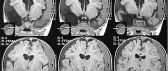
In addition, an MRI of the brain is done, which helps to determine high intracranial pressure, as well as various consequences of traumatic injuries, hypoxia, and tumor-type formations.
Studies are carried out to determine the level of hormones in the blood and determine the biochemical data of the blood composition, this is necessary to determine endocrine and metabolic disorders.
A number of studies are mandatory to determine a syndrome of organic origin:
- Ultrasound of the adrenal glands;
- Ultrasound of the thyroid gland and internal organs;
- MRI and CT scan of the adrenal glands.
A particularly severe and frequent violation of the reproduction of knowledge acquired before the disease was found in patients with aresorptive hydrocephalus (in 12 out of 21 patients). All 12 patients with hydrocephalus had typical Korsakoff syndrome with a pronounced frontal radical.
Unlike patients in other groups, when updating “old” knowledge in patients with hydrocephalus, the violation of trace selectivity with contamination, perseverations, or random responses came to the fore. This is what led to the peculiarity of selective retrograde amnesia in patients with hydrocephalus. Almost all patients with hydrocephalus (unlike the other groups examined) had a violation of updating the dates and events of their own biography. In some patients after bypass operations, partial restoration of “old” knowledge was noted, which indicated a predominant difficulty in the reproduction department.
Thus, in 58 of the 326 patients we studied, impairments of varying degrees in the reproduction of knowledge acquired long before the manifestation of the disease were found. It is very important that all patients had similar selective amnesia syndrome.
All patients with selective retrograde amnesia had similar localization of brain damage. In all cases, there was involvement of the medio-basal parts of the brain of bilateral localization with a predominance of damage to the structures of the right hemisphere. The identification of a wide interest in the medio-basal (periventricular) parts of the brain in all our patients with selective retrograde amnesia makes us think about the significance of combined damage to these very areas of the brain, where the fibers that connect many structures pass.
Disruption of the functions of many parts of the brain located periventricularly leads to the fact that not one, but several factors at once cause a violation of the reproduction of knowledge acquired long before the disease. The time factor necessary for reproducing both “old” knowledge and current information is currently being discussed especially widely.
It is believed that past images exist in the mind in their own “space-time marker.” The right hemisphere is considered as the brain substrate for time perception (Balonov et al., 1980; Bragina, Dobrokhotova, 1980. 1988; Dobrokhotova, Bragina, 1977). Recently, it has been shown that impaired perception of time duration is noted in patients with damage to the frontal and parietal lobes of the right hemisphere (more often with combined brain damage) (Harrington et al., 1996).
Now let us remember that in almost all of our patients with impaired updating of knowledge acquired before illness, the worst situation was with the reproduction of the most important historical dates. It can be assumed that the “time stamp” plays a leading role not only in recording information (this happened long before the disease), but also in reproducing, and, possibly, consolidating information.
It was noteworthy that in all our patients with selective retrograde amnesia, neuropsychological examination always revealed dysfunction of the frontal and parietal lobes of the brain, predominantly in the right hemisphere. It is important that violations were almost always found in tests with schematic clocks.
The perception of time also includes the ability to evaluate time sequences - images, events, facts (“earlier-later”). The perception of a sequence of stimuli is now more often associated with the right or left frontal lobes (Shimamura et al., 1990). It is interesting that in our patients, difficulties in listing the sequence (using the example of non-working holidays) were not always detected.
When analyzing our own and literary data, a rare occurrence of violations of the reproduction of one’s own biography (“autobiographical amnesia”) was noted. In the patients we studied, defects in reproducing the dates of their biography (less often, facts) were noted only in patients with hydrocephalus and craniopharyngiomas. In this case, the factor of violation of the selectivity of traces was always dominant.
The role of the diencephalic region in disrupting the reproduction of consolidated knowledge seems perhaps less clear than that of the structures of the right hemisphere. However, its anatomical involvement was found in all cases (intraventricular hemorrhages, craniopharyngiomas, hydrocephalus). Perhaps the diencephalic region (along with other parts of the brainstem) provides the necessary level of wakefulness to reproduce “old” knowledge. In addition, the diencephalic region of the brain is directly involved in memory processes and is “responsible” for the delayed reproduction of current information.
Thus, all of the above substantiates the important role of the right hemisphere, especially the medio-basal parts of the frontal and parietal lobes, and the diencephalic region in the reproduction (probably also in fixation and consolidation) of information received before the disease. And the assumption about the predominant connection of the diencephalic region with the right hemisphere has been around for a long time (Korsakova, Moskovichiute. 1985).
However, the analysis of the topic of brain damage in patients with selective retrograde amnesia studied by us and the literature data do not allow us to exclude the participation of the medio-basal parts of the left hemisphere in the reproduction (probably in the storage and consolidation) of “old” information. It can be assumed, taking into account the specifics of information processing by the two hemispheres, that when fixing and encoding semantic information, the right hemisphere is “responsible” for the formation of a specific “temporal” (and spatial) “mark” of the stimulus, and the left hemisphere ensures its classification. One might think that a similar specialization of the hemispheres occurs when reproducing semantic knowledge acquired long ago. When “decoding” information, searching for it in individual semantic “fields” (for example, the history of the country, autobiography, etc.) is carried out primarily with the participation of the left hemisphere. The right hemisphere ensures the reproduction of specific characteristics, including the dates of events. The content side of events may be updated with the participation of both hemispheres (logical sequence and specific spatio-temporal “binding” of the event). Such joint work of the hemispheres in the future makes it possible to voluntarily (consciously) reproduce the necessary information received a long time ago, in contrast to the unconscious (forced) reproduction of different episodes of life, for example, during epileptic seizures of the type “outbursts of experiences in the past.”
selective retrograde amnesia – previous | the following is the internal picture of the disease
A. R. Luria and psychology of the 21st century. Content
Treatment method for diencephalic syndrome
The invention relates to medicine and can be used in the treatment of central autonomic disorders. In addition to drug therapy with tranquilizers and symptomatic drugs, patients with diencephalic syndrome undergo anodic micropolarization of the solar plexus ganglia, exposing them to a constant electric current of 800-1000 μA for 35-45 minutes daily for 10-15 days. In this case, from the 5-6th day of treatment, the amount of administered drugs is gradually reduced until they are completely discontinued by the end of treatment. Such effects are repeated, if necessary, no earlier than after 2 months. The method allows you to shorten the treatment time to 2-2.5 weeks, reduce the number of administered medications and their dosages until complete withdrawal.
The invention relates to medicine, namely to neurology, and can find application in the treatment of central autonomic disorders.
Diencephalic syndrome (hereinafter referred to as DS) is one of the most common types of neurological pathology. The most common form of DS, known as vegetative-vascular dystonia, occurs in 31% of urban residents over 10 years of age, with a significant predominance among women. Its frequency increases during the period of greatest working capacity. The consequences of DS in the form of vascular cardiac and cerebral accidents, severe endocrine pathology in more than 40% of cases lead to permanent disability. The incidence of DS shows a clear upward trend, especially in industrialized countries. A characteristic feature of this disease is its pronounced resistance to all currently available treatment methods. DS is a polyetiological disease with polymorphic symptoms. The identification of DS into an independent clinical and diagnostic form is due to a general pathogenetic mechanism, the core of which is a persistent increase in the excitability of the diencephalic (hypothalamic) structures of the brain, leading to dysregulation of various visceral, neurohumoral and psycho-emotional functions. Currently, conservative treatment methods are used to treat DS, such as drug therapy, physiotherapy, radiotherapy, diet and balneotherapy. Drug therapy is predominantly symptomatic in nature and is aimed primarily at normalizing the functions impaired in this form of DS: blood circulation, digestion, internal secretion, psycho-emotional tone. The closest to the proposed method is the method of medicinal therapy of DS through the administration of medicinal benzodiazepine group (Relanium), which reduce excitability of the nervous substrate, in particular brain stem structures, as well as symptomatic remedies. We took this method as a prototype (Vein A.M., Solovyova A.D., Kolosova O.A. Autonomic-vascular dystonia. - M.: Medicine, 1981, p. 276; Shefer D.G. Hypothalamic syndromes. - M.: Medicine, 1971, p. 340).It consists in the fact that patients with DS are administered orally Relanium in an amount of up to 10 mg per day, and the following medications are administered as symptomatic therapy, depending on the prevailing symptoms: 1 ) vasodilators: cinnarizine 25 mg 3 times a day, Cavinton 5 mg 3 times a day or other vasodilators; 2) antihypertensive: clonidine 0.075-0.15 mg 2-3 times a day; 3) antispasmodics: no-spa 40-80 mg 1-2 times a day; 4) regulators of peristalsis: cerucal 10-20 mg 1-2 times a day; 5) adrenergic blockers: anaprilin 10-40 mg 2-3 times a day, pyrroxan 15-30 mg 2-3 times a day; 6) antidepressants: ametriptyline 25 mg 2-3 times a day; 7) vitamins B, C; electrolytes and microelements: panangin 1 tablet 3 times a day, potassium orotate 0.5-1 g 3 times a day; 9) antihistamines: suprastin 25 mg 1-2 times a day; 10) diuretics: furosemide 40 mg once a day. Vegetative paroxysms in patients with DS with this method of treatment are stopped by parenteral administration of a 0.5% solution of Relanium in a dose of up to 4-6 ml. This method of treating DS allows in most cases to achieve positive effect in the form of improvement in the general well-being of patients, some weakening of the main symptoms of the disease, and in some cases - a decrease in vegetative paroxysms and a decrease in the degree of their severity. In isolated cases, short-term remission lasting up to 1-2 months is possible. Relapses of the disease are usually associated with the influence of exogenous factors (geomagnetic fluctuations, weather dependence, stress), as well as with cessation of medication. With the constant use of individually selected medications, the course of the disease is wave-like, periods of deterioration in the condition and well-being of patients can also be associated with addiction to the drugs used, which forces either increasing doses to acceptable limits, or switching to the use of analogue drugs. The length of hospital stay for such patients ranges from 5 weeks to 2-3 months, depending on the severity of the condition; outpatient treatment is carried out for life. There is no cure for DS. As you can see, the disadvantages of this method of drug therapy for DS are: 1) the indiscriminate, diffuse nature of the effect on the body; 2) long periods of hospital stay, which is associated with individual selection of therapy; 3) the need for constant, almost lifelong, medication; 4) the possibility of side effects from potent drugs, 5) the possibility of addiction to certain drugs, primarily tranquilizers; 6) polypromasia; 7) the obligatory need for repeated hospital treatment to change the drug therapy regimen. The use of other methods of treating DS, known to us from the literature and approved (physiotherapy, balneotherapy, spa treatment, etc.) does not significantly affect the condition of the patients. Until now, methods local conservative effects on the solar ganglia have not been used in diencephalic syndrome. The technical result of the present invention is to reduce the length of hospital stay to 10-14 days, reduce the number of drugs used to 1-2 drugs and reduce their dosages by 2-3 times, up to until they are completely cancelled. This result is achieved by the fact that, in addition to the described method of drug therapy, patients with DS undergo anodic micropolarization of the solar plexus ganglia by applying a direct current of 800-1000 μA for 35-45 minutes daily for 10-15 days, moreover, starting from the 5-6th day, the amount of administered drugs is gradually reduced until they are completely discontinued by the end of treatment, and this effect, if necessary, is repeated no earlier than after 2 months. Having been treating patients with focal brain lesions for a number of years, We, along with surgical treatment and drug therapy, tried to use anodic polarization using the transcranial micropolarization technique - TCMP (Bogdanov O.V. and others. RF Patent 2122443 dated 07/01/1997), applying electrodes to the scalp in areas of projection of acute lesions (Naryshkin A.G., Shelyakin A.M., Tyulkin O.N., Gorelik A.L. Method for the treatment of acute focal brain lesions. RF Patent 2188674 dated September 10, 2002).At the same time, we observed the high effectiveness of such effects and the absence of side effects. We are not aware of any attempts to apply this method for the treatment of DS. Having recently observed patient O., 50 years old, for post-ascariasis toxic solaritis with daily, violent, vegetative crises and the lack of effect from the use of drug therapy with ganglion blockers, tranquilizers and antispasmodics, we tried to give him anodic polarization of the solar plexus, applying electrodes to the solar points of the anterior surface of the abdomen and using the same parameters of influence as with TCM. After the fifth procedure, it unexpectedly turned out that within three days our patient had no solar vegetative crises. This prompted us to extend the course of polarization to 10 procedures, which led to regression of autonomic symptoms. This observation gave us the idea to clinically examine the state of the solar plexus ganglia in patients with DS, since most of them had similar symptoms. It was found that almost all patients with DS have painful solar points, and also have objective clinical signs of vegetative irritation of varying degrees of severity. Then we tried to use a similar method for the treatment of patients with DS, empirically selecting the optimal parameters of the effect. Depending on the specific situation and the severity of pathological manifestations, the current strength ranged from 800 to 1000 μA, and the duration of one procedure was 35-45 minutes. The criteria for the optimal strength of direct current when exposed to the solar ganglia and its duration were subjective feelings of drowsiness, the appearance of unpleasant sensations under the electrodes. With a direct current strength of less than 500-600 μA, these changes were not observed. When the current increased above 1000 μA (1 mA), patients experienced unpleasant sensations under the electrodes, accompanied by redness of the skin. When the exposure time was less than 20 minutes, characteristic changes did not have time to develop, and when the exposure was prolonged beyond 45 minutes, some patients experienced unpleasant sensations in the form of heaviness in the head and in the epigastric region, feelings of anxiety, and weakness. As our experience shows, this method of influencing the solar plexus ganglia in DS can effectively stop vegetative paroxysms, significantly reduce, even completely eliminate, the dose of administered medications, and also achieve stable and long-term remissions. Moreover, we achieve this result in 1-2 weeks, depending on the severity of the disease. The essence and effectiveness of the method is illustrated by the following examples. Example 1. Patient M., born in 1950, IB 4698/2001, was admitted on September 10, 2001 with a diagnosis : “Cerebral arachnoiditis with diencephalic syndrome.” Upon receipt of a complaint of intense headaches in the parietotemporal regions, more on the right, constant, compressive in nature, a feeling of interruptions in heart rate, a feeling of constant discomfort in the abdomen, attacks of tachycardia with chills, trembling and a feeling of tightness in the limbs, a rise in temperature to subfebrile values, lasting up to 5-6 hours, upon resolution of which pronounced pollakiuria and diarrhea are noted. Real deterioration within 6 months, attacks became daily, anorexia appeared, and therefore the patient lost 17 kg of weight, as well as insomnia, signs of a depressive state. From medical history: similar symptoms since the age of 20, with a frequency of attacks up to two times a month, rheumatism, cerebral rheumovasculitis, minor chorea in puberty, blunt abdominal trauma without surgical intervention at the age of 13 years, at the age of 36 years - spontaneously developed severe pain syndrome in the lumbar-thoracic spine, which forced the patient to use crutches for 4 months, without convincing objective disorders. Annual depressive episodes. Observed by a therapist with a diagnosis of coronary heart disease: atherosclerotic cardiosclerosis, paroxysmal tachyarrhythmia, urolithiasis, 2-sided nephroptosis of the 2nd degree, stones of both kidneys. Constantly receives Relanium up to 20-25 mg per day, for paroxysms - in the form of injections, amitriptyline 25 mg 2 times a day, ranitidine 0.3 g 3 times a day, anaprilin up to 40 mg per day, papazole and no-shpu for elevated blood pressure, as well as potassium orotate and multivitamin complexes. On examination: skin pale, dry, skin turgor is reduced. Heart sounds are muffled, heart rate 76 per minute, occasionally - increase to 110 per minute with frequent extrasystoles, blood pressure 130/80 mm, vesicular breathing, abdomen is soft, painful in all parts, with deep palpation - unbearable pain in solar points, with perverted pupillary reaction, against the background of which, at the time of examination, a reduced autonomic paroxysm developed with chills, tremor, and redness of the skin of the upper body. Severe pain on sliding palpation of large arteries, more on the right. Index of vegetative irritation according to Markelov - 3. Neurologically: transient, mild anisocoria on the right, deep reflexes are somewhat strengthened on the right, coordination tests with slight intention tremor, predominant on the left. Blood and urine tests - without any features. ECG - signs of left ventricular hypertrophy. Ophthalmologist: low degree myopia. Computed tomography of the abdominal cavity - without pathological changes. Electroencephalogram: pronounced cerebral changes of a diffuse-organic type with signs of deep brainstem dysfunction, predominantly at the diencephalic level. Based on clinical examination data, the diagnosis was made: “Diencephalic syndrome of toxic-metabolic etiology with vegetative paroxysms mixed type and generalized vegetative irritation.” The patient was prescribed the following therapy: 1) Relanium - 20 mg per day; 2) anaprilin - 40 mg per day; 3) pyrroxan - 30 mg 2 times a day; 4) Cavinton - 5 mg 3 times a day; 5) panangin - 2 tablets 2 times a day. With this therapy for three days, the patient’s headaches decreased somewhat, but the rest of the symptoms persisted, vegetative paroxysms were observed daily. Then she was offered a course of anodic polarization of the solar plexus ganglia. The current strength was 800 μA; as the current increased, the patient developed a burning sensation under the electrodes, and as it decreased, irritability and anxiety appeared. When the procedure lasted more than 45 minutes, weakness and drowsiness occurred; the effect in the form of a feeling of general comfort and relaxation occurred at the 30th minute of exposure. After the third procedure, the patient’s paroxysms stopped, and after the fifth, the pain and discomfort completely regressed. From that moment on, the patient independently stopped taking any medications, primarily tranquilizers. Upon completion of a course of 10 procedures, complaints, neurological and somatic symptoms completely disappeared, the patient was discharged after spending 15 days in the hospital. Two months later, she sought outpatient treatment due to renewed discomfort in the epigastric region and dyspeptic complaints. The patient underwent a repeated course of anodic micropolarization on an outpatient basis. The subsequent remission has persisted to the present day. Thus, the use of anodic micropolarization on the area of projection of the solar ganglia made it possible not only for the first time and to fully alleviate the condition of a chronic patient, but also to eliminate dependence on drugs. Example 2. Patient I., born in 1926. , IB 1012/2002, received 02/18/2002, has been observed by us (A.G. Naryshkin) for more than 20 years. Complains of frequent, or even daily, attacks in the form of intense burning-squeezing headache, pollakiuria, accompanied by nagging pain in the left half of the abdomen, a rise in blood pressure to 250/120 mm, myasthenic-like symptoms, coldness and numbness of the extremities. The duration of the attack is usually 2-3 hours, stopped by the introduction of 2 to 6 ml of relanium intramuscularly. History: malaria, tuberculous bronchadenitis, frequent stressful situations, strumectomy at the age of 7, measles with neurological complications, purulent appendicitis complicated by peritonitis, dysentery, tonsillocardial syndrome. Since 1978, attacks of burning pain in the left half of the abdomen began to bother me, and in 1980, the attack described above developed for the first time. She was examined and treated many times in various medical institutions. In 1984, in the clinic of the Institute of Emergency Medicine of the USSR Academy of Sciences, complex therapy was selected, including ganglerone and relanium, against the background of which the frequency of attacks decreased by 2 times. Subsequently, she continued to use this regimen, independently adjusting the dosage of the drugs. She applied due to an increase in attacks after the discontinuation of the drug ganglerone. Upon admission: the skin was pale yellowish, dry, turgor was reduced, pronounced pastiness of the legs, elements of alopecia. Slight trembling of the head and hands at rest. Sharp pain on palpation of the upper, middle, lower solar points, ribs, iliac crests. Pain during sliding palpation of the vessels of the upper and lower extremities up to a. tibialis on the feet and a. radialis on the wrists, also - pain on palpation a. temporalis on both sides, more on the left. Emotionally unstable, tearful. Ophthalmologist: beginning cataracts, retinal atherosclerosis. Therapist: chronic bronchitis, pneumosclerosis, emphysema, respiratory failure grade 0-1, hypothyroidism, coronary heart disease: atherosclerotic cardiosclerosis, circulatory failure grade 1-2. X-ray of the spine: widespread involutive osteochondrosis, L5 pseudospondylolisthesis. Computed tomography of the abdominal cavity: no pathological changes in the retroperitoneal tissue were detected. Atherosclerosis of the abdominal aorta. ECG: migration of the supraventricular pacemaker, incomplete blockade of the right bundle branch, deviation of the electrical axis to the left, left ventricular hypertrophy, repolarization disorder. EEG: pronounced diffuse changes in bioelectrical activity with signs of deep brainstem dysfunction. Based on anamnesis and clinical results examination confirmed the diagnosis: “Diencephalic syndrome of infectious-metabolic origin with vegetative paroxysms.” Treatment was prescribed according to a regimen similar to that described in example 1, with the addition of aminophylline 15 mg 3 times a day, clonidine 0.075 mg per day and L-thyroxine, which The patient takes it constantly. Additionally, the patient was proposed to the polarization of the solar ganglias with the parameters of the impact, similar to the 4th day in the hospital in the hospital and after two sessions of micropolarization, paroxysms stopped and no longer resumed. After 6 procedures, overall well -being improved, the emotional background increased, the signs of autonomic irritation were reduced to the index - 1 according to Markelov. From this moment, a gradual abolition of drugs has begun: on the 5th day - relanium, on the 6th day - sympatholytics, on the 7th day - vascular drugs. From the 8th day of treatment, the patient did not take any drugs except L-thyroxine. A total of 12 sessions were held. Designed from the hospital on the 18th day. After discharge and to this day, he feels good, the only attack in a reduced form was noted in August 2002. The example of 3. Golol M., 53 years old, a disabled person of the 2nd group, applied on an outpatient basis, on the recommendation of the attending physician, in June 2002, in June 2002 With complaints of constant weakness and high fatigue, frequent polymorphic headaches with dizziness and nausea, sleep disorders, various dyspeptic disorders, frequent rise of blood pressure with a picture of hypertensive crises (from 2 to 6 times a month), stopped only by ambulance brigades, allergic manifestations In the form of urticaria, rhinitis, bronchial asthma with an increase in suffocation attacks, accompanied by chills, paresthesia, cooling and numbness of the limbs. Constantly takes a relanium 5 mg up to 4 times a day, various anti-allergic and antihypertensive drugs. It takes a real disease from the age of 26, when it has undergone a hypertoxic form of influenza. The disease is progressive. It was repeatedly examined and treated in hospitals in the city with diagnoses: diencephalmic syndrome, neurasthenia, infectious-allergic bronchial asthma, coronary heart disease: atherosclerotic cardiosclerosis, hypertension 2 tbsp., Gallstoneous disease, chronic pancreatitis, chronic enterocolitis. The last seven years have been the 2nd disability. In the anamnesis: measles, scarlet fever, dysentery, in youth, frequent tonsillitis, tonsillectomy, vegetovascular dystonia, twice shackles of the brain, there is also a chronic psycho-traumatic situation in the family: the son suffers from a mental illness. When examining: examination: The skin is pale, wet, pasty, pronounced red dermographism. Heart tones are muffled, AD -150/90, in the lungs -hard breathing. The stomach is soft, painful in all departments. There is a sharp pain at the solar points, the autonomic irritation index - 3 according to Markelov.eag: a pronounced stem dysfunction with a predominance at the level of the diencephalic region, with a decrease in the threshold of convulsive readiness. The generational micropolarization of the ganglias of solar plexus with a current force of 1000 mka was carried out, so. As with less current strength, a characteristic effect in the form of drowsiness and relaxation did not occur. The duration of the procedure was 35 minutes, since with an increase in the exposure time, the patient had discomfort and anxiety. Improving well -being was noted with the fifth procedure in the account, the attacks of suffocation ceased, the blood pressure was established on numbers 130/80 mm. From this moment, the rethum has been canceled, a gradual decrease in doses of cloofeline according to the generally accepted scheme (in order to avoid cancellation syndrome) has begun. A total of 12 procedures were carried out. At the end of the course, there is a regression of pain and unpleasant sensations, manifestations of allergies, improving the psychoemotional state, restoration of night sleep, stabilization of blood pressure in numbers 125/80 mm. The intake of medicines is completely discontinued. In the future, after a two-month remission, a partial resumption of the previous symptoms in a reduced form was noted, the patient again began to take a relaxium-1-2 tablets per week. Currently, a repeated course of micropolarization is being carried out. Currently, 6 patients with a diagnosis of “diencephalus syndrome” have been treated: 4 women and 2 men. Age - from 26 to 73 years, the prescription of the disease from 5 to 33 years. All patients had a pronounced and persistent improvement in well -being, a positive dynamics of clinical and laboratory tests. Four of them completely stopped taking medications, two crossed from the constant to the episodic intake of tranquilizers with rare and impressive attacks of the disease. Three patients of the retirement age moved to an active lifestyle, two, having a disability of the 2nd group, returned to labor activity. The number of courses from 1 to 3 ranged, the terms of the currently observed remissions - from 2 to 8 months. The existing observations, thus, indicate the high efficiency of the proposed method, which, in comparison with known other methods, has several advantages: 1) significantly reduces treatment periods-to 2-2.5 weeks, while the treatment periods of patients with other ways are from 5 to 6 weeks; 2) reduces the amount of drugs administered to 1-2 drugs and allows you to reduce their dosages, up to complete cancellation, while generally accepted drug therapy includes a large (up to 5-7 drugs for a separate patient) the amount of drugs and often admits The increase in dosages. The change was developed and passed clinical testing in the neurosurgical department of the city hospital of 23 St. Petersburg.
Formula of the invention:
A method for treating diencephalic syndrome using tranquilizers and symptomatic drugs, characterized in that they additionally carry out anodic micropolarization of the solar plexus ganglia, influencing them with a direct electric current of 800-1000 μA for 35-45 minutes daily for 10-15 days, Moreover, starting from the 5-6th day, the amount of administered drugs is gradually reduced until they are completely discontinued by the end of treatment, and such effects are repeated, if necessary, no earlier than after 2 months.