MRI of the veins of the brain is a modern diagnostic technique that allows you to obtain an accurate picture of the veins and determine the slightest deviations in the functioning of the vascular system.
Magnetic resonance imaging of veins is used to diagnose a wide range of diseases. The method is based on the effect of magnetic radiation on human tissue, which is absolutely safe and makes it possible to accurately visualize the structure of brain vessels.
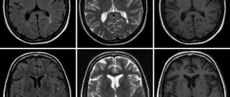
Diagram of cerebral circulation on an MRI scan
What is MRI of the brain based on?
The diagnostic effect of brain MRI is based on nuclear magnetic resonance. In response to the powerful radiation created by the generator, the hydrogen atomic nuclei contained in the tissues line up along the electromagnetic field lines and begin to vibrate. Each atom becomes like a spinning mini-spinning top, emitting energy waves.
Different structures emit different amounts of energy - some give it out more intensely, while others less so. The difference is recorded by a device that takes pictures (slices) in different projections.
To do this, the patient is placed inside a tomograph in which generators maintain a high-frequency electromagnetic field. Special radio emitters generate pulses, and coils record the energy sent by vibrating atoms.
The resulting slice images are combined into a three-dimensional matrix using a special computer program, in which dark or light unhealthy areas are visualized against a gray background.
Anatomy of cerebral veins, what is visible on MRI?
Veins are blood vessels that are designed to drain blood from the brain. Oxygenated blood enters the nerve center through the arteries, and after giving nutrients and oxygen to the cells, the blood returns to the heart through the veins.
Disturbances in the functioning of the cerebral veins can contribute to poor health, fatigue and the gradual development of cerebral edema. This is why timely and accurate diagnosis is so important.
Examination of the condition of the venous bed using a magnetic resonance imaging scanner is called MR venography.
In terms of information content, MRI of the arteries and veins of the brain is one step ahead of traditional diagnostic methods using X-rays or ultrasound. Using MR images, diagnosticians receive visual information about the functioning of the veins of the brain.
MR venography allows you to accurately determine:
- anatomical pattern of the venous system;
- congenital and acquired abnormalities in the structure and functioning of blood vessels;
- disturbances in trophism and blood flow from parts of the brain;
- possible damage to blood vessels and their consequences;
- the presence of tumor formations blocking the vascular bed.
A tomography examination is a completely safe procedure that allows not only detailed and high-quality visualization of the structural features of the venous network of the brain, but also scanning for preventive and diagnostic purposes with any frequency without negative impact on health.
Advantages of magnetic resonance imaging over other methods
An MRI examination gives results much more accurately than x-rays, echoencephalography (EchoEG), ultrasound and other diagnostic options. It allows you to obtain maximum data about existing tumors, diseases, post-traumatic and post-stroke changes. Unlike CT and X-rays, in this case the body is not irradiated.
Only soft tissues are visualized in the finished images. The bones of the skull are not visible, so they do not interfere with analysis and decoding.
The contrast agent used in MRI diagnostics is much less likely to cause allergic reactions compared to X-ray contrast agents used for radiography.
Features of the procedure
The price of a head MRI is slightly higher than a computed tomography or radiography. But this method, unlike the other two, has virtually no contraindications. The exception is the presence in the body of magnetic metal-containing implants or a cardiac stimulator. In addition, MRI is the most harmless method, since it does not involve instrumental invasion or radiation exposure.
The diagnostic procedure lasts from 45 minutes to an hour. It goes unnoticed by the patient. An MRI of the head and neck, the price of which varies depending on the type of device used, can be done under anesthesia or in a state of medicated sleep, if it is difficult for the patient to remain motionless for this period of time. Under different types of equipment, open and closed type units are assumed. Open tomographs do not have one of the walls, which allows people with claustrophobia, overweight, and small children who cannot remain alone in the chamber to undergo diagnostics. Open machines are more expensive, so the price of a head MRI will cost more than a procedure using standard equipment.
Price
Dear patients, on October 9 and 10 there is a 20% discount on MRI!
| Description | Price | Until October 12 | From 21:00-2:00 7:00-9:00 |
| MRI of the brain | RUB 5,400 | RUB 4,590 | RUB 4,050 |
| MRI of cerebral vessels (angiography) | RUB 5,400 | RUB 4,590 | RUB 4,050 |
| Brain MRI with contrast | RUR 11,000 | RUB 10,250 | — |
| Issue of film with photograph | 500 rub.r. | 400 rub.r. | 400 rub.r. |
| Recording a photo on flash | RUR 1,000 | RUR 650 | 600 rub. rub. |
All MRI prices >>>
Decoding the received data
Decoding of the results is carried out immediately after the study and takes about half an hour. To accurately determine where to get an MRI of the brain, you should check the availability of licenses and other permits of the selected diagnostic center. After the examination, the decrypted results are given to the patient or transferred to his attending physician.
The transcript of the image contains information about:
- the amount of fluid in the spinal cord canal;
- blood flow speed;
- activity of the cerebral cortex when exposed to various stimuli;
- level of tissue diffusion.
MRI of the brain is a painless and harmless procedure that cannot cause headaches or other ailments in patients. However, with the help of this type of diagnosis, the doctor can get a clear clinical picture, determine the cause of pain and establish the correct diagnosis.
How does the procedure work?
The patient removes all metal jewelry and takes out removable dentures containing metal.
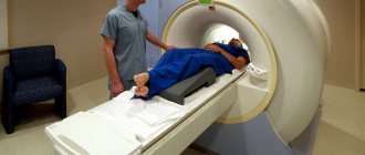
The patient is placed on a movable table and secured with special belts. This measure is necessary because you will have to lie inside the tomograph, remaining motionless, for a long time.
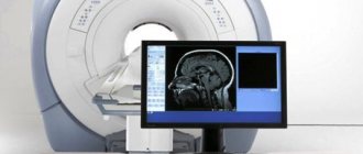
A device equipped with wires that transmit and receive radio signals is placed on the head. The equipment is quite noisy and tires with constant clicks and whistles. Therefore, the patient’s ears are protected with earplugs. After this, the table slides into the device, and the specialist sits down at the computer, in which the transmitted data is analyzed and processed.
The technique takes pictures, the quality of which depends on the characteristics of a particular MRI scanner. The thinner the visual slices the equipment makes, the more accurate the final images will be. The duration of diagnosis is 20-30 minutes, and when contrast is used - up to an hour.
After MRI diagnostics, you can immediately return to your normal life. No side effects subsequently or during the MRI examination occur, with the exception of an extremely rare allergy to gadolinium salts.
Finished photographs are handed out printed or recorded on a magnetic medium - disk or flash card. It can be sent to email with SMS notification.
- Standard - done without the introduction of contrasting solutions, but at the same time provides a sufficient amount of information.
- With contrast, before which drugs containing gadolinium salts are injected into the vein - gadopentetic and gadoteric acids, Omniscan, Magnevist, etc. These solutions penetrate into the bloodstream and, once in the rays of the MRI scanner, highlight the resulting “picture”. At the same time, the changed areas become better visible, which simplifies decoding. The technique is most often used to detect vascular abnormalities, multiple sclerosis and tumor formations. The dose of contrast agent is selected individually, taking into account weight.
- MR angiography – is performed to assess the condition of blood vessels in atherosclerosis, aneurysms, blood clots and pre-stroke conditions. It is done with gadolinium contrast to show blood flow problems in detail.
- MR imaging of the pituitary gland, an appendage that is an endocrine gland. The pituitary gland secretes hormones responsible for reproductive function, tissue metabolism and the regulation of human growth. An examination is prescribed if an adenoma is suspected - a benign tumor that causes migraine-like pain, hormonal imbalances, gigantism, infertility, obesity and sexual dysfunction. The same method is used to identify malignant pituitary formations that have similar symptoms and are accompanied by a pronounced deterioration in health.
How long does an MRI procedure take?
Magnetic resonance scanning of one part of the body lasts 20-30 minutes. If contrast is used, the duration can increase to 50 minutes (excluding preparation time), and when examining several parts of the body - up to 1-1.5 hours.
How long does an MRI examination take? The duration of the procedure depends on several factors:
- type of tomograph and its power;
- contrasting;
- scope of examination;
- compliance with the study rules (patient immobility).
For example, examining the knee takes about 30 minutes, the cervical, lumbar or lumbosacral spine without contrast takes about 40 minutes, and cerebral vessels with contrast takes up to 1 hour.
How long should I wait for test results? Typically, the entire procedure, from issuing a medical card to obtaining a doctor’s report, lasts no more than 2 hours. In cases where the issuance of a conclusion requires consultation with related specialists or a consultation, the wait for results may take up to 2-3 days.
How is MRI performed on children?
MRI is considered a safe examination method, so it can be performed on patients of any age, including children. The only condition for high-quality diagnosis is complete immobility of the examined child during the procedure.
The approach to examining each little patient must be individual:
- If it is necessary to perform an MRI of soft tissues or the brain on a child of conscious age who can lie motionless for up to an hour but feels restless, a relative of the child may be allowed to be present in the room during the procedure.
- For a balanced school-age child, it is often enough to explain how the procedure works.
- If there are indications for MRI, but it is not possible to ensure the child’s immobility (for example, up to 5 years), then the examination can be carried out after preliminary sedation, that is, under anesthesia.
Examinations under anesthesia require the presence of an anesthesiologist and special equipment, so they are not carried out in every diagnostic center, which must be taken into account when registering small children for the procedure.
Can pregnant women undergo an MRI?
No teratogenic effect from the magnetic field created by the tomograph has been clinically identified. However, doctors recommend refraining from prescribing this procedure during pregnancy without compelling reasons, for example, to diagnose acute cerebrovascular accidents or if a tumor process is suspected. When prescribing an MRI for pregnant women, the doctor always compares the expected benefits of the study and the possible harm from it.
Among the many hardware diagnostic methods, MRI is considered one of the most preferable (after ultrasound) during pregnancy. How is this diagnostic procedure performed in pregnant women? They try to conduct the study in the second or third trimester of pregnancy, when the formation of all organs in the fetus has already occurred. In this case, procedures with contrast are avoided.
Gadolinium rarely causes allergic or anaphylactic reactions. The contrast is excreted unchanged by healthy kidneys within 12 hours. In pregnant women, the kidneys experience increased stress, which can lead to a retention of the drug in the blood of the expectant mother. Prolonged circulation of gadolinium contrast in the blood can theoretically lead to its sedimentation in tissues, which can lead to unpredictable consequences.
There is no information in official medical sources about the dangers of gadolinium-based contrast agents for the intrauterine development of the fetus. And the point is not that the contrast is absolutely safe, but that its clinical trials have not been conducted on pregnant women. Given the lack of reliable data on the possible danger of gadolinium contrast for the fetus, contrast should not be used during forced MRI during pregnancy.
MRI of the brain MRI of the hip MRI of the shoulder MRI of the knee MRI of the kidneys MRI of the abdominal cavity MRI of the pancreas MRI of the paranasal sinuses MRI of the eye orbits MRI of the hippocampus MRI of the pituitary gland MRI of the adrenal glands MRI of the liver MRI of the spleen MRI of the salivary glands MRI of the inner ear MRI of the heart MRI of the gallbladder MRI of the thyroid gland MRI of the maxillary sinuses MRI of the sella turcica MRI of the spinal cord MRI of the whole body MRI of the retroperitoneum MRI of the hand MRI of the pelvis MRI of the prostate MRI of the bladder MRI of neck vessels MRI of brain vessels MRI of the temporomandibular joints MRI of the soft tissues of the hip MRI of the soft tissues of the neck MRI of the sacral spine MRI entire spine MRI lumbosacral spine MRI thoracic spine MRI cervical spine MRI sacroiliac joints MRI wrist joint MRI foot MRI ankle joint MRI elbow joint MRI auditory nerve MRI optic nerve MRI coccyx MRI mammary glands MRI jaw MRI stomach MRI mediastinal organs MRI of the lymph nodes MRI of the brachial plexuses MRI of the intestine MRI of the trigeminal nerve MRI of the facial nerve MRI of the sciatic nerve MRI of the teeth MRI of the lungs and bronchi MRI of the uterus
Indications for general anesthesia for MRI
Intravenous or inhalational sedation is only necessary for patients who are unable to hold their body still for long periods of time. Main indications for general anesthesia:
- Claustrophobia is the fear of closed spaces. Such patients, while inside the device, experience panic, which negatively affects their health and makes MRI diagnostics impossible.
- Mental disorders accompanied by unpredictability of behavior and high excitability.
- Uncontrollable involuntary head movements (swaying, shaking, tics).
- Epilepsy and other types of convulsive readiness and seizures - anesthesia is given only intravenously due to the risk of provoking a seizure attack.
- Early childhood. Small children cannot lie still for a long time in an MRI scanner, so light mask anesthesia is indicated for them.
- Severe pain syndrome, in which prolonged stay in one position causes discomfort, cramps, pain and spasms.
Indications for magnetic resonance imaging of the brain
- Neoplasms or their metastases. Diagnosis is prescribed for persistent migraine-like pain, sudden loss of vision and hearing, auditory, olfactory and visual hallucinations, attacks of confusion, sudden reading and writing disorders, often accompanying cancer pathologies.
- Epilepsy and other diseases manifested by fainting, confusion and convulsions.
- Suspicion of single or multiple cystic cavities filled with fluid, bloody or other contents.
- Possible presence of parasites (cysticercus and echinococci) carried through the vascular bed with the bloodstream into the head.
- Inflammations – meningitis, encephalitis, arachnoiditis, myelitis. Lesions caused by infections - measles, herpes, tuberculosis, toxoplasmosis, tick-borne encephalitis.
- Rehabilitation after stroke, traumatic brain injury and surgery. Using magnetic resonance diagnostics, the doctor evaluates the effectiveness of the treatment and predicts long-term results.
- The likelihood of developing multiple sclerosis, Alzheimer's disease and other degenerative processes.
- Children are examined for congenital pathologies and hydrocephalus.
With all these diseases, life and health directly depend on a timely diagnosis. Therefore, at the slightest suspicion of brain disorders in yourself or your child, you need to come to the clinic and be examined.
Indications and contraindications for MRI of cerebral veins
Indications for magnetic resonance imaging of cerebral veins may include the following symptoms and conditions:
- frequent headaches;
- sudden dull headache;
- traumatic brain injuries;
- tinnitus, dizziness, fainting;
- a feeling of pressure and fullness in a certain area of the head;
- feeling of pressure on the eyeballs;
- coordination problems;
- problems with memory, speech;
- a sharp decline in cognitive abilities and the ability to concentrate in young patients;
- cognitive impairment in older people;
- single or repeated epileptic seizures.
If one or more of the above symptoms occur, patients should consult a doctor who will give a referral for an MRI of the veins of the brain.
Diagnostician prepares patient for examination
MR venography can be indicated not only in the presence of any ailment, but also for preventive purposes for the early detection of vascular diseases.
Contraindications to the procedure
MRI is considered a safer method of visual diagnosis of internal body structures than CT. However, MR scanning also has a number of contraindications, which can be divided into absolute and relative.
Absolute contraindications include:
- the presence of metal objects in the body (fragments from wounds, shot);
- a pacemaker or neurostimulator installed in the patient;
- cochlear implants, vascular clips;
- the patient’s weight category is over 130 kilograms and body girth from 150 centimeters;
- first trimester of pregnancy;
- age up to 5 years.
Before prescribing a referral for an MRI of the cerebral veins, the doctor must check the patient for the presence of risk factors.
Relative contraindications do not negate the diagnosis, but require careful preliminary preparation. The procedure is not indicated in the case of:
- severe damage to the cardiovascular system in the acute stage;
- presence of tattoos on the body containing paint with metal particles;
- mental disorders in the acute stage;
- fear of being in a confined space;
- inability to maintain long-term immobility.
Prostheses made of non-ferromagnetic materials do not prohibit MRI screening if the following requirements are met:
- availability of documents for the prosthesis;
- the prosthesis was installed no later than 3 months before the diagnosis;
In addition, if the prosthesis is located outside the scanning area, its presence will not interfere with the screening.
What the results show
MRI studies, especially those performed with contrast, reveal numerous pathological processes. In the sections, compactions, cystic cavities, and hematomas (collections of blood) are visible in detail. Scars, parasites and their cysts, foci of degeneration, sclerosis and inflammation are identified.
Vascular changes are diagnosed, manifested by impaired patency, narrowing or dilation of blood vessels, the appearance of aneurysms (protrusion of walls) and thrombosis.
The degree of tissue damage in traumatic brain injuries, hemorrhagic and ischemic strokes is determined. The affected areas look lighter and are visible even if they are small in size and have sparse neurological symptoms.
Congenital defects are determined - underdevelopment and hypertrophy of the organ, small and incorrectly located gyri, cysts, holoprosencephaly - lack of division into hemispheres. Hydrocephalus is detected - accumulation of fluid in the ventricles, which with this anomaly are greatly enlarged.
Pathological areas and neoplasms appear as dark or lightish spots of various sizes and shapes, standing out against a grayish background. Oncological seals, especially malignant ones, have blurred, uneven edges and surrounding areas of necrosis.
MR diagnostics is recommended periodically for everyone who has been treated for oncological pathologies of any location. It detects metastases, which usually accompany cancer recurrence.
Will a head MRI show the causes of headaches?
An MRI of the head can reveal anatomical causes of headaches. Tomographic images will clearly show:
- neoplasms of benign and malignant nature;
- degree of tumor spread and metastasis;
- neurological lesions of the eyes, inner ear;
- signs of inflammatory processes in the sinuses;
- vascular pathologies;
- lesions of the membranes of the brain;
- conduction disturbance in nerve fibers (demyelination).
Using tomography data, a neurologist will be able to use exclusion to find the true cause of headaches and decide on treatment tactics.
| Service | Price according to Price | Discount Price at Night | Price by Discounts During the Day |
| from 23.00 to 8.00 | from 8.00 to 23.00 | ||
| MRI of cerebral vessels (MR angiography) | 3300 rub. | 2690 rub. | 2990 rub. |
| MRI of neck vessels (MR angiography of the neck) | 3500 rub. | 2690 rub. | 2990 rub. |
| MRI of the brain and cerebral vessels | 6800 rub. | 5380 rub. | 5980 rub. |
| Comprehensive MRI examination of brain and neck vessels | 6600 rub. | 5380 rub. | 5980 rub. |
| MR arteriography | 3300 rub. | 2690 rub. | 2990 rub. |
Contraindications
- Installed pacemakers and other electronic devices that are disrupted by the surrounding electromagnetic field.
- Fixed dentures with metal elements present in the mouth, crowns containing metal, braces and other orthodontic structures. The metal they contain is heated by a magnet and deteriorates, damaging surrounding tissue.
- Tattoos applied to the skin, made with metal-containing ink. Due to the heat caused by the electromagnets, burns may occur in these areas. In the absence of information about the pigment. used when applying a tattoo, it is better not to risk it and do a CT scan, ultrasound or x-ray. Examination is also prohibited for any metal piercing that cannot be removed.
- MRI examinations with contrast are not performed during pregnancy or if contrast agents are intolerant. Such an examination is not prescribed for severe kidney pathologies that make it difficult to eliminate gadolinium.
Magnetic resonance imaging is a safe and highly informative procedure that detects pathologies in the early stages. Therefore, in case of migraine-like phenomena, coordination problems, a sharp decrease in hearing and vision, fainting, or progressive memory deterioration, you must definitely go to the clinic and be examined. The price of MRI diagnostics is low and quite affordable for Muscovites and residents of Moscow Region.
Get an MRI of the brain >>>
Preparing for an MRI of the head and neck?
If the patient is not taking drugs that affect the vascular system, special preparation is not needed. Another category of patients is recommended to stop taking medications 6-8 hours before the procedure. If refusal of drug therapy threatens complications, you should inform your doctor.
It is not recommended to take alcohol before an MRI of the brain, as it affects metabolic processes in the brain, which can distort the results of the study.
After the MRI diagnosis, if the patient did not take sedatives, you can immediately return to your normal lifestyle. If the procedure was performed under anesthesia, the patient will have to stay in the clinic for some time.