More about the encephalogram
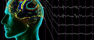
The essence of the examination is to record the electrical activity of neurons in the structural formations of the brain.
An electroencephalogram is a kind of recording of neural activity on a special tape using electrodes. The latter are attached to areas of the head and record the activity of a certain area of the brain. The activity of the human brain is directly determined by the work of its midline formations - the forebrain and the reticular formation (connecting neural complex), which determine the dynamics, rhythm and construction of the EEG. The connecting function of the formation determines the symmetry and relative identity of signals between all brain structures.
The study is prescribed if there are suspicions of various disorders of the structure and activity of the central nervous system (central nervous system) - neuroinfections such as meningitis, encephalitis, poliomyelitis. With these pathologies, the activity of the brain changes, and this can be immediately diagnosed on the EEG, and in addition, the localization of the affected area can be established. An EEG is carried out on the basis of a standard protocol, which records measurements taken while awake or asleep (in infants), as well as using specialized tests.
The main tests include:
- photostimulation - exposure of closed eyes to bright flashes of light;
- hyperventilation - deep, rare breathing for 3-5 minutes;
- opening and closing eyes.
These tests are considered standard and are used for encephalograms of the brain in adults and children of any age, and for various pathologies. There are several additional tests prescribed in individual cases, such as: clenching your fingers into a so-called fist, staying in the dark for 40 minutes, depriving yourself of sleep for a certain period, monitoring night sleep, and passing psychological tests.
These tests are determined by a neurologist and are added to the main tests performed during the examination when the doctor needs to evaluate specific brain functions.
What does an electroencephalogram show in epilepsy?
- Registration of EEG during an epileptic seizure allows us to record high-amplitude paroxysmal activity in the form of peak waves and sharp waves
- Outside of an attack, the convulsive readiness of the brain may not appear, so various tests are used to provoke epileptic activity. Often evidence of paroxysmal activity is the presence of high-voltage theta and delta waves
- For long-term recording of the brain encephalogram, you can use EEG monitoring or video-EEG monitoring (registration of an electroencephalogram and video recording of the patient’s behavior for 3-8 hours, sometimes throughout the day) with subsequent decoding.
What can be assessed with an EEG?
This type of examination allows you to determine the functioning of parts of the brain in different states of the body - sleep, wakefulness, active physical, mental activity and others. EEG is a simple, absolutely harmless and safe method that does not require disruption of the skin and mucous membrane of the organ.
Currently, it is widely in demand in neurological practice, since it makes it possible to diagnose epilepsy and highly identify inflammatory, degenerative and vascular disorders in the brain. The study also provides identification of the specific location of neoplasms, cystic growths and structural damage as a result of trauma.
EEG using light and sound stimuli makes it possible to distinguish hysterical pathologies from true ones, or to identify simulation of the latter. The study has become almost indispensable for intensive care units, providing dynamic monitoring of comatose patients.
EEG interpretation for other neurological disorders
- The most common sign of organic brain diseases - tumors, traumatic brain injuries, vascular disorders - is the presence of interhemispheric asymmetry, a slowdown in the electroencephalogram rhythm frequency, as well as the appearance of signs of paroxysmal activity in certain areas of the brain
- To diagnose sleep disorders and related problems (snoring, insomnia, obstructive sleep apnea syndrome), polysomnography is often necessary (EEG, ECG, neuromuscular conduction, blood oxygen saturation, severity of snoring, breathing, movements of legs, arms, eyes are studied... )
- Dynamic analysis of the encephalogram is widely used in cases of consequences of birth injuries in a child, and in cases of delayed mental, motor and speech development in children. In this case, the decoding is based on the study of various indirect signs (slowdown in the formation of the alpha rhythm with low amplitude and disorganization, the predominance of slow waves in a state of wakefulness at the age of 5-7 years and older, a shift in the focus of activity to the anterior parts of the brain, etc.).
Process of studying the results
The analysis of the results obtained is carried out in parallel during the study, and during the recording of indicators, and continues after its completion. When recording, the presence of artifacts is taken into account - mechanical movement of electrodes, electrocardiograms, electromyograms, and induction of mains current fields. The amplitude and frequency are assessed, the most characteristic graphic elements are identified, and their temporal and spatial distribution is determined.
Upon completion, a patho- and physiological interpretation of the materials is made, and on its basis an EEG conclusion is formulated. Upon completion, the main medical form for this study, called the “clinical electroencephalographic report”, is filled out, compiled by the diagnostician based on the analyzed data from the “raw” recording.
The transcript of the EEG conclusion is formed on the basis of a set of rules and consists of three sections:
- Description of the leading types of activity and graphic elements.
- Conclusion after description with interpreted pathophysiological materials.
- Correlation of indicators of the first two parts with clinical materials.
The main descriptive term in EEG is “activity”, it evaluates any sequence of waves (sharp wave activity, alpha activity, etc.).
Types of human brain activity recorded during EEG recording
The main types of activity that are recorded during the study and subsequently subjected to interpretation and further study are wave frequency, amplitude and phase.
Frequency
The indicator is estimated by the number of wave oscillations per second, recorded in numbers, and expressed in a unit of measurement - hertz (Hz). The description indicates the average frequency of the activity being studied. As a rule, 4-5 recording sections of duration are taken, and the number of waves in each time interval is calculated.
Amplitude
This indicator is the range of wave oscillations of the eclectic potential. It is measured by the distance between the peaks of waves in opposite phases and is expressed in microvolts (µV). A calibration signal is used to measure the amplitude. If, for example, a calibration signal at a voltage of 50 µV is determined on a record with a height of 10 mm, then 1 mm will correspond to 5 µV. In deciphering the results, interpretations are given to the most common meanings, completely excluding rare ones.
Phase
The value of this indicator evaluates the current state of the process and determines its vector changes. On the electroencephalogram, some phenomena are assessed by the number of phases they contain. Oscillations are divided into monophasic, biphasic and polyphasic (containing more than two phases).
Rhythms of brain activity
The concept of “rhythm” in the electroencephalogram is considered to be a type of electrical activity related to a certain state of the brain, coordinated by appropriate mechanisms. When deciphering the EEG rhythm indicators of the brain, its frequency corresponding to the state of the brain region, amplitude, and its characteristic changes during functional changes in activity are entered.
Indications
A course of eye photostimulation procedures is recommended for the following conditions:
- Chronic eye fatigue due to prolonged hard work;
- Pathologies of the optic nerve.
- Amblyopia.
- Astigmatism.
- Glaucoma.
- Myopia of varying degrees.
As a preventive measure to maintain and improve the level of vision, photostimulation is indicated for those whose work activities involve constant, prolonged eye strain. Thanks to photostimulation, eye tone is maintained and the condition of the local circulatory system improves.
Rhythms of a waking person
Brain activity recorded on the EEG in an adult has several types of rhythms, characterized by certain indicators and states of the body.
- Alpha rhythm. Its frequency remains in the range of 8–14 Hz and is present in most healthy individuals – more than 90%. The highest amplitude values are observed when the subject is at rest, in a dark room with his eyes closed. It is best identified in the occipital region. It is fragmentarily blocked or completely subsides during mental activity or visual attention.
- Beta rhythm. Its wave frequency fluctuates in the range of 13–30 Hz, and the main changes are observed when the subject is active. Pronounced fluctuations can be diagnosed in the frontal lobes under the obligatory condition of active activity, for example, mental or emotional arousal and others. The amplitude of beta oscillations is much less than alpha.
- Lambda rhythm. It has a small range - 4–5 Hz, and is triggered in the occipital region when it is necessary to make visual decisions, for example, when searching for something with open eyes. The vibrations disappear completely after concentrating your gaze on one point.
- Mu rhythm. Defined by the interval 8–13 Hz. It starts in the back of the head, and is best observed in a calm state. Suppressed when starting any activity, not excluding mental activity.
The importance of electroencephalography in neurological diagnostics
Electroencephalography (EEG) is a method for studying the activity of the brain of animals and humans; is based on the summary registration of bioelectrical activity of individual zones, regions, and lobes of the brain. EEG is used in modern neurophysiology, as well as in neurology and psychiatry. Brain activity is accompanied by electrical activity, which can be recorded in the form of electroencephalograms. EEG provides an integral record of brain activity; It is naive to expect, say, the content of a thought from deciphering such a recording. For example, specialists involved in setting up and repairing computers perfectly understand the “language” of pulse diagrams or potential diagrams taken at various points in electronic circuits. But even such a specialist, no matter how subtly he feels the “pulse” of the computer, can only say one thing - the computer is working correctly or incorrectly. And not one of them, looking only at the oscilloscope screen, at the readings of other measuring instruments, without knowing the circuit diagram of the machine, the principles of its operation, will be able to say what problem it solves: whether it calculates the root of a quadratic equation, or processes the payroll .
The electrical activity of the brain is small and is expressed in millionths of a volt; it can be registered only with the help of special highly sensitive instruments and amplifiers, which are called electroencephalographs.
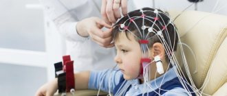
EEG registration is carried out by placing metal plates (electrodes) on the head, which are connected by wires to the input of the device. The output is a graphical representation of the oscillations of the difference in bioelectric potentials of a living brain.
Normal EEG
EEG is a complex curve consisting of waves of different frequencies and amplitudes. Depending on the frequency, EEG waves are distinguished, denoted by the Greek letters “alpha”, “beta”, “delta”, etc.
Alpha rhythm Beta rhythm Delta rhythm
In a healthy person, EEGs may vary depending on the physiological state (sleep and wakefulness, perception of visual or auditory signals, various emotions, etc.). The EEG of a healthy adult in a state of relative rest reveals two main types of rhythms: the α rhythm, characterized by an oscillation frequency of 8-13 Hz, and the β rhythm, manifested by a frequency of 14-30 Hz.
“The Father of Cybernetics” N. Wiener believed that alpha ri groups incoming information within a certain period of time, within which different events are perceived as simultaneous.
With various brain diseases, more or less severe disturbances of the normal EEG pattern occur, which can be used to determine the severity and location of the lesion, for example, to identify the location of a tumor or hemorrhage.
Features of EEG in various brain diseases
The beginning of the clinical use of EEG is considered to be the mid-30s, when in the USA Devis, Jasper and Gibbs discovered specific manifestations on the EEG in patients with petit mal seizures.
EEG in the diagnosis of epilepsy
The most informative is the registration of EEG of patients with epileptic seizures. EEG is the first and often the only neurological outpatient study that is performed during epileptic seizures.
First of all, EEG helps to distinguish epileptic seizures from non-epileptic ones and classify them.
With EEG you can:
- identify the areas of the brain involved in provoking seizures;
- monitor the dynamics of the action of drugs;
— resolve the issue of stopping drug therapy;
— identify the degree of brain dysfunction in interictal periods.
The best time to conduct an EEG is no earlier than a week after the attack. An electroencephalogram taken shortly after the attack may show no changes.
To understand this, we can use an analogy with a capacitor: a brain that has disturbances in the functioning of anticonvulsant systems accumulates changes, condenses them, which manifests itself in increasing disturbances on the EEG. During an attack, the brain experiences a kind of short circuit, discharging the changes accumulated in the brain.
Several percent of practically healthy adults experience disturbances in the bioelectrical activity of the brain in the form of various “epiphenomena”, conditioned epileptiform activity.
Perhaps this type of reaction is an innate feature that gives carriers of the corresponding genes some biological advantages. This, for example, is evidenced by the fact that in top-class pilots, who have the fastest reactions, EEG often shows discharges of the epileptiform type.
In children without clinical manifestations of epilepsy, but with psychopathy, with an aggressive character, and even just neurotics, “epiphenomena” are detected on the EEG even more often. This reaction usually disappears in older age without any treatment. However, 14-15% of children subsequently develop epileptic disease.
During grand mal seizures with loss of consciousness, the EEG may show peak-wave complexes in all areas of the brain (true paroxysmal activity - see figure),
and with focal epilepsy, changes are detected only in limited areas of the brain, more often in the temporal regions.
In people with alcoholic epilepsy, it is not always possible to detect convulsive activity on the EEG.
Conversely, changes similar to epileptic discharges can be caused by movements of the eyes and muscles of the head, pulsation of blood vessels, respiratory movements, heart function, chewing, swallowing or touching the electrode.
The results of the EEG depend on the age of the patient, the medications he is taking, the time of the last attack, the presence of tremor (shaking) of the head and limbs, visual impairment, and skull defects. All of these factors may influence the correct interpretation and use of EEG data.
Correctly interpreting EEG signals is somewhat of an art.
Functional tests are of great importance in the diagnosis of brain lesions: intermittent light stimulation (photostimulation), intense deep breathing for 2-3 minutes (hyperventilation), sound stimulation, research after a sleepless night (sleep deprivation), etc.
When using functional tests, EEG changes can be detected in 90% of patients with epilepsy.
The number of EEG examinations and their frequency depends on what the attending physician needs to identify. If there are no attacks (for example, if they are successfully treated), then an EEG can be done approximately 1-2 times a year. In the presence of attacks, changes in treatment or dosage of drugs, the frequency of EEG increases.
The diagnosis of epilepsy cannot be made in the absence of clinical manifestations of the disease and, conversely, this diagnosis cannot be excluded with a normal EEG if there are epileptic seizures. An EEG only helps the doctor clarify the diagnosis and determine the form of the attacks. Well, accordingly, it is not changes in the EEG pattern that are subject to treatment, but the attacks themselves.
EEG in the diagnosis of neoplasms
If the tumor is located close to the surface of the brain and affects mainly the cortex and subcortical structures, changes in the EEG occur on the affected side. Local pathological changes are noted in the area of tumor projection - inhibition of the alpha rhythm, increase in the amplitude of delta waves.
Intracerebral tumors cause significant general changes in the EEG, masking focal disturbances of biopotentials. To more clearly identify focal pathology, EEG studies are indicated after dehydration and hormonal therapy, leading to a decrease in diffuse slow waves.
For tumors of temporal localization, EEG diagnosis indicating the focus of pathological electrical activity in the temporal region is most accurate (up to 90%). As a rule, focal beta activity is observed.
According to modern standards, an EEG study can be recommended as a screening test for suspected tumors. Due to its harmlessness, relative accessibility and speed of implementation, if the doctor is unsure of the diagnosis, the EEG can tell him whether it is worth referring the patient for an additional (usually tomographic) study or not. EEG for vascular diseases and after injuries
The early period after a concussion is characterized by the presence of irritative changes, similar to disturbances in vascular diseases (see Fig.).
In the long-term period of TBI, a feature of the EEG is the presence of synchronicity of rhythms in different leads, often the low-amplitude nature of the EEG. A decrease or inversion of the fronto-occipital gradient of alpha activity is characteristic.
With EEG you can:
- monitor the dynamics of the action of drugs;
- assess the degree of brain dysfunction;
- examine the functional state of the brain in people in whom structural studies (for example, magnetic resonance imaging) show that the brain is “normal”, but brain dysfunction is obvious clinically (for example, in metabolic encephalopathy).
In these conditions, the greatest value of an EEG is not in confirming the diagnosis - the injury itself is “not visible” during examination. With repeated studies, EEG helps to assess the speed and completeness of the disappearance of signs of brain dysfunction. Further treatment depends on this.
Conducting research
EEG is completely harmless and painless. During the examination, the patient sits in a chair or lies on a couch with his eyes closed. To conduct an EEG, small electrodes are attached to the head using a special helmet, which are connected by wires to an electroencephalograph. The device amplifies the potentials received from the sensors hundreds of thousands of times and records them on paper or in computer memory.
If the examination is carried out on a child, then he needs to be explained what awaits him during the examination and convinced that it is painless. The patient should not feel hungry before the study, as this may cause changes in the EEG. The head should be washed clean before the EEG - this will allow for better contact of the electrodes with the scalp and obtain more reliable research results. With preschool children, it is necessary to practice putting on a “helmet” (playing as an astronaut, tank driver, etc.) and remaining motionless with eyes closed, and also teach them to breathe deeply and frequently.
If during an EEG the patient has a seizure, the effectiveness of the study increases significantly, since it will be possible to more accurately identify the location of the disturbance in the electrical activity of the brain. However, taking into account the interests of patient safety, seizures are not specifically provoked. Sometimes patients do not take medications before an EEG study. This should not be done.
An EEG study is performed by a specially trained neurologist, sometimes called an electroencephalographist or neurophysiologist. He describes the results of the study and gives his conclusion. However, a neurophysiologist cannot make a final diagnosis without more complete clinical data. Many EEG changes may be nonspecific, i.e. their accurate interpretation is possible only taking into account the clinical picture of the disease and sometimes after additional examination. Diagnostic value of EEG
Recently, electroencephalography has often been contrasted with new, high-tech methods for imaging brain activity, such as positron emission or functional magnetic resonance imaging (PET and fMRI). These methods provide a detailed image of the brain structures involved in functioning normally or when damaged by pathological processes.
What are the benefits of EEG? Some of them are obvious: EEG is quite easy to use, cheap and does not involve exposure to the subject (non-invasive). EEG can be recorded near the patient's bed and used to monitor the stage of epilepsy and long-term monitoring of brain activity.
But there is another, not so obvious, but very valuable advantage of EEG. In fact, PET and fMRI are based on measuring secondary metabolic changes in brain tissue, not primary ones (that is, electrical processes in nerve cells). An EEG can show one of the main parameters of the nervous system - the property of rhythm, which reflects the consistency of the work of different brain structures. Consequently, by recording an electrical (as well as magnetic) encephalogram, the neurophysiologist has access to the actual information processing mechanisms of the brain. This helps reveal the pattern of processes involved in the brain, showing not only “where” but also “how” information is processed in the brain. It is this possibility that makes EEG a unique and certainly valuable diagnostic method.
Electroencephalographic examinations reveal how the human brain uses its functional reserves.
Rhythms in sleep
A separate category of types of rhythms that manifest themselves either in sleep conditions or in pathological conditions includes three varieties of this indicator.
- Delta rhythm. Characteristic of the deep sleep phase and for comatose patients. It is also recorded when recording signals from areas of the cerebral cortex located on the border with areas affected by oncological processes. Sometimes it can be recorded in children 4–6 years old.
- Theta rhythm. The frequency interval is within 4–8 Hz. These waves are triggered by the hippocampus (information filter) and appear during sleep. Responsible for high-quality assimilation of information and forms the basis of self-learning.
Based on the results obtained during EEG recording, an indicator is determined that characterizes a complete all-encompassing assessment of the waves - bioelectrical activity of the brain (BEA). The diagnostician checks the EEG parameters - frequency, rhythm and the presence of sharp flashes that provoke characteristic manifestations, and on these grounds makes a final conclusion.
Contraindications
The main contraindication for this procedure is the patient’s intolerance to pulsed flashing light. Such intolerance is usually observed in patients with epilepsy, people with unstable mental health, and with cancerous tumors in the brain. You should talk to your ophthalmologist about the possibility of other contraindications.
MGK specialists have developed and implemented special eye photostimulation programs. The procedures are carried out using the most modern equipment and can be prescribed to adults and children. After carrying out the necessary diagnostic studies, an individual hardware treatment regimen is selected for each patient, which is then adjusted in accordance with the current results. The procedures are carried out under the supervision of highly qualified specialists, do not cause discomfort and do not require any restrictions in the usual way of life.
Decoding of electroencephalogram indicators
In order to decipher the EEG and not miss any of the smallest manifestations in the recording, the specialist needs to take into account all the important points that may affect the indicators being studied. These include age, the presence of certain diseases, possible contraindications and other factors.
Upon completion of the collection of all research data and their processing, the analysis is completed and then a final conclusion is formed, which will be provided for making a further decision on the choice of therapy method. Any disturbance in activity may be a symptom of diseases caused by certain factors.
Alpha rhythm
The normal frequency is determined in the range of 8–13 Hz, and its amplitude does not go beyond 100 μV. Such characteristics indicate a healthy state of a person and the absence of any pathologies. The following are considered violations:
- constant fixation of the alpha rhythm in the frontal lobe;
- exceeding the difference between the hemispheres by up to 35%;
- constant violation of wave sinusoidality;
- presence of frequency dispersion;
- amplitude below 25 μV and above 95 μV.
The presence of disturbances in this indicator indicates a possible asymmetry of the hemispheres, which may be the result of oncological tumors or pathologies of cerebral circulation, for example, stroke or hemorrhage. A high frequency indicates brain damage or TBI (traumatic brain injury).
A complete absence of the alpha rhythm is often observed in dementia, and in children, deviations from the norm are directly related to mental retardation (MDD). Such a delay in children is evidenced by: disorganization of alpha waves, shift of focus from the occipital region, increased synchrony, short activation reaction, overreaction to intense breathing.
These manifestations can be caused by inhibitory psychopathy, epileptic seizures, and a short reaction is considered one of the primary signs of neurotic disorders.
Beta rhythm
In the accepted norm, these waves are clearly detected in the frontal lobes of the brain with a symmetrical amplitude in the range of 3–5 μV, recorded in both hemispheres. A high amplitude leads doctors to think about the presence of a concussion, and when short spindles appear, to the occurrence of encephalitis. An increase in the frequency and duration of spindles indicates the development of inflammation.
In children, the pathological manifestations of beta oscillations are considered to be a frequency of 15-16 Hz and a high amplitude present - 40-50 µV, and if its localization is the central or anterior part of the brain, then this should alert the doctor. Such characteristics indicate a high probability of delayed development of the baby.
Delta and theta rhythms
An increase in the amplitude of these indicators above 45 μV on a constant basis is characteristic of functional brain disorders. If the indicators are increased in all brain regions, then this may indicate severe dysfunction of the central nervous system.
If a high amplitude of the delta rhythm is detected, a tumor is suspected. Inflated values of the theta and delta rhythm recorded in the occipital region indicate a child’s lethargy and a delay in his development, as well as impaired circulatory function.
Classification by E.A. Zhirmunskaya
A system for describing EEG types (patterns) and groups was proposed by E.A. Zhirmunskaya in 1984 and is still the basis for visual, verbal assessment of EEG. It should be noted that according to the classification of E.A. Zhirmunskaya's (1991) concept of EEG type refers only to resting EEG recorded in subjects in a state of passive wakefulness, with their eyes closed, and does not include changes that occur in response to efferent stimuli.
The advantage of this classification is the ability to give a general description of the EEG. This creates a certain convenience for the subsequent formation of a conclusion by a medical specialist. At the same time, when making a judgment about the type of EEG, the nature of the EEG pattern in various types of epilepsy is not taken into account, and the focus of pathological activity is not highlighted. According to the classification of E.A. Zhirmunskaya distinguishes 5 types of EEG.
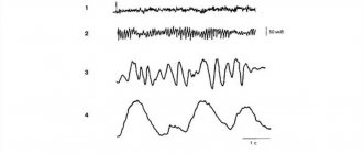
Type I – organized (normal EEG)
The main component of the EEG is the α rhythm, regular in frequency, clearly modulated into spindles, with a medium and high index, with well-defined zonal differences. The α rhythm has the highest amplitude (50-70 μV) in the occipital leads. Towards the anterior parts of the cortex, the severity of the α rhythm decreases. The wave shape is usually smooth. In the temporal regions of the cortex, the α rhythm is presented in the form of fragmentary low-amplitude spindles.
Slow waves are almost not pronounced. δ-activity can also be recorded in the form of single diffuse waves of small amplitude (Fig. 1).
It is advisable to classify EEG with a less ordered structural and spatial organization as this type, the main criterion of which is pronounced α-activity (Fig. 2).
EEGs belonging to type I are interpreted as the ideal norm or as slight changes within the acceptable range of the norm.
Rice. 1. Male, 46 years old.
Electroencephalogram within the age norm. The α-rhythm is modulated, zonal differences are clearly expressed, fragmentary θ- and δ-waves are recorded in the anterior sections
Rice. 2. Man, 72 years old.
Electroencephalogram within the age norm. The α-rhythm is weakly modulated, zonal differences are clearly expressed. Diffuse θ- and δ-activity is recorded in the fronto-central regions
Type II – hypersynchronous (monorhythmic)
It may have variants with a predominance in all areas of the brain or only the α-rhythm, low-frequency ß-activity, or rhythmic θ-activity. The main thing in the structure of this type is the high index of regular fluctuations of biopotentials with the loss of their zonal differences.
Various options for such enhancement of activity synchronization are possible: with preservation and even enhancement of α-range oscillations; with the disappearance of α-activity and its replacement by low-frequency ß-activity or θ-activity. The main characteristic of this type of EEG is the increased regularity of oscillations in the α- (less often ß- or θ-) ranges with the absence of zonal differences.
The option when the α-rhythm dominates, unmodulated or weakly modulated with mild or distorted zonal differences and the absence of fast and slow activity, refers to a moderately disturbed EEG (Fig. 3, 4).
When regular ß-activity with a frequency of 14-25 Hz of small amplitude predominates, the EEG is also considered moderately impaired. In the presence of moderately high or high ß-activity, the EEG is characterized as significantly impaired (Fig. 5).
Rice. 3. Woman, 60 years old.
Encephalopathy. The electroencephalogram shows a hypersynchronous weakly modulated α-rhythm, enhanced in all leads, zonal differences are reduced
Rice. 4. Man, 50 years old.
Symptomatic partial epilepsy. The electroencephalogram shows a hypersynchronous weakly modulated α-rhythm, enhanced in the frontal-central-parietal regions of the brain, zonal differences are reduced, synchronous bilateral discharges of α- and δ-wave complexes periodically occur
Rice. 5. Male, 45 years old.
A complex of functional disorders in the intestines, a feeling of discomfort, increased emotionality. The electroencephalogram shows hypersynchronous high-amplitude ß-activity, enhanced in all leads
Type III – desynchronous (normal variant)
EEG is characterized by low-amplitude bioelectrical activity with the absence or sharp decrease in the number of α-waves. ß-Activity is moderate, diffuse θ- and δ-waves of small amplitude are noted (Fig. 6).
EEGs classified as types II and III reflect regulatory changes in brain activity. In type II, there is a weakening of the activating influences on the cortex from the reticular formation of the brain stem and an increase in deactivating influences from other parts of the limbic-reticular complex.
In type III, on the contrary, there is an increase in activating influences from the reticular formation of the brain stem, which is expressed in desynchronization of α-activity on the EEG. EEGs classified as types II and III reflect regulatory changes in brain activity. Types II and III EEG can occur in patients with neuroses, vegetative-vascular dystonia, cervical osteochondrosis and a number of other diseases.
Rice. 6. Male, 19 years old.
There are no neurological complaints. The electroencephalogram shows low-amplitude bioelectrical activity with the presence of irregular diffuse Θ- and δ-waves. α-Activity is fragmentary and low-amplitude. Variant of the norm
Type IV – disorganized (with a predominance of α-activity)
On the EEG, the main activity is α-activity. In this case, there is significant disorganization of the α-rhythm in all parameters: frequency, amplitude, waveform, distortion of zonal differences. This disorganized α rhythm can dominate all areas of the brain. ß-Activity is also often enhanced, often represented by low-frequency oscillations and increased amplitude; there is an increase in the index of θ- and δ-waves with a fairly high amplitude, the appearance of peaks, sharp waves, flashes and complexes in different areas of the brain. This type of EEG reflects changes in biopotentials in many diseases, which can be associated with microstructural lesions in different parts of the brain, including the cerebral cortex. This category includes atherosclerosis, transient ischemic attacks, neuroinfections, and closed craniocerebral injuries (Fig. 7; 8; 9). Disturbances of the walls of brain vessels and microscopic lesions of brain tissue that occur in these diseases are accompanied by changes in metabolism and neurodynamic disorders. This is accompanied by dysfunction in the activity of the regulatory systems of the brain.
Rice. 7. Male, 38 years old.
5 years ago there was a severe traumatic brain injury with loss of consciousness, loss of ability to work: impaired coordination of movement, slowness of speech. The electroencephalogram was recorded against the background of restoration of working capacity, disorganization of the α-rhythm in shape and frequency, the presence of θ-, δ-rhythm and sharp waves were noted
Rice. 8. Male, 31 years old.
Rice. 9. Woman, 22 years old.
Prolactinemia. The electroencephalogram shows disorganization of the α-rhythm, the presence of sharp waves, and groups of θ-activity
Type V – disorganized (with a predominance of θ- and δ-activity)
δ-Activity is poorly expressed. Amplitude level is medium or high. The type is characterized by the weak presence of α-activity. Fluctuations in biopotentials in the α-, ß-, θ- and δ-ranges are recorded without a clear sequence (Fig. 10).
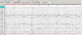
Type V means that in patients it is no longer regulatory, but microstructural lesions in the cerebral cortex that come to the fore. If there is an EEG of this type, there is every reason to talk about organic brain damage. If an EEG of this type shows a clear focus of pathological activity or a distinct interhemispheric asymmetry, we can assume the presence of a gross macrofocal lesion in one of the brain hemispheres (Fig. 11). In the presence of local pathological activity, it is recommended to view the EEG in bipolar montage mode, which will clarify the localization of the lesion (Fig. 12, 13). The appearance of both local and diffuse pathological phenomena and paroxysmal discharges most often accompanies this particular type of EEG, although it can also occur against the background of any other type of EEG.
Rice. 10. Woman, 66 years old. Secondary generalized convulsive attack.
Cyst in the right anterior frontal region. On the electroencephalogram, fluctuations in the biopotentials of the α-, ß-, Θ- and δ-ranges are recorded without any clear sequence. A clear focus of δ activity is detected in the right temporal region
Rice. 11. Male, 24 years old.
At the age of 8 years he was operated on for a tumor in the left frontotemporal region. At the age of 10, a tumor of the optic chiasm was discovered; bone grafting was not performed. Currently there are generalized seizures at night. The electroencephalogram shows disorganized bioelectrical activity and the presence of local δ-activity in the fronto-parietal-temporal region of the left hemisphere
Rice. 12. Woman, 19 years old.
Partial epilepsy. The electroencephalogram shows disorganized bioelectrical activity, the presence of local δ-activity in the central region of the left hemisphere and the temporal region of the right hemisphere, and a focus of ß-activity in the parietal regions of the right hemisphere
Rice. 13. Woman, 19 years old. Partial epilepsy.
With bipolar montage, the same segment of the electroencephalogram is presented as in Fig. 2.43 δ-activity in leads F3-C3, SZ-RZ and in leads F8-T4 is recorded in antiphase, which suggests the presence of lesions in the left central region (C3) and in the right temporal region (T4) respectively.
Decoding values in different age intervals
An EEG recording of a premature baby at 25–28 gestational weeks looks like a curve in the form of slow flashes of delta and theta rhythms, periodically combined with sharp wave peaks 3–15 seconds long with a decrease in amplitude to 25 μV. In full-term infants, these values are clearly divided into three types of indicators. During wakefulness (with a periodic frequency of 5 Hz and an amplitude of 55–60 Hz), the active phase of sleep (with a stable frequency of 5–7 Hz and a fast low amplitude) and quiet sleep with flashes of delta oscillations at a high amplitude.
Over the course of 3-6 months of a child’s life, the number of theta oscillations is constantly growing, while the delta rhythm, on the contrary, is characterized by a decline. Further, from 7 months to a year, the child develops alpha waves, and delta and theta gradually fade away. Over the next 8 years, the EEG shows a gradual replacement of slow waves with fast ones - alpha and beta oscillations.
Until the age of 15, alpha waves predominate, and by the age of 18, the BEA transformation is complete. Over the period from 21 to 50 years, stable indicators remain almost unchanged. And from 50, the next phase of rhythmicity restructuring begins, which is characterized by a decrease in the amplitude of alpha oscillations and an increase in beta and delta.
After 60 years, the frequency also begins to gradually fade, and in a healthy person, manifestations of delta and theta oscillations are noticed on the EEG. According to statistics, age indicators from 1 to 21 years, considered “healthy,” are determined in subjects 1–15 years old, reaching 70%, and in the range of 16–21 – about 80%.
EEG classification according to Cohn
Some authors propose to classify EEGs according to the severity of the anomalies encountered in them. Thus, Cohn (1949) distinguishes four types of EEG: 1) normal and borderline altered; 2) easily changed; 3) moderately altered and 4) severely altered.
- Borderline altered EEG. EEGs are said to be borderline normal when there are slight deviations from an unchanged EEG. This means that the degree of alpha activity shows fluctuations, reaching the point of irregular activity. The amplitude of alpha waves can be very large, and interhemispheric amplitude differences can reach 30%. Theta waves often reach the voltage of alpha waves. Beta activity may be recorded more clearly than in the beta EEG group. Such changes are often observed in autonomic and vasomotor disorders, in psychopaths, and sometimes as an electrographic expression of those residual effects of damage to the central nervous system that were present in early childhood. The appearance of central arc-like waves (rhythms), high alpha activity and the splitting of theta frequencies into harmonics can be assessed as an indicator of increased excitability. Similar changes in the EEG are often found in various chronic diseases, states of internal tension, circulatory disorders and developmental delays.
- Easily modified EEGs. Alpha activity is irregular or very labile, that is, its frequency fluctuates more than ± 1.5 fluctuations per second from the average value. Amplitude interhemispheric differences exceed 30%. The effect of closing the eyes is poorly expressed or absent. Diffuse or local theta activity is higher than normal. High beta activity, small sharp waves, and paroxysmal groups of theta waves may occur.
- Moderately altered EEG. The alpha rhythm slows down to 8–7/s or is absent altogether. There is a clear interhemispheric asymmetry. Diffuse theta activity predominates. There are mid-group delta waves and peaks. Such changes are an expression of pathological disorders of brain function.
- Severely altered EEG. The alpha rhythm is absent or there are small groups of it with a sharply slowed frequency. Diffuse theta and delta waves are recorded. High amplitude trains of beta waves may occur. Polymorphic slow activity can be periodic or continuous, causing the presence of dysrhythmia with significant fluctuations in frequency and amplitude. Paroxysmal types of activity are common23.
The most common diagnosed pathologies
Thanks to the electroencephalogram, diseases such as epilepsy or various types of traumatic brain injury (TBI) are quite easily diagnosed.
Epilepsy
The study allows you to determine the localization of the pathological area, as well as the specific type of epileptic disease. At the time of a convulsive syndrome, the EEG recording has a number of specific manifestations:
- pointed waves (peaks) - suddenly rising and falling can appear in one or several areas;
- the combination of slow pointed waves during an attack becomes even more pronounced;
- sudden increase in amplitude in the form of flashes.
The use of stimulating artificial signals helps in determining the form of epileptic disease, since they provide visibility of hidden activity that is difficult to diagnose with EEG. For example, intense breathing, requiring hyperventilation, leads to a decrease in the lumen of blood vessels.
Photostimulation is also used, carried out using a strobe (a powerful light source), and if there is no reaction to the stimulus, then most likely there is a pathology associated with the conduction of visual impulses. The appearance of non-standard vibrations indicates pathological changes in the brain. The doctor should not forget that exposure to powerful light can lead to an epileptic seizure.
What is EEG?
Electroencephalography (EEG) is a diagnostic method that allows you to assess the functional activity of various parts of the brain, identify signs of pathological changes in the cerebral cortex in various diseases in children and adults (convulsive syndrome and epilepsy, tumors, vascular disorders, consequences of injuries, neurotic and mental disorders ).
EEG allows you to record the bioelectrical activity (electrical potentials) of various parts of the brain and record the results on paper or on a computer monitor screen. The result is an encephalogram - a graphic curve in the form of rhythms of different heights, amplitudes, and durations, among which pathological elements can be found. The results obtained are analyzed.