- home
- Functional diagnostics
- Ultrasound of the head
Ultrasound of the head and neck is an important and very informative type of examination. In addition to providing excellent visualization of stationary objects such as lymph nodes or mucous membranes, the method also provides information about dynamic environments such as the bloodstream. The combination of these characteristics makes sonography a mass screening method for a wide variety of patient groups. Moreover, ultrasound of the head is so safe that it is performed even on infants just a few days old.
Ultrasound of cerebral vessels
Dopplerography of cerebral vessels is prescribed for suspected neurological diseases. The following symptoms may indicate this:
- Panic attacks.
- Difficulty speaking.
- Difficulties in perceiving information.
- Impaired coordination of movements.
- Decreased visual, auditory, kinesthetic sensations.
Based on the results of the ultrasound, the doctor can determine what changes in the structure of the blood vessels could cause these symptoms and prescribe treatment, if necessary.
The procedure is simple and safe and has no age restrictions. It can be carried out at home using a portable scanner.
The essence of Doppler ultrasound
Based on the name of the procedure, it is performed using ultrasonic waves. Their work is based on the Doppler effect. The procedure differs from a standard ultrasound: a vessel with blood passing through it is displayed on the monitor screen. Looking at the resulting image, the specialist assesses the state of blood flow and vascular patency, which affects the quality of blood supply to the brain.
Dopplerography of cerebral vessels makes it possible to study the work of the veins, which “supply” blood to the brain, and the arteries, which are responsible for the outflow of waste products from it. The vessels being examined are located in the cervical and cranial regions. The type of ultrasound will depend on their location:
- Dopplerography of extracranial vessels located on the surface of the neck (jugular veins, carotid, vertebral and subclavian arteries). Any disturbances in their structure affect the state of the brain, which is why these vessels are called “maternal” vessels.
- Dopplerography of transcranial, or “daughter” vessels. The doctor installs an ultrasound sensor in the thinnest parts of the skull and evaluates the functioning of the veins and arteries that supply the brain.
When and why is duplex scanning of the vessels of the neck and head performed?
Indications for the examination are varied, primarily dizziness, headaches, lightheadedness, episodes of loss of consciousness, swaying when walking, osteochondrosis of the cervical spine, numbness or weakness in the arm or leg, asymmetry of blood pressure and pulse in the arms, decreased memory and concentration attention, speech impairment, previous stroke or ischemic attack, condition after surgery on the carotid arteries, increased blood pressure (arterial hypertension), coronary heart disease, diabetes mellitus. In case of cervical osteochondrosis, duplex scanning can reveal signs of compression of the vertebral arteries, including when performing tests with head turns.
Duplex scanning is recommended for people over 40 years of age
, even if they feel healthy, but have risk factors (high cholesterol levels in the blood; family history of cardiovascular diseases; smoking; sedentary lifestyle, etc.). The purpose of the examination is to determine the patency of blood vessels, assess the presence, size and structure of intraluminal formations (atherosclerotic plaques), study the condition of the vascular wall, the geometry of the vessels (the presence of deformations, tortuosities), and assess the quantitative and qualitative characteristics of blood flow in the vessels under study. Detection of the pathological process is possible in almost any area of the vascular system. That is why it is important to conduct a detailed study of extracranial and intracranial vessels along the entire length available for study.
Up
How is the procedure done?
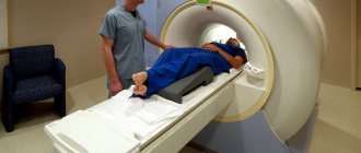
Ultrasound duplex scanning of the vessels of the neck and head does not require special preparation. Before the procedure, it is advisable to refrain from smoking, drinking strong tea, coffee, alcohol, and energy drinks. If you are taking medications that improve cerebral circulation, they should not be discontinued without the consent of your doctor. The examination is carried out lying on your back, with your head slightly turned to the side opposite to the side being examined. The doctor conducting the duplex scan moves a sensor on the surface of the neck, on which a special gel is applied to improve contact between the sensor and the skin. When performing the examination, slight pressure from the sensor may be applied; most people tolerate this easily. The monitor screen of an ultrasound scanner displays the information received by the sensor using ultrasound and converted by the device into the form of a digital signal. During the test, you may also hear special noises that help assess the flow of blood in your body.
Thus, the doctor assesses the condition of all the main vessels of the neck - the carotid, vertebral and subclavian arteries, internal jugular and vertebral veins on the left and right. Then transcranial duplex scanning (TCDS) is performed,
that is, the study of intracranial vessels. The doctor alternately installs a special sensor on different areas of the head: temporal, occipital, and sometimes supraorbital. Additionally, rotational and other functional tests are carried out. A comprehensive study of the vessels of the neck and brain is optimal for problems solved by neurologists, chiropractors, cardiologists, and cardiovascular surgeons. This scope of research corresponds to modern diagnostic standards. The whole procedure lasts about 30-40 minutes. The protocol, which is handed to the patient, records all blood flow indicators with a full description of the detected pathology, and gives a general conclusion on the study. If you have had a similar study done before, it is advisable to take the previous protocol with you for comparison.
Up
Are there any contraindications to duplex scanning?
Unlike other instrumental research methods, ultrasound duplex scanning of blood vessels has no contraindications. Ultrasound examination is completely harmless for the patient. The safety of the ultrasound diagnostic method is one of its advantages, which allows repeated examinations over time and evaluation of the effectiveness of the treatment.
What pathology can duplex scanning detect?
Up
The most common pathology detected by duplex scanning is atherosclerosis. This is the deposition of fatty substances (lipids) and the proliferation of connective tissue in the walls of the arteries. Currently, atherosclerosis is considered as a complex, multicomponent pathological process affecting the arterial wall, lipid metabolism, blood coagulation system, and cellular elements. Although the prevalence of atherosclerosis increases with increasing age, it is a disease and not an age-related change. Atherosclerosis also occurs in young people, and may be absent in old people. Atherosclerosis of the main arteries of the head and neck most often develops in the third or fourth decade of life. An early symptom of atherosclerotic lesions of the carotid arteries is an increase in the thickness of the vessel wall (intima-media). The ultrasound method of duplex scanning will reveal signs of atherosclerotic lesions of the brachiocephalic arteries (thickening of the intima-media, changes in its structure, the presence of atherosclerotic plaques, their nature, extent, degree of narrowing of the vessel and the nature of blood flow at the site of narrowing). Detection of atherosclerotic vascular changes in a patient with arterial hypertension is associated with a high risk of adverse cardiovascular events (acute coronary syndrome, myocardial infarction, stroke, etc.). Early detection of stenosis (narrowing) of the carotid arteries helps prevent such a serious and dangerous disease as stroke. In a number of foreign countries that have achieved success in preventing stroke, an annual examination of the entire population over 40 years of age is used, using ultrasound techniques (duplex scanning of the vessels of the head and neck with color mapping of blood flow). This allows you to determine the risk group.
In addition to atherosclerotic changes, duplex scanning reveals various congenital vascular anomalies: deformations of the carotid and vertebral arteries, pathological tortuosity; hypoplasia (congenital reduction in the diameter of the vessel), anomalies of entry and exit of the vertebral arteries. In pathology of the cervical spine, disturbances in the course of the vertebral arteries, asymmetry of blood flow and signs of compression of the vertebral arteries in the canals of the transverse processes of the cervical vertebrae are most often detected.
Thus, ultrasound duplex scanning with color flow is a highly informative and completely safe diagnostic method; it helps to identify vascular pathology at the initial stage. The results of the examination will help your doctor create an individual plan of treatment and preventive measures in order to maintain your health.
Calcified atherosclerotic plaque at the mouth of the internal carotid artery (two-dimensional mode).
Characteristics of blood flow in the common carotid artery (two-dimensional echography, spectral Doppler and color flow mode).
Characteristics of blood flow in the middle cerebral artery (two-dimensional echography, spectral Doppler and color flow mode).
In what cases is ultrasound ultrasound prescribed?
The main symptoms signaling a cerebrovascular accident are:
- Impaired coordination of movements.
- Deterioration of hearing and memory.
- Frequent attacks of headache.
- Impairment or partial loss of taste.
- Insomnia, difficulty falling asleep.
- Noise, whistling and ringing in the ears.
- Deterioration of visual function, manifested in loss of visual fields.
- Difficulty concentrating.
- Decreased sensitivity and motor activity of the arms and legs.
A doctor may prescribe a procedure in the following cases:
- The patient has diabetes mellitus.
- For arrhythmia, as this increases the risk of cardiac clot rupture and artery blockage.
- The patient has suffered a heart attack or stroke.
- Osteochondrosis of the cervical spine.
- Before planned heart surgery.
- If the patient is a smoker with many years of experience.
- An area with strong pulsation is visible on the neck (in this case, an ultrasound of the neck is prescribed).
Based on the above symptoms, the doctor can determine the location and name of the vessel in which the patency is impaired. Several pronounced symptoms indicate poor blood flow in a large vessel. In this case, extracranial Doppler ultrasound is prescribed. Violation of only one function will be the reason for conducting transcranial Doppler ultrasound.
Difference between ultrasound, duplex and triplex examination of the vessels of the head and neck
Ultrasound Dopplerography (Doppler ultrasound) of the vessels of the head and neck —
an outdated method that is sometimes still used. During Doppler sonography, only the patency of the vessel and the speed of blood flow can be determined. If a blockage is detected, it is impossible to clarify the cause, since with this method the doctor does not see the vessel. During an ultrasound examination, the doctor can only guess about the cause of the disease based on the speed of blood flow, pulse sinusoid frequency, and blood pressure.
Duplex scanning of vessels (USDS) allows you to visualize the vessel in black and white. It is possible to evaluate not only the patency of the vessel and the speed of blood flow, but also to find the reasons that led to the violation: narrowing, tortuosity, abnormal development of the vessel; blood clots; atherosclerotic plaques, etc. During a duplex ultrasound, the doctor uses two ultrasound modes: ultrasound visualization of the wall and lumen of the vessel in gray tones and spectral Doppler ultrasound (USDG).
During a triplex study of the vessels of the head and neck, in addition to the above modes, a third mode is additionally used - color Doppler mapping of blood flow (CDB). Triplex vascular study is sometimes called a more complex name - duplex vascular study using color coding of blood flow (CDC).
Triplex ultrasound of head and neck vessels expands the capabilities of duplex scanning by displaying the direction of blood flow in red and blue. The color indicates to the doctor the direction of blood flow: shades of red - towards the ultrasound sensor, shades of blue - away from the sensor. Higher blood flow speeds are colored in lighter tones of red or blue, and low ones are colored in darker tones.
Fig 1. Carotid artery stenosis (triplex ultrasound of neck vessels)
With triplex ultrasound, a more accurate diagnosis is achieved, since using three different scanning modes it is easier for the doctor to see areas of vessel narrowing, turbulence in blood flow, areas of lack of blood flow, and identify atherosclerotic plaques, blood clots, abnormalities in vascular development and their significance. In addition, the color mode allows you to detect small vessels that cannot be detected with black and white imaging
Rules for preparing for research
On the day of the procedure, you must refrain from:
- Taking antispasmodics such as “Riabal”, “No-shpa”, “Drotaverine”, “Papaverine”, “Baralgin”, “Cinnarizine”.
- Smoking.
- Drinking black tea and drinks containing caffeine.
- Staying in unventilated, stuffy rooms with large crowds of people, which can have a detrimental effect on vascular tone.
If the patient is undergoing treatment with cardiovascular medications, then the advisability of discontinuing them before the procedure must be agreed with a neurologist. In any case, the sonologist must be informed about the treatment being performed before the ultrasound.
Stages of extracranial Dopplerography:
1. In the office, remove all jewelry from your neck and head.
2. Remove clothing from those parts of the body through which the sensor will be passed: neck, collarbones, shoulder blades.
3. Lie on the couch with your head facing the monitor. The doctor will apply acoustic gel to open areas of the body and begin to move the device over them, placing it in areas where medium and large arteries and veins are located. To obtain more accurate data on the state of vascular tone, the patient will need to hold his breath at the doctor’s request, take special medications and change body position.
Doppler ultrasound of vessels located in the cranial area has a different specificity. Acoustic gel is applied to the temples, the back of the head and the area above the eyes. The device is installed there because in these places the bones of the skull are the thinnest and are able to transmit ultrasonic waves.
Preparation for ultrasound of the brachiocephalic arteries
The procedure does not require special preparation from the patient, however, to increase its accuracy, it is recommended to adhere to several rules:
- On the day of the examination, avoid drinking tea, coffee, energy and alcoholic drinks.
- Coordinate your medication intake with your doctor.
- 2-3 hours before the diagnosis, refrain from hot baths and smoking.
Methodology
Ultrasound of the brachiocephalic arteries and vessels consists of examining the arterial vascular wall and analyzing changes in its diameter. During the procedure, the IMM (intimate media complex) of the carotid artery is examined.
The patient is in a supine position, and a special conductive gel is applied to the skin in the neck area. The duration of the manipulation is approximately 30-45 minutes.
The patient is prohibited from turning his head or talking during the examination. To obtain the most accurate picture, the doctor can conduct several tests: examination in an upright position, holding your breath and ultrasound using antianginal agents.
The doctor immediately writes down the test results, and at the end of the ultrasound diagnosis the patient receives a conclusion with all the necessary data.
What does ultrasound of the brachiocephalic arteries show?
During diagnosis, the specialist receives the following type of information:
- The degree of patency of blood vessels and arteries.
- State of the intimate-media complex of the carotid artery.
- The presence of stenosis (narrowing) and the degree of its severity.
- Blood flow speed.
- Damage to vascular atherosclerosis.
Separately, during ultrasound, the extracranial section of the brachiocephalic arteries is studied. The data obtained make it possible to identify a number of chronic processes in the body and determine the risk of ischemic stroke.
By what principle is the received data decrypted?
Each vessel has its own parameters, on the basis of which calculations are made and the degree of their discrepancy with the norm is assessed.
Transcranial and extracranial Dopplerography of arteries compares digital data in each vessel segment:
- Artery wall thickness.
- Vessel diameter.
- Phases of blood flow, its features.
- The degree of symmetry of blood flow through identical arteries located on different sides.
- Diastolic blood velocity.
- Maximum systolic blood flow velocity.
- Pulsation and resistive indices, systole and diastole ratio.
- The degree of pathological narrowing of blood vessels (stenosis), the condition of the artery beyond its limits.
Dopplerography of veins evaluates the following parameters:
- Condition of the venous wall of the vessel.
- The nature of blood movement through the studied vessels.
- Vein diameter
Data obtained at rest and after functional tests are compared with normal values. Any deviations are marked with a special abbreviation of letters and numbers. Based on them, the neurologist prescribes further studies or forms a course of treatment.
Where can I get Doppler ultrasound of cerebral vessels?
The study is carried out in municipal clinics and public hospitals, specialized clinics, and general medical centers. Depending on the location of the ultrasound, the cost of each type of Doppler ultrasound will be in the range of 500-6000 rubles. The procedure has many advantages, including painlessness and the absence of side effects. Within 20 minutes, a sonologist will be able to determine the presence of anomalies in venous or arterial inflow and assess the degree of development of danger to blood vessels.
However, the procedure will not help determine the cause of compression of the thin arteries, since the capabilities of the device do not allow them to be seen from the outside.
How to prepare a child for a brain ultrasound
There are no special requirements for performing an ultrasound. You will not need to adhere to a special diet or regimen. The main thing is psychological preparation.
If a child has reached a conscious age and is afraid of visiting a doctor, you need to talk to him and tell him about what will happen in the office. Particularly emphasize that it does not hurt at all, and the baby will be able to look at the large monitor and see how the doctor works.
It is also best to take your medical history and medical record from the clinic with you to your appointment, as well as a referral from your attending physician.