10.10.2014
Maltseva Marina Arnoldovna
Head of the consultation department - neurologist, specialist in the field of extrapyramidal pathologies, doctor of the highest category
A fracture of the base of the skull is one of the most severe traumatic brain injuries. This condition is accompanied by damage to one or even several bones that form the base of the skull. Damaged in this case may be:
- occipital bone;
- temporal bone;
- ethmoid bone;
- sphenoid bone.
A fracture of the base of the skull can occur from a variety of serious physical injuries: car accidents;
- when a person falls from a great height;
- when hitting the face in the area of the lower jaw or base of the nose.
In most patients, the base fracture occurs from the arch. Statistics say that the number of such patients reaches 59%.
Symptoms of a basal skull fracture
A fracture of the base of the skull is an open traumatic brain injury. It is worth noting that injuries accompanied by the release of blood or cerebrospinal fluid from the ear canals or nose are classified as open penetrating head injuries.
Based on location, injuries are divided into fractures:
- middle cranial fossa;
- anterior cranial fossa;
- posterior cranial fossa;
The most common fractures encountered in medical practice are middle cranial fossa fractures. Such fractures can be longitudinal, transverse or oblique. Most often, doctors fix longitudinal fissures in the temporal lobes. In this case, fractures can spread through various holes, crevices and thinning of the skull bones.
Among the main manifestations of fractures:
- bleeding from the ear canal;
- decreased hearing acuity;
- release of cerebrospinal fluid;
- the appearance of bruises in the area of the temporal muscle.
All of the above damage occurs when there is a blow to the occipital region.
It is worth noting that transverse fractures are clinically characterized by the patient being completely deaf. The patient may also exhibit disturbances in vestibular function, loss of taste in the two anterior thirds of the tongue, and may also experience peripheral facial palsy.
Causes of skull fracture
All causes of skull fracture are inherently mechanical injuries. In such cases, injury to the bone tissue of the head may occur:
- Falling from a great height or high speed;
- Hit your head hard with a heavy object;
- As a result of a traffic accident (car accident).
All these situations can arise as a result of non-compliance with safety rules on the road and at work, as a result of street fights and aggressive sports in sports clubs, or extreme sports.
When assessing age and social categories, groups at increased risk of skull fracture include children, active teenagers and middle-aged people, as well as people dependent on drugs and alcohol.
Consequences of skull base fractures and prognosis
Experts note that the future quality of life of patients directly depends on the quality of rehabilitation after TBI, as well as on the nature of this injury, pathologies and possible infections. In the absence of purulent inflammation, in most cases, doctors give a favorable prognosis.
If there are infectious complications, doctors talk about the possibility of developing complications such as encephalopathy, frequent headaches, uncontrolled increase in blood pressure, etc. in the future. This condition may also be accompanied by recurrent epileptic seizures.
Some traumatic brain injuries can cause excessive bleeding, coma, and even death. With such injuries, the prognosis of doctors is extremely unfavorable.
Bleeding of small volumes, intracerebral hematomas, etc. can form. It is worth noting that in this situation, the patient’s future directly depends on the timeliness and adequacy of the treatment.
Clinical Brain Institute Rating: 3/5 — 10 votes
Share article on social networks
Skull fracture: diagnostic methods
Diagnostics is carried out comprehensively and includes the following activities:
- Medical examination and collection of information about the patient’s ailments. During the examination, the doctor assesses the general condition of the victim, the reaction of the pupils, and measures the pulse and blood pressure. The position of the tongue and the symmetry of the patient's jaw are also assessed. Neurological reactions are studied;
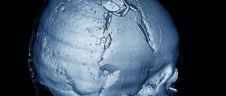
- X-rays of the skull are taken in two projections;
- A computed tomography (CT) or magnetic resonance imaging (MRI) scan is performed.
In extremely severe cases, when it is impossible to make a complete diagnosis, treatment of the patient is prescribed based on the external symptoms of the pathology.
First aid
For a fracture of the base of the skull, the most important factor is proper first aid. First of all, you need to call an ambulance, and then begin the necessary manipulations to alleviate his condition.
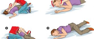
First of all, the victim must be positioned correctly. If he is conscious, they put him on his back; if he is unconscious, he must be turned on his side and placed under a cushion so that in case of nausea he does not choke on vomit. The head should be at rest. To do this, you can use folded clothes or a pillow, whatever you have at hand. Then a sterile tight bandage with an antiseptic should be applied to the wound.
Be sure to unbutton your clothes to ensure free access of air. It is also necessary to remove dentures (if any) and remove glasses.
Before the ambulance arrives, if breathing is impaired, artificial respiration is performed. The patient should be given heart medications.
If a long time may pass before the ambulance arrives, ice should be applied to the injury site and, if possible, taken to the hospital yourself. Further manipulations are carried out by medical workers.
Causes of the pathological process
Fracture of the bones forming the base of the skull can only occur as a result of brute mechanical force. In this case, several bones may be fractured at once (ethmoid, sphenoid, temporal and occipital).
The danger of a fracture lies in the close proximity of the brain and spinal cord, which can result in serious complications.
A fracture of the base of the skull can occur in the following situations:
- Traffic accident , especially when the accident occurs as a result of a head-on collision of a vehicle. Driving at high speeds and while drunk increases the likelihood of injury,
- Falling upside down
- Fights with direct blows to the lower jaw and bridge of the nose. Most of these brawls are committed under the influence of alcohol or drugs,
- Hitting your head on hard objects and surfaces.
Pathogenesis of injury
The mechanism of action on the bones of the skull can be either direct or indirect.
If the bone breaks at the point where the blow occurred, the fracture is called a direct fracture. If the force of the shock wave was transferred from other bones through inertia, we are talking about an indirect mechanism of injury.
A calvarial fracture is usually the result of a direct blow. Bones bend under force.
Fractures of the base of the skull are often indirect. The injury occurs from the impact of a shock wave, which is formed when falling from a great height onto the legs or pelvis and is transmitted through the bones of the spine.
Mechanism of damage development
As a result of a fracture of the base of the skull, the membrane of the brain is ruptured, resulting in a communication with the external environment. The integrity of the skull is compromised, and reliable protection is lost. This is one of the factors contributing to the penetration of microbes into the skull. Microbes cause the development of infections that are dangerous to the body.
When the anterior cranial fossa is fractured, hemorrhage occurs in the periorbital tissue (a symptom of glasses), and nosebleeds occur. If the plate of the ethmoid bone is damaged, cerebrospinal fluid (CSF) may leak through the nasal passages. Possible impairment of sense of smell, oculomotor or visual function.
Treatment options
Patient treatment methods are selected based on the nature of the injuries received. Therapy can be carried out through surgery or treated conservatively. Conservative methods are used mainly for linear skull fractures. They are also acceptable for use in mild to moderate injuries, when the leakage of cerebrospinal fluid can be stopped without surgical intervention.
Important: Brain injury in a child: what medications are used
Conservative treatment includes:
- Strict adherence to bed rest.
- Carrying out lumbar punctures 2-3 times a day every day. At the same time, oxygen is introduced into the subarachnoid space of the spinal cord.
- Taking diuretics.
- Daily disinfection of the oral cavity, middle ear and nasopharynx to prevent the development of purulent inflammation.
Surgical intervention is necessary in the following cases:
- Depressed fracture of the skull;
- Linear fracture of the skull bones with the formation of a large number of fragments;
- Damage to bone tissue, resulting in compression of the brain, as well as rupture of blood vessels and nerve endings;
- Recurrent purulent inflammations.
In case of a depressed skull fracture, surgical intervention is necessary.
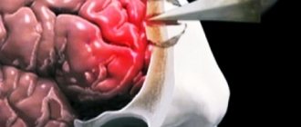
Treatment of linear, as well as other types of fractures, with the help of surgical intervention is carried out by craniotomy. After removing all fragments and purulent formations, the skull is closed with the previously removed bone or a special titanium plate. The most commonly used prosthetics are plates.
Main manifestations
Symptoms of a fracture reflect the location and extent of damage to the skull.
- A fracture of the anterior fossa is indicated by nosebleeds and liquorrhea, as well as bruising of the eyelids around the eyes, which appear on days 2–3, and in the mastoid area.
- Symptoms of damage to the middle fossa are expressed by ear bleeding and liquorrhea, rupture of the eardrum, hearing loss, bruising behind the ear or in the temporal region.
- Injury to the posterior fossa is most common when a fracture of the base of the skull occurs (in 70% of cases). There are bruises in the ear area. Paralysis of the tongue and larynx may occur.
When an extensive impact occurs - during a fall from a height, in an accident - injury occurs to all the pits at the same time.
Other symptoms that may indicate a fracture has occurred:
- very severe bursting headache and vomiting;
- loss of consciousness or lethargy, slow speech;
- decreased vision;
- paralysis or excessive movement;
- different pupil sizes;
- drop or increase in blood pressure;
- tachycardia, bradycardia;
- unilateral facial distortion.
A fracture in the area of the foramen magnum threatens damage to the medulla oblongata, which contains the vital centers of the body - digestive, respiratory, cardiovascular. Severe injuries to this area become incompatible with life.
Providing assistance to the victim
First aid is the key to saving life. Before the ambulance arrives, the person must be placed on his back - when he is conscious. If the state is unconscious, place it in a half-turn position with the head to one side so that the patient’s tongue does not stick and he himself does not choke on vomit. The victim should lie on a hard surface without a pillow.
It is necessary to fix the head and upper body, and then apply an antiseptic bandage to the wound - if the injury is penetrating, with bone displacement, there is a high probability of developing a brain infection. If there is a suspicion of a combined injury (cranial vault, spine), the victim must not be moved or his head turned. Also, one should not obstruct the flow of blood and cerebrospinal fluid by applying bandages to the ears or packing the nose to avoid the development of high intracranial pressure.
It is not recommended to give painkillers before the ambulance arrives - this may increase bleeding.
First medical aid, after the initial diagnosis has been carried out (examination and checking of reflexes, pulse and breathing), consists of removing mucus and blood from the respiratory tract, applying a pressure bandage, and performing artificial ventilation. If a person experiences symptoms of painful shock or a sharp drop in blood pressure, the doctor performs intravenous injections. The victim is transported only on a stretcher.
Diagnosis in a hospital consists of CT scanning (most often) or MRI - conventional radiography cannot provide complete information about the condition of the injury, since there are many bone partitions in this area that block the view. In addition, tomography immediately provides information not only about bone damage, but also diagnoses damage to the brain centers and the presence of hematomas.
Therapy
A fracture of the base of the skull can have consequences that develop very quickly and can lead to death or serious complications that affect the entire subsequent lifestyle of a person - edema (fluid accumulation in the tissues) and infection of the brain.
Clinical symptoms of edema are expressed in severe headaches and vomiting, impaired consciousness, hallucinations and coma. It is difficult to determine this disorder from them, since they themselves can characterize a fracture - it is important to establish signs of edema based on tomography results. It threatens with critically increased intracranial pressure, due to which the blood supply to the brain may stop - this threatens his death. It is relieved by dehydration using diuretics intravenously.
To prevent infection from entering the brain, which threatens the development of meningitis (inflammation of the membranes) or encephalitis (inflammation of brain tissue), antibiotics are administered preventively.
After injury, histamine-like substances (for example, serotonin) are formed in the body due to brain hypoxia, vascular disorders, and resorption of hemorrhages. In order to prevent this phenomenon, antihistamines are prescribed.
If the fracture occurs without displacement of the bones, conservative treatment is sufficient, which consists of complete bed rest and prevention of infectious consequences. If symptoms and diagnostics indicate not only an open penetrating injury, a depressed or comminuted fracture, but also the development of complications (intracerebral hematomas), surgery is performed. A craniotomy is performed followed by bone grafting (most often special plates are installed).
Rehabilitation
For any skull fractures in an adult or child, in addition to treatment, a long period of rehabilitation is carried out. During the healing period of injuries, as well as for a period of at least 6 months after that, the patient is prohibited from any type of physical activity.
During the recovery period, patients are advised to periodically wear a Shants collar. It is also possible to attend sessions of magnetic and acupuncture therapy, massages and electrophoresis. The victim is recommended to attend sessions with a psychologist and psychiatrist, and in some cases, sessions with a speech therapist are necessary.
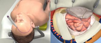
Surgery
The type of surgical treatment is selected individually by the neurosurgeon in each situation and is indicated in the following cases:
- Compression of the brain by bone fragments,
- Infectious and inflammatory complications (meningitis),
- Formation of extensive intracranial hematomas and hemorrhages,
- Traumatic damage to the meninges and blood vessels,
- Excessive nasal or ear liquorrhea (leakage of cerebrospinal fluid).
The tactics and extent of surgical intervention depend on the severity of the injury, the nature of the injury, the presence of concomitant diseases and complications.