One of the most important human organs is the brain. There is no life without it, because it is the coordinator of the work of all departments of the body. Therefore, it is necessary to take timely care of its health and normal functioning. Today, MRI of the brain with contrast is considered the most highly informative method. Where is it better to do this scan and for what?
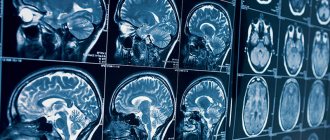
Magnetic resonance imaging is based on the principle of nuclear magnetic resonance, that is, the ability of the nuclei of certain substances to emit and absorb the energy of radio frequency pulses. But MRI of the brain with contrast is even more accurate. It helps to visualize pathologies that cannot be determined using X-rays, CT scans, or even conventional tomography. The advantages of this method are painlessness, non-invasiveness, safety, and the absence of a recovery period between checks.
MRI of the brain with contrast, which shows
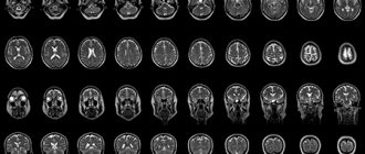
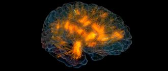
Magnetic resonance imaging allows you to visualize an accurate three-dimensional image of the brain; it can be used to examine in detail the deep layers of tissue and blood vessels of the head. And at the same time avoid overlapping the image of the skull bones. It shows disturbances in brain activity, helps detect tumors, aneurysms, and diseases of the nervous system. The quality of the images reveals a complete picture of all structures. The advantage of this technique is the ability to track dynamics, so it is often used before and after surgery.
MRI of the brain with and without contrast: differences
The main difference is the administration of a substance containing gadolinium ions or salts into the body. Whereas the usual procedure does not require the use of special drugs.
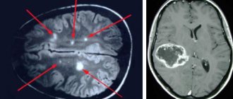
The contrast is a coloring liquid. Once injected, it spreads through the bloodstream and illuminates the tissues, making them clearer. The more intense the blood flow, the faster the contrast agent disperses. The magnetic field of the tomograph visualizes the organ being examined. Damaged tissues change color under its influence, allowing the specialist to determine the boundaries of the pathology as accurately as possible.
MRI of the brain with contrast helps to reveal a detailed picture of the state of the blood vessels, the processes occurring in the brain and its reaction to external stimuli. When examining neoplasms, metastases and their locations can be detected. In addition, this method makes it possible to diagnose tumors no larger than 1 millimeter in size. All changes that occur are monitored in real time.
The survey has two subtypes:
- The first involves gradual drip administration of the drug and is prescribed for diagnosing disorders of the vascular system.
- The second requires a bolus injection of dye. This method allows for a large volume of the drug to enter the vascular bed in one injection. It is designed to detect cancer and vascular accidents.
Since no preparation is required for a brain MRI with contrast, you can decide that examinations can be done as often as you like. In fact, there are a number of limiting factors:
- Contrast tolerance by the patient;
- Identifying side effects;
- Subjective well-being of the patient.
If tolerance is good, then adults can be examined once every seven days. In the event of side effects or a negative reaction, the procedure is carried out only if serious indicators are present.
It is worth remembering that the contrast substance itself cannot have a harmful effect, since it is excreted from the body in the urine during the day, which means it cannot accumulate and affect a person. Unpleasant sensations cause side effects, which include allergic reactions, shortness of breath, dizziness, itching in the eye area.
Immediately after scanning, the received data is decrypted. This takes about thirty to forty minutes, after which the radiologist hands over the received data or sends it to the treating doctor. Either X-ray paper or CDs are used as media. It contains indicators such as the characteristics of fluid flow in the spinal canal, the activity of the cortex under the influence of stimuli, the speed of blood flow, and the degree of tissue diffusion.
Contrast agents for MRI
MRI with contrast
During the procedure, drugs based on gadolinium salt are administered intravenously - Magnevist, Dotarem, Primovist, Omniscan. They are well tolerated and rarely cause intolerance. After being introduced into the bloodstream, the contrast spreads throughout the body, penetrating all tissues and structures and “illuminating” them.
As a result, the image obtained during MRI becomes brighter, more contrasty and more detailed. This allows the doctor to see the smallest pathological foci that are difficult to detect during a routine examination.
MRI of the brain with contrast for children
For children and adults, the indications for scanning are the same. Due to the impossibility of complete immobility for children under six years of age, they most often undergo the procedure under anesthesia with a laryngeal mask. You should not worry about the possible negative consequences of using anesthesia; according to statistics, complications occur no more often than in 1-2 percent of cases. In some cases, the anesthesiologist may recommend sedation, in which sleep is not as deep, which means breathing function is not impaired.
When performing brain MRIs with contrast on children, centers suggest replacing anesthesia with the presence of a significant adult who will reassure the baby, hold his hand, tell him a story, and ensure he remains still.
How does a head MRI with contrast work?
Nuclear magnetic resonance technology helps study organs based on the level of tissue saturation with hydrogen elements. Additional importance is given to their magnetic properties. This type of screening is prescribed in difficult clinical cases:
- patients with cancer for a clear and informative study of the tumor and the affected area;
- persons with demyelinating disorders;
- patients with certain types of strokes.
During the study, doctors can monitor the condition of all parts of the brain, including the cerebellum, nuclei of the basal type, ventricles, the ratio of white and gray matter and other structural characteristics. Screening with an MRI scanner is harmless to humans. This is due to the lack of radiation exposure during tomography.
A contrast agent is used to improve data visualization. The drug is administered intravenously using a catheter. The product penetrates into blood vessels and tissues after 10–30 seconds. It highlights pathological areas, clearly highlighting them in the images, which is not always possible to see in the native examination mode. Diagnostics using an additional amplifier goes like this:
- before introducing contrast, the laboratory assistant scans according to the native protocol without the use of a dye;
- then the patient is given an injection with contrast in a previously established amount;
- the tomograph restarts.
Most of the subjects do not report any symptoms or health problems.
Indications for MRI of the brain with contrast
It is used to make or clarify a diagnosis. Among the indications:
- Frequent fainting;
- Headaches of unknown etiology;
- Tumors of the brain and cerebellopontine ganglion;
- Epilepsy;
- Abscess;
- Sinusitis;
- Hearing damage;
- Visual impairment;
- Aphasia;
- Anomalies and pathologies of blood vessels;
- Infectious diseases of the nervous system;
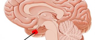
- Pituitary adenoma;
- Paroxysmal states;
- For head injuries and contusions accompanied by internal bleeding, an MRI of the brain with contrast is required;
- Neurodegenerative conditions;
- Multiple sclerosis;
- Senile dementia.
Features of using contrast in MRI
According to statistics, at least 20% of the total number of studies on an MRI machine are carried out using contrast. It is especially often prescribed if the task is to find even small tumors, to understand what condition they are in, and the differences in structure.
The main purpose of using a contrast agent is to improve image clarity. Thus, diagnostics become even more effective, and the task of making the correct diagnosis for your doctor is greatly simplified.
Contraindications
It is worth considering that intravenous administration of any substance has contraindications, so before agreeing to it, you need to check with your doctor about the possible risks. Be sure to inform if the patient has any types of allergies or kidney failure. This examination can be prescribed even to children; long-term observations have shown its safety for children. Of course, the price for an MRI of the brain with contrast in Moscow is higher than for a regular one, but health is not something you can save on.
Contraindications are absolute, these include:
- Wearing permanent implants made of metal or foreign metal bodies;
- Use of electronic pacemakers;
- The patient's weight is above 120 kilograms;
- Hematopoietic anemia.
Relative contraindications that will require additional preparation for MRI of the brain with contrast include:
- State of drug or alcohol intoxication;
- Heart failure;
- Tattoos with dyes containing metals;
- Panic attacks, claustrophobia, anxiety disorders;
- Availability of insulin pumps, prosthetic heart valves, non-ferromagnetic implants;
- Allergy;
- Pregnancy up to twelve weeks.
In the presence of these contraindications, scanning is done only on the direction of a doctor.
Features of preparation and implementation of the procedure
Initially, you should make sure that there are no metal elements on your clothes. The same goes for jewelry, that is, chains, piercings, rings. The last meal should be two hours before the procedure. Before the examination, the doctor asks questions regarding the patient's condition.
This allows us to identify the presence of implants, plates and wires, and other contraindications. It is also important to find out whether the person is allergic to the substance used and whether a previous test has been carried out.
After preparation, the patient is placed on a sliding table. The laboratory technician installs a catheter to administer contrast. After the substance is delivered, a tingling sensation may occur. This is a normal reaction and should not be alarmed. After this, the table moves into the tomograph tunnel and scanning is carried out. On average, the procedure lasts about an hour. During this time, it is prohibited to move or talk. After the examination is completed, the patient changes clothes and awaits the results. As a rule, they are handed over immediately.
Price
For an MRI of the brain with contrast, the price in Moscow is about 6,000 rubles. If the procedure takes place under sedation or anesthesia, you will have to pay for the services of an anesthesiologist. The cost is also affected by the model of the device - more modern high-field silent devices will cost more. But the quality of images taken with such a tomograph is many times higher. Some medical centers offer the opportunity to do magnetic resonance imaging at night. This service will cost at an increased rate.
Indications for MRI with contrast
The decision that a patient needs to use MRI with contrast is made by the attending physician based on an analysis of the patient’s condition. Not all types of examinations are carried out using such a tool.
Among the most common indications are:
- Suspicion of the localization of tumors of various types in the human pituitary gland.
- Going through the rehabilitation period after many types of operations, including removal of intervertebral hernias.
- The likelihood of developing tumors in the spinal cord, both benign and malignant.
- Identification of multiple sclerosis, the degree of its activity, assessment of the effectiveness of the treatment methods used.
- Tracking metastases in the early stages of tumor lesions of the brain and spinal cord.
Contrast is also necessary to determine the condition of the spinal cord or brain structures themselves.
MRI of the brain with contrast: where is the best place to do it?
There are more than six dozen medical centers where this procedure is performed in the capital alone. Therefore, one must approach their choice responsibly. And focus not only on cost, but also on the quality and thickness of the sections. In order for them to be as thin as possible, it is worth giving preference to tomographs with a field of at least 1.5 Tesla. Such devices take photographs of tissues sequentially, obtaining at least 20 photographs at a time. The thickness of the sections is 4-5 millimeters. The leaders in the number of truly competent doctors who can perform MRI of the brain with contrast are Moscow centers. You can contact most of these clinics not only if you have an appointment, but also on your own.
Side effects and contraindications
Most patients tolerate the procedures easily, but mild side effects are possible. It can be:
- The appearance of a metallic taste in the mouth.
- Feeling of warmth throughout the body.
- The appearance of blisters and itching.
- The appearance of pink spots in the area where the contrast goes beyond the vessels.
A more pronounced reaction manifests itself in lack of air, urticaria, swelling of the face and neck, shortness of breath, and decreased urine volume. It is extremely rare that an allergic reaction to contrast may cause anaphylactic shock.
MRI is not performed for blood diseases, individual intolerance to contrast agents, chronic renal failure and during pregnancy.