WHAT IS THE SURGICAL INTERVENTION?
It is possible to perform an open biopsy using craniotomy, as when removing a tumor. In this case, a window is opened in the skull bone by removing a fragment, which is subsequently replaced in its original place. However, this approach is not always practiced and is limited to superficial lesions in prominent areas of the brain or when samples need to be taken from the cranial bone or meninges.
If possible, partial resection of the lesion is performed and a sample of this tissue is taken for analysis. Minimally invasive biopsy methods are chosen: for diffuse damage to brain tissue without clear boundaries for small and deep lesions for significant lesions in a very visible area of the brain with a high risk of serious neurological consequences To do this, a trepanation or a single small hole is made as an entry point into the skull, through which a cannula is inserted and directed to specific, pre-planned coordinates.
The classic method, which is still used in many centers and for other types of operations, is stereotaxy. Stereotaxy involves obtaining target coordinates by performing a CT scan of the skull immediately before entering the operating room. A metal frame is attached to the patient's skull to serve as a reference for obtaining coordinates, which will later be attached to another component on the operating table.
Because this procedure is not particularly pleasant for the patient, has higher logistical requirements, and takes longer to prepare, an alternative technique has been developed. The cannula is inserted into specific coordinates in the brain using neuronavigation technology (frameless stereotactic brain tumor biopsy), which is used at the Instituto Clavel. This method provides maximum accuracy and safety.
During neuronavigation biopsy of brain tumors, the patient's head must be in a fixed position relative to the landmark of the navigation device. An accurate, high-quality MRI of the patient's skull is loaded onto the console of this device and a target is determined to hit.
The neuronavigator combines the downloaded image with the marked target and shows on the screen the position of the biopsy cannula in relation to the target, allowing neurosurgeons to reach it safely. The path is selected in the image in such a way as to avoid risk areas inside the brain and reach the desired depth.
Each time biopsy samples are taken from brain tumors, a fresh sample is sent to the laboratory to be analyzed and assessed for viability and the presence of abnormal tissue before sending additional samples for final examination and wound closure.
Material and methods
Anatomical study
To determine the morphometric parameters of endoscopic transorbital approaches, measurements were taken on 5 dry preparations of the skull of deceased adults without any pathology of the head. Using a caliper, the distances between entry access points and key anatomical landmarks in the area of surgical interest were measured. With the superolateral approach, the distance from the attachment of the lateral ligament of the eyelids to the anterior edge of the inferior orbital fissure, the medial and lateral edges of the superior orbital fissure, the anterior edge of the foramen rotundum, the foramen ovale, the foramen spinosum, and the internal carotid artery in the foramen lacerum was determined. Using the retrocaruncular approach, the distances from the anterior lacrimal crest to the anterior and posterior ethmoidal foramina, medial and lateral edges of the optic canal were measured both from the side of the orbit and from the side of the cranial cavity.
Characteristics of a series of operated patients
In 2017-2018 At the National Medical Research Center for Neurosurgery, 12 patients (10 women and 2 men aged 24–78 years) with tumors of the skull base were operated on using transorbital endoscopic approaches (Table 1).
Table 1. Characteristics of the analyzed series of patients Note. MPNST (malignant peripheral nerve sheath tumor) is a malignant tumor of the peripheral nerve sheaths, V1 is the first portion of the trigeminal nerve. Tumor removal operations are highlighted in bold. All patients were primary. In ½ cases a biopsy was performed, in the remaining 6 cases the tumor was removed. In 8 of 12 patients, a superolateral transorbital approach with an incision under the eyebrow was used. In 2 patients a retrocaruncular approach was used, in 1 case a lateral retrocanthal approach was used, and in 1 case a superomedial approach with an incision under the eyebrow was used.
Equipment used
Endoscopic manipulations were carried out under the control of rigid endoscopes with a diameter of 4 mm with a viewing angle of 0 and 45°, Image1 Full HD endovideo cameras, LED and a light source; they were performed using specialized instruments and a high-speed bur for endoscopic surgery of the nasal cavity, paranasal sinuses and skull base; Photo and video documentation was made using an AIDA DVD-M photo and video capture device (all devices manufactured by Karl Storz GmbH & Co. KG, Tuttlingen, Germany).
Description of surgical technique
The operations were performed under general anesthesia (endotracheal + intravenous anesthesia), the operating team included 2 surgeons to work with 4 hands. The assistant held a retractor for displacing the tissues of the orbit and an endoscope, and the main surgeon used two instruments to manipulate the wound. In Fig. 1 shown
Rice. 1. Layout of the operating team when performing transorbital endoscopic operations. operating team layout diagram.
To perform a superolateral approach, after additional local infiltration anesthesia of soft tissues with a 2% lidocaine solution, a skin incision was made along the fold below the eyebrow in the projection of the outer 2/3 of the upper edge of the orbit (Fig. 2, a).
Rice. 2. Schemes of incisions for transorbital endoscopic approaches. a — skin incision along the skin fold below the eyebrow in the projection of the outer 2/3 of the upper edge of the orbit with a superolateral approach; b - retrocaruncular incision of the conjunctiva (the dotted line indicates the continuation of the incision line hidden behind the eyelids; CL - caruncula lacrimalis). The subcutaneous tissue, the orbital part of the orbicularis oculi muscle and the periosteum were sharply dissected, after which the edges of the wound were sutured and stretched and the supraorbital edge of the frontal bone was skeletonized along with the outer and lateral walls of the orbit to create a space between them and the periosteum of the orbit. After this, a retractor and endoscope were inserted. Peeling the periosteum towards the apex of the orbit, the superior and inferior orbital fissures were identified and, using a bur with irrigation, the large wing of the sphenoid bone between them was resected until the dura mater was exposed (Fig. 3, a).
Rice. 3. Zones of bone resection during transorbital endoscopic approaches. a — area of resection of the greater wing of the sphenoid bone using the superolateral approach; b — area of resection of the medial wall of the orbit using retrocaruncular access. The area of the bone resection zones is approximately 4.5 cm2. (EF - ethmoidal foramina, FR - foramen rotundum, GSW - greater wing of the sphenoid bone, IOF - inferior orbital fissure, LP - lamina papyracea, OC - optic canal, SOF - superior orbital fissure). In case of intradural localization of the pathological process, the latter was opened with a linear or angular incision and manipulations continued in the middle cranial fossa. The skull base defect was reconstructed using free fragments of subcutaneous fat and fascia lata with additional sealing with synthetic adhesive compositions. The wound was closed in layers: first, the orbicularis oculi muscle was restored with separate sutures, then the subcutaneous tissue was connected with a continuous suture, and finally, an intradermal suture was made with non-absorbable material 6−0 with the ends of the thread secured using a patch. The initial and final stages of the operation were performed under the control of a binocular loupe with an autonomous light source.
With retrocaruncular access, the upper and lower eyelids were sutured slightly outward from the lacrimal openings and caruncula lacrimalis
.
The threads were stretched using clamps, then the conjunctiva was vertically dissected posterior to the caruncula lacrimalis
(see Fig. 2, b). The tissue of the orbit was sharply dissected in the direction of its medial wall and, having reached it, a retractor was installed and manipulations continued in the space between the periosteum of the orbit and the medial wall under the control of an endoscope. If necessary, the ethmoidal arteries were coagulated and transected and the medial wall of the orbit was resected (see Fig. 3, b). Upon completion of the operation, a continuous suture was placed on the conjunctiva with non-absorbable material 6−0, bringing the ends of the thread through the skin and fixing them with a plaster. The technique for performing the lateral retrocanthal approach was similar to that described in the work of K. Moe et al. [1].
At the end of the operation, a temporary tarsorrhaphy was performed and a pressure bandage was applied to the eye for 4 or 5 days. Sutures were removed on the 6th or 7th day after surgery.
RECOVERY AND REHABILITATION AFTER BIOPSY OF BRAIN TUMORS
Generally, a one-night hospital stay is required for a minimally invasive biopsy unless complications occur and the patient's condition is worse than when admitted to the clinic.
The patient's recovery is usually influenced not so much by the operation itself as by the course of the disease, age and previous condition of the patient. It is a painless and short procedure (about an hour), allowing the patient to almost immediately regain his autonomy for basic daily activities.
If surgery with craniotomy is required, the patient usually spends the first night in the intensive care unit for observation. Since the aggression on brain tissue is minimal, post-operative symptoms are not noticeable and the risks are limited, so the patient can leave the hospital after two nights.
What can you see
With the help of a biopsy we can solve a very wide range of problems:
- Obtain complete information about the characteristics of the pathology;
- Determine the nature of the tumor;
- Confirmation or refutation of a diagnosis made earlier;
- Diagnostics for the purpose of differentiation;
- Determine the stage of pathology;
- Eliminate the source of pathology;
- Evaluation of treatment effectiveness, etc.
Prostate biopsy
RISKS OF BIOPSY OF BRAIN TUMORS
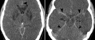
The main risk with biopsies of brain tumors is bleeding, although most are not large enough to be of major concern. The risk of moderate or severe bleeding is less than 1%, and the lesions with the highest risk of bleeding are considered to be high-grade gliomas.
Potential neurological deficits depend on the point of entry into the brain, but especially on the location of the lesion. Depending on where the target is located, one of the safe entry points into the cerebral cortex will be determined. For this purpose, not the most noticeable areas are selected, that is, those whose functions are less significant, and damage to their neurons will be invisible to the patient (unlike other, more noticeable areas, for example, those that control movement).
The main risks of neurological complications are associated with voluntary movements that are controlled in certain areas of the brain, such injuries usually require a minimally invasive procedure, and are less likely to affect the sensory organs or swallowing.
Some possible post-operative symptoms that may not be related to complications are headache, dizziness and nausea, provided they are mild and respond well to medications. In some cases, the patient may be somewhat disoriented and unstable in the first hours. The latter usually occurs in older people.