What is the brain, encephalon, its meaning?
The brain is the anterior part of the central nervous system. Brain
located in the cranial cavity, it interacts with the human body (men, women) with the external environment, integrating the functioning of all body systems.
The brain has the ability to assimilate, organize, store, and retrieve information about past experience. The brain is the material substrate of higher nervous activity. Phylogenetically, the brain is the anterior end of the neural tube. Ontogenetically, the brain is a derivative of the cerebral vesicles, from which parts of the brain are formed: the telencephalon, the diencephalon, the midbrain, the hindbrain, which is represented by formations such as the pons, cerebellum, and medulla oblongata. The cavities of the brain vesicles develop into the ventricles of the brain.
Functions
Each shell has an individual structure and functional purpose:
- The dura mater (DRM) forms processes through which the brain is divided into several important areas. They stabilize and fix the organ in the cavity of the cranium.
- The arachnoid supports the circulation of cerebrospinal fluid and normalizes metabolic reactions in the brain. Liquor washes it on both sides: in the subdural and subarachnoid space.
- The pia mater of the brain is rich in blood vessels through which arterial blood flows to the structures of the central nervous system.
In addition to protection, they provide nutrition to brain cells and take part in removing waste products from the body.
Brain structure
The cerebrum, or telencephalon, is represented by two hemispheres, which are connected to each other by the corpus callosum, corpus callosum. It consists of nerve fibers running transversely from one hemisphere to the other. The corpus callosum ensures the unity of functioning of both hemispheres. When the corpus callosum is cut, each hemisphere of the brain begins to function independently of each other. Under the corpus callosum is the fornix, fornix. Anterior to the pillars is the anterior commissure, comissura anterior. Between the anterior part of the columns of the fornix and the knee of the corpus callosum there is a thin vertical plate of brain tissue - the transparent septum. Between the plates of the septum there is a slit-like cavity that does not have an ependymal lining. A number of authors call it the 5th ventricle.
The surface of the hemispheres is covered with a layer of gray matter - this is the cerebral cortex. Underneath it is the white matter and subcortical nuclei: striopallidal system, extrapyramidal system.
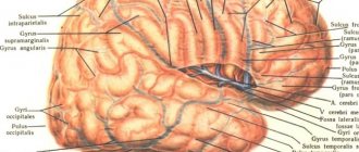
If you make a horizontal section of the brain through the cerebral hemispheres at the level of the thalamus and subthalamic nuclei, you can see the following formations: pineal body, superior colliculus, thalamus, frenulum, posterior limb of the internal capsule, globus pallidus of the lenticular nucleus, putamen of the lenticular nucleus, lateral sulcus, fence , anterior part of the internal capsule, head of the caudate nucleus, column of the fornix, anterior horn of the lateral ventricle, genu of the corpus callosum, septum pellucidum, interthalamic commissure, lentiform nucleus, external capsule, extreme capsule, convolutions of the insula, lateral sulcus, internal capsule, subthalamic nucleus, tail caudate nucleus, lateral geniculate nucleus, red nucleus, gray matter of the superior colliculus, cerebellar vermis.
| Registration number: | LS-001577-180711 |
| Trade name of the drug: | Noopept® |
| Chemical name: | N-phenylacetyl-L-prolylglycine ethyl ester |
| Dosage form: | pills |
Compound
Each tablet contains: active substance: noopept (N-phenylacetyl-L-prolylglycine ethyl ester) – 10 mg,
excipients: potato starch – 13.5 mg, lactose monohydrate – 55 mg, microcrystalline cellulose – 21.2 mg, magnesium stearate – 0.3 mg, povidone – 0.0008 mg.
Description
Tablets are white, round, flat-cylindrical with a bevel. Pharmacotherapeutic group: nootropic agent. ATX code: .
pharmachologic effect
Noopept® has nootropic and neuroprotective properties. Improves learning ability and memory, acting on all phases of processing: initial information processing, consolidation, retrieval. Prevents the development of amnesia caused by electric shock, blockade of central cholinergic structures, glutamatergic receptor systems, and deprivation of the paradoxical phase of sleep. The neuroprotective (protective) effect of Noopept® is manifested in increasing the resistance of brain tissue to damaging influences (trauma, hypoxia, electroconvulsive, toxic) and reducing the degree of damage to brain neurons. The drug reduces the volume of the lesion in a thrombotic stroke model and prevents the death of neurons in cultured tissue of the cerebral cortex and cerebellum exposed to neurotoxic concentrations of glutamate and free radical oxygen.
Noopept® has an antioxidant effect, an antagonistic effect on the effects of excess calcium, improves the rheological properties of blood, having antiaggregation, fibrinolytic, and anticoagulant properties. The nootropic effect of the drug is associated with the formation of cycloprolylglycine, which is similar in structure to the endogenous cyclic dipeptide, which has antiamnestic activity, as well as the presence of a choline-positive effect. Noopept® increases the amplitude of the transcallosal response, facilitating associative connections between the cerebral hemispheres at the level of cortical structures.
Promotes the restoration of memory and other cognitive functions impaired as a result of damaging influences - brain injury, local and global ischemia, prenatal damage (alcohol, hypoxia). The therapeutic effect of the drug in patients with organic disorders of the central nervous system is manifested starting from 5-7 days of treatment. Initially, the anxiolytic and mild stimulating effects present in the spectrum of activity of Noopept® are realized, manifested in the reduction or disappearance of anxiety, increased irritability, affective lability, and sleep disturbances. After 14-20 days of therapy, a positive effect of the drug on cognitive functions, parameters of attention and memory is revealed.
Noopept® has a vegetative-normalizing effect, helps reduce headaches, orthostatic disorders, and tachycardia.
When discontinuing the drug, no withdrawal syndrome is observed.
Does not have a damaging effect on internal organs; does not lead to changes in the cellular composition of the blood and biochemical parameters of blood and urine; does not have immunotoxic, teratogenic effects, does not exhibit mutagenic properties.
Pharmacokinetics
N-phenylacetyl-L-prolylglycine ethyl ester, absorbed in the gastrointestinal tract, enters the systemic circulation unchanged, penetrates the blood-brain barrier, and is determined in the brain in higher concentrations than in the blood. The time to reach maximum concentration is on average 15 minutes. The half-life from blood plasma is 0.38 hours. Partially remains unchanged, partially metabolized to form phenylacetic acid, phenylacetylproline and cycloprolylglycine. It has high relative bioavailability (99.7%), does not accumulate in the body, and does not cause drug dependence.
Indications for use
Impaired memory, attention and other cognitive functions and emotionally labile disorders, including in elderly patients, with:
- consequences of traumatic brain injury
- post-concussion syndrome
- cerebrovascular insufficiency (encephalopathies of various origins)
- asthenic disorders
- other conditions with signs of decreased intellectual productivity
Contraindications
Pregnancy, lactation period. Age up to 18 years. Hypersensitivity to the components of the drug. Lactase deficiency, lactose intolerance, glucose-galactose malabsorption. Severe dysfunction of the liver and kidneys.
Directions for use and doses
Noopept® is used orally, after meals. Treatment begins with the use of the drug with a dose of 20 mg, divided into two doses of 10 mg throughout the day (morning and afternoon). If the therapy is insufficiently effective and the drug is well tolerated, the dose is increased to 30 mg (see “Special Instructions”), distributed into three doses of 10 mg each day. You should not take the drug later than 18 hours. The duration of the course of treatment is 1.5 – 3 months. A second course of treatment, if necessary, can be carried out after 1 month.
special instructions
If it is necessary to increase the dose of the drug (up to 30 mg/day), with long-term use, as well as with simultaneous use with other drugs, the occurrence of adverse reactions or worsening of the condition, you should consult a doctor.
Overdose
Specific manifestations of overdose have not been established.
Side effects
Allergic reactions are possible. In patients with arterial hypertension, mostly severe, while taking the drug, a rise in blood pressure may be observed.
Interaction with other drugs Noopept® has not been found to interact with alcohol, sleeping pills, antihypertensive drugs, and psychostimulant drugs.
Release form
Tablets 10 mg. 25 tablets each in a blister pack made of polyvinyl chloride film and printed varnished aluminum foil. 2 blister packs along with instructions for use in a cardboard pack.
Storage conditions
In a dry place, protected from light, at a temperature not exceeding 25 ° C. Keep out of the reach of children.
Best before date
3 years. Do not use after the expiration date indicated on the package.
Conditions for dispensing from pharmacies
Without a doctor's prescription.
, Russia. 601125, Vladimir region, Petushinsky district, pos. Volginsky, t/f (49243) 71-5-52.
Send consumer complaints to JSC "LEKKO": 601125, Vladimir region, Petushinsky district, pos. Volginsky, t/f (49243) 71-5-52.
Brain of a newborn, children, child, human: structure, anatomy
The brain of a newborn baby is shorter and wider than that of school-age children and adults. It is devoid of all tertiary and a number of secondary grooves. By the end of the child’s first year of life, the brain increases in size by 2–2.6 times. By 3 years it increases 3 times. From birth to adulthood, brain weight increases 4 times, and body weight increases 21 times.
The mass of the right hemisphere is most often greater than the mass of the left hemisphere. After birth, the parietal and frontal lobes develop most intensively. And because of this, the overall configuration of the brain changes. Unlike the adult brain, in a newborn the neurons of different layers are located closely next to each other, because of this, the radial striation of the cortex may be absent. Single neurons may be located in the subcortical white matter. In the substantia nigra of the brain stem, neurons do not yet have the pigment melanin, which usually appears by 3–4 years of age. Up to 3–6 months of extrauterine life, the outer embryonic layer, which is called “Obersteiner’s layer,” remains in the cerebellar cortex. Obersteiner's layer consists of medulloblasts and spongioblasts. The surface of the inferior olives of the fetal medulla oblongata is smooth. After the birth of a child, the olives acquire elevations and then noticeably increase in size with age. Almost always in newborns, immature cellular elements are found in the subependymal parts of the ventricular system of the lateral ventricles, the presence of which erroneously resembles manifestations of Virchow's local encephalitis. Immature cells are located in the subependymal layer diffusely or in the form of separate foci. Sometimes they can be traced along the blood vessels over a significant extent of the white matter. By 3–6 months of a child’s life, these cells gradually disappear. The presence of a large number of immature cells in the subependymal parts of the ventricular system is an additional morphological sign of fetal prematurity.
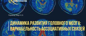
The second hypostasis of the gene
In the early 50s of the last century, the idea arose that memory cannot be limited only to electrical processes - for long-term storage of information in the brain, it must be preserved in a chemical form. Although at that time there were still very general ideas about the cell genome, the idea arose that it not only stores hereditary information, but also participates in the storage of information acquired during life.
To test this, we needed to see whether learning triggered the synthesis of nucleic acids and proteins in the brain. After the principle of the genome—DNA → RNA → protein—became known, experiments became more targeted. And this is what turned out. Immediately after animals are taught a skill, RNA synthesis increases in their brains. (In order to detect this, they were injected with radioactively labeled RNA precursors.) This happened with mice that were trained to avoid electric shock in response to a sound signal, and with chickens that developed an imprint on an object, and with goldfish that were trained to swim with a raft attached to their abdomen. And if RNA synthesis is slowed down, then animals make many mistakes or are not able to learn the skill at all.
At the same time, new proteins are also synthesized in the brain - this was also determined by the inclusion of radioactive isotopes. Protein synthesis blockers disrupt long-term memory without affecting short-term memory. From this it becomes clear how genes work: when trained on a DNA template, RNA is synthesized, which, in turn, generates new proteins. These proteins come into action a few hours after the acquisition of information, and they ensure its storage. And the initiators of all these events are electrical processes occurring on the membrane of the nerve cell.
A group of researchers from the Department of Systemogenesis of the Institute of Normal Physiology of the Russian Academy of Medical Sciences, led by Doctor of Medical Sciences, Corresponding Member of the Russian Academy of Medical Sciences K.V. Anokhina set herself the task of finding research methods that would make it possible to simultaneously study the activity of nerve cells throughout the brain in connection with any behavior or cognitive activity. “When we started our work, we were convinced that information from synapses is transmitted to another, deeper level - it penetrates the cell nucleus and somehow changes the functioning of genes,” says Konstantin Vladimirovich. “It remains to find these genes.”
It must be said that a myriad of genes work in brain cells—in humans, half of all studied genes are expressed only there. The task was to find from all their multitude the key ones involved in storing new information. The search was successful in the mid-1980s, when K.V. Anokhin and his colleagues paid attention to the so-called “immediate early genes.” They received this name for their ability to be the first to respond to extracellular stimuli. The role of “early” genes is to “wake up” other - late genes. Their products—regulatory proteins—transcription factors—act on sections of the DNA molecule and trigger the process of transcription—copying information from DNA into RNA. Ultimately, the “late” genes synthesize their proteins, which cause the necessary changes in the cell, for example, the formation of new neuron connections.
How does a child's brain mass change with age?
If you monitor how the mass of a child’s brain changes depending on age, you will notice the following picture. If the child’s age is from 3 to 8 days, body length is 49 – 50 cm, then the brain weight will be 336 grams. At 1 month, the child’s height is 52 cm, brain weight is 360 grams. At 3 months, the child’s height is 56 cm, brain weight is 520 grams. At 6 months, with a height of 62 cm, GM weight is 670 g. At 9 months, with a height of 67 cm, GM weight is 760 g. At 1 year of age, the child’s height is 73 cm, brain weight is 960 g. At 1.5 years, with a height of 79 cm, the weight of the GM is 1045 g. At 2 years old, with a height of 85 cm, the weight of the GM is 1070 g. At 3 years old, the child’s height is 89 cm, and the brain weight is 1150 g. At 5 years old, height is 106 cm, weight is 1240 g. At 10 years old, height 132 cm, brain weight 1300 g. At 12 years old, height 145 cm, brain weight 1370 grams.
From grammatical outposts to labyrinths of creativity
Using PET scans, researchers continued to study human speech using the whole brain. They saw where speech information is processed: individual words, the meaning of the text, where it is memorized. They showed that the medial extrastriate cortex is involved in processing the orthographic structure of words, and a significant part of the left superior temporal cortex (Wernicke's area) is probably involved in semantic analysis. Word order is analyzed by the anterior superior temporal cortex. When a person is shown a coherent text without even being asked to read it (they just had to count the number of times a letter appears), cerebral blood flow increases, which means the brain is involved in linguistic work. (If presented with words mixed in random order, the brain does not react this way.)
|
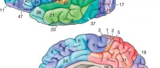
Even the “divine” process of creativity turned out to be decipherable, at least by physiologists in the laboratory of N.P. Bekhtereva came close to this. A person is given a creative task, for example, to compose a story from a set of words, and in real time they see which areas of the brain begin to work actively. It turned out that creative activity is accompanied mainly by changes in connections between different areas of the brain. Most of the new connections appear in the left anterior temporal zone with the anterior zones of the cortex, and with the posterior zones, on the contrary, the connection is weakened. The connections between the parietal and occipital structures are lost. And all this happens precisely when performing a creative task, but if the task is devoid of creative elements, there are no such changes. Local cerebral blood flow increases in the right prefrontal cortex during a more creative task compared to a less creative one. From this, scientists conclude that this particular area is directly related to “creativity.”
| Brain organization of creative thinking. Shows the area of the brain in which local blood flow increases during a more creative task compared to a less creative one (right prefrontal cortex). |
Researchers are also interested in the phenomenon of involuntary attention: for example, a person is driving a car, listening to the radio, talking and suddenly instantly reacts to the knocking of the engine, indicating that something is wrong with the engine. In two laboratories using two different methods: S.V. Medvedev using the PET method and Yu.D. Kropotov, using the method of implanted electrodes, discovered the same zones where activation occurs at such moments - in the temporal and frontal cortex. Activation occurs in response to a mismatch between expected and actual stimuli, for example when the sound from a motor is not what it should be. Another phenomenon is selective attention, which helps a person, in the continuous hum of voices at a cocktail reception, to follow the speech of one interlocutor, the one who is interesting to him. Apparently, the prefrontal cortex is responsible for focusing spatial attention in this case. It tunes either the right or left auditory cortex, depending on which ear receives important information.
When talking about brain mapping, it is important to understand that the brain, strictly speaking, is not divided into clearly demarcated areas, each of which is responsible only for its own function. Everything is much more complicated, since in the process of performing any function, neurons from different areas interact with each other, making up a neural network. Studying how individual neurons are combined into a structure, and the structure into a system and whole brain, is a task for the future.
“PET is a powerful tool for studying almost any function, but it alone is not enough,” says S.V. Medvedev. — The purpose of PET is to answer the question “where?”, and to answer the question “what is happening?”, PET should be combined with electrophysiological methods. Together with British physiologists, we have created a system for parallel analysis of PET and EEG, which complement each other. This approach is probably the future.”
A year ago (the article was published in 2004 - P.Z. ), a group of scientists from six countries announced the creation of a three-dimensional computer map of the human brain, which can be used to determine a person’s predisposition to certain diseases. The creators of the map believe that they can already associate certain diseases, such as Alzheimer's disease or autism, with different parts of the cerebral cortex. Now they are busy clarifying the details of their invention.
Physiology, brain function
Physiologically, all the work of the brain is built on the principles of hierarchy, integrity, systematicity and plasticity. These are the principles of functioning that carry out all conditioned and unconditioned reflexes. They contribute to the flow of conscious mental activity of a person. The principle of hierarchy is that phylogenetically younger parts of the brain carry out higher-order control, complementing but not replacing the function of phylogenetically more ancient parts. As a result of this, the body’s capabilities in more subtle differentiation of each stimulus by each analyzer are expanded, and a more adequate perception of the overall picture of the world is achieved based on the correlation of the results of the activities of many analyzers.
The highest form of expression of the hierarchical principle is the process of corticalization of functions. The principle of hierarchy is combined with the principles of integrity and systematicity, which consist in the fact that the brain functions as a single whole with the entire nervous system, while receiving afferent impulses, carries out its analysis and synthesis, forms a flow of efferent impulses that determine the adequate activity of all peripheral organs . As a result, a stable system is formed that provides continuous information communication: center - periphery - environment - periphery - center. Plasticity refers to the functional variability of nerve centers, which is clearly manifested in the process of compensation for impaired brain functions.
Irradiation of excitation plays an important role in the normal functioning of the brain. The feedback mechanism consists of closing the input and output of the same element or system. The dominant mechanism regulates the relationships between nerve centers.
Neurons are “literate” and “creative”
With the help of implanted subcortical electrodes, physiologists from the Institute of Human Brain of the Russian Academy of Sciences managed to learn a lot about how the brain copes with speech. As already mentioned, Broca's and Wernicke's areas related to speech have been known for a long time. “It would be more correct to limit ourselves to the definition of “related to speech” and not to use the expression “speech zone,” emphasizes S.V. Medvedev. — Remember the joke about a cockroach, which, it turns out, has “ears on its legs”? We need to realize that Broca’s and Wernicke’s areas may not be the center of speech, but some kind of interface.”
| Test for distinguishing semantic and grammatical features of speech. A group of neurons that changes electrical activity depending on the nature of the response. |
In a completely different place in the cerebral cortex, researchers found a detector for the grammatical correctness of a meaningful phrase. A group of neurons increases their electrical activity if the phrase the subject hears is grammatically correct, and decreases it when it is grammatically incorrect. If a subject is presented with the phrases “blue ribbon” and “blue ribbon,” these “literate” neurons will immediately notice the difference. Another group of neurons distinguishes between words of the native language, words that are phonetically similar to them, and foreign words. “This means that the neural population almost instantly analyzes the phonetic structure of the word and classifies it into the following types: “I understand,” “I don’t understand, but something familiar” and “I don’t understand at all,” says S.V. Medvedev. In this regard, the question arises whether these neurons work the same or differently in people gifted with innate literacy and in those who have problems with this. Most likely, there are differences, but in order to give an accurate answer, you need to recruit quite a lot of subjects.
“We found groups of neurons that distinguish between concrete and abstract words, neurons that appear to be responsible for counting,” says Svyatoslav Vsevolodovich further. “We have identified areas of the brain that are associated with generalization and decision making. All neuronal systems are characterized by polyfunctionality: this means that the same cells can participate in different functions. The specialization of neurons is relative - depending on the situation, they can take on different responsibilities. For example, when the captain of a ship dies, a navigator or someone else takes his place. Therefore, the brain is a very flexible system.” Neurons lose their ability to be interchangeable over time and become more specialized. A small child cannot walk and talk at the same time; if you call him, he will stumble and fall. The fact is that his entire cortex is occupied by either one or the other. The student should not be distracted during the lesson, otherwise he will not learn the material. Over time, more and more division of brain territories occurs, so an adult can simultaneously drive a car and carry on a conversation, talk on the phone and look at documents, etc.
N.P. Bekhtereva and her collaborators found neurons in the brain that work as an error detector. What is their role? They react to any violation of the stereotypical sequence of actions. “You leave home and on the street you feel: “Something is wrong...” explains S.V. Medvedev. “That’s right - they forgot to turn off the light in the bathroom.” Error detector neurons are located in different parts of the brain - in the parietal cortex of the right hemisphere, in the Rolandic sulcus, in the superior parietal and parietotemporal areas of the cortex, in the cingulate gyrus.
But the method of implanted electrodes also has limitations. Electrodes, of course, are not implanted everywhere where physiologists would like them, but only where necessary for clinical indications. Doesn't this mean that we are looking where it is brighter, and not where we have lost?
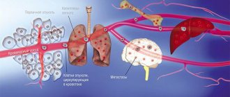
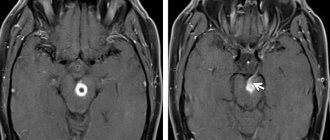
Diseases, disorders, brain lesions
Diseases, disorders, and brain lesions are varied. In further articles, we will focus on pathologies such as brain tumor, brain cyst (including arachnoid, retrocerebellar, liquor), trauma, concussion or bruise of the brain, brain cancer, hydrocephalus (hydropsis), vascular atherosclerosis, aneurysm, encephalopathy, demyelination, ischemia, ischemic or hemorrhagic stroke, heart attack, atrophy, spasm or vasoconstriction, glioblastoma, meningioma, dysfunction, dystonia, diffuse changes, hypoxia (oxygen starvation), encephalitis, inflammation, vascular diseases, atrophic changes. The clinical picture of such diseases depends on the type of pathology.
Brain treatment in Saratov, Russia
Sarklinik provides treatment for a number of diseases, diseases of the central and peripheral nervous system in Saratov, in Russia for children, adolescents, adults, boys, girls, guys, girls, men, women, brain treatment in Saratov . Hardware and non-hardware treatment methods make it possible to restore the functioning of the human nervous system.
Sign up for a consultation. There are contraindications. Specialist consultation is required.
Text: ® SARCLINIC | Sarclinic.com \ Sarlcinic.ru Photo: © pixologic / Photobank Photogenica / photogenica.ru The people depicted in the photo are models, do not suffer from the diseases described and/or all coincidences are excluded.
Related posts:
Urination, urination mechanism, why does it happen, does urinary incontinence happen?
Dysmorphomania, treatment of dysmorphomania in Saratov
Dysmorphophobia, treatment in Saratov, how to treat dysmorphophobia
Where in Saratov you can cure your nerves
Nocturnal enuresis code according to ICD 10 F 98.0
Comments ()
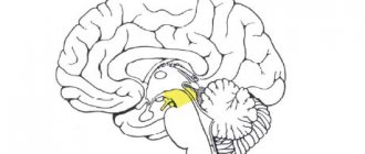
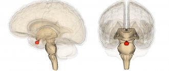
Understanding the Brain Stem
The brain stem is considered the most ancient element of the human central nervous system. Compared to other structures, it is relatively small in size - about 7 cm in length. It is formed by the following formations: the pons, the midbrain and the medulla oblongata. In some sources, the trunk also includes the intermediate section and the cerebellum, since they also contain the nuclei of the nerve centers.
Brain stem physiology
All components of the central nervous system are interconnected by bundles of long processes of neurons. In the trunk they form an extensive network: some of them transmit impulses to the trunk nuclear formations, others send them to the organs of the body. These formations are a cluster of neuron bodies - the main structure of gray matter.
There are several groups of nuclei in the trunk:
- Motor;
- Vegetative;
- Sensitive.
The motor nuclei control muscle function. These include: gray matter of the cranial nerves, vestibular nuclei, red nuclei, reticular formation, neurons of the tegmentum of the quadrigeminal, as well as the substantia nigra.
Through descending pathways from them, conditioned and unconditioned reflexes are realized. Also, thanks to them, the human body corrects the tone of the body muscles in the process of maintaining a posture, both at rest and during directed movement.
Vegetative nuclear formations control the functioning of internal organs. With their help, the constancy of the internal environment is maintained in the human body.
Since the same processes of neurons cannot receive and transmit impulses, the ANS in the brain stem is represented by the structures of the sympathetic and parasympathetic NS. The first activates the activity of internal organs and accelerates metabolism in cells, while the other, on the contrary, inhibits them.
Sensitive nuclei of the trunk are involved in the perception of information from the environment through the senses. Their presence allows a person to navigate the environment. They are also used to perform reflex actions: coughing, sneezing, etc.
The nuclei of the cranial nerves of the trunk are responsible for the work of 10 pairs of corresponding nerves: there are olfactory, visual, oculomotor, glossopharyngeal, etc. They control the activity of muscles similar to the name, with the help of which this organ is controlled.
In addition to them, the trunk contains structures of the reticular formation. They are responsible for activating the cerebral cortex and controlling the reflex activity of the spinal part of the central nervous system. This developed network of accumulation of neuron cell bodies originates in the lower part of the medulla oblongata and extends to the lower boundaries of the thalamic formations.
The red nucleus is located in the middle part of the brain. It takes a direct part in the processes of coordination of movements: nerve fibers are sent to it from the “small brain”, ensuring the connection of the latter with the subcortical structures. Thanks to this link, a person carries out unconscious reflex movements.
In the quadrigeminal region of the middle section lies the substantia nigra. It and the red nucleus belong to the stem part of the extrapyramidal system. Like previous structures, the substantia nigra is formed by neurons, the surface of which is covered with neuromelanin. This is what gives it its characteristic dark color. The black substance is responsible for the motor function of the body, muscle tone, breathing, and cardiac activity.
The structures of the quadrigeminal plate are responsible for the transmission of visual and auditory impulses to the brain, that is, they take part in the perception of information by a person through the organs of hearing and vision.
Physiologically, the trunk and its structures ensure the proper functioning of the entire NS. Thanks to such a complex organization of this part of the central nervous system, a person is able to perceive information about the environment: feel, hear, smell and see. Since the trunk contains nuclei responsible for the functioning of vital systems of the body, damage to it threatens the victim with disability, and in the worst case, death.