General information
This part of the brain arose long before the front part became responsible for the functions of smell.
It consists of various parts of the entire analyzer, which is responsible for the functions of human senses. It is worth noting that the fish brain is almost entirely composed of ancient structure. But in humans, the limbic system, part of which is the olfactory brain, is responsible for these functions. It is localized at the junction of the inferior and medial surfaces of both hemispheres of the brain. If we consider its phylogenetic path of development, we can distinguish 3 main stages:
- Paleopallium, is one of the components of the temporal lobe of the cerebral cortex;
- Archipallium is located next to the so-called bulbus olfactorius area, covered with bark of old origin;
- Neopallium is a modern cloak; the highest centers of olfactory reflexes are formed in it - these are unique analyzers of the cortex.
Animals in ancient times used only this system to operate their olfactory functions. As we evolved, the olfactory brain became part of the limbic system, which is responsible for human sensory sensations.
Average
The midbrain has a relatively simple structure, small in size, and includes two main parts: the roof (the centers of hearing and vision are located in the subcortical part); legs (contain conducting pathways). It is also customary to include black matter and red nuclei in the structure of the cloth.
The subcortical centers, which are part of this department, work to maintain the normal functioning of the hearing and vision centers. Also located here are the nerve nuclei that ensure the functioning of the eye muscles, the temporal lobes that process various auditory sensations, turning them into sound images familiar to humans, and the temporoparietal node.
The following functions of the brain are also distinguished: controlling (together with the medulla oblongata) the reflexes that arise when exposed to a stimulus, helping with orientation in space, forming an appropriate reaction to stimuli, turning the body in the desired direction.
The gray matter in this part is a high concentration of nerve cells that form the nerve nuclei inside the skull.
This department is small in size and simple in structure, consisting of parts:
- roofs – visual and auditory centers are included;
- legs - includes pathways.
The midbrain is 2 cm long and is a narrow canal that circulates cerebrospinal fluid. The rate of renewal of cerebrospinal fluid is approximately 5 times a day.
The main functionality of the midbrain:
- Sensory. The contained subcortical centers are responsible for the auditory and visual departments.
- Motor. Along with the oblongata, it ensures the functioning of the reflex actions of the body, helps to navigate in space, and is also responsible for the reaction to surrounding stimuli: the volume of sound or the brightness of light. Responsible for controlling automatic actions: swallowing, chewing, walking, breathing.
- Ensures the functioning of the body's motor system, coordination and muscle tone.
- Conductor. Provides conscious work of body movements.
The midbrain provides control over the work of muscles, giving instructions for straightening or bending, i.e. allows a person to move.
Midbrain nuclei
The nuclei play a special role in the functioning of the body:
- The colliculus nuclei in the upper part belong to the visual centers of the brain. Signals are sent from the retina to the brain, and an orientation reflex occurs - turning the head towards the light. The pupils dilate, the lens changes curvature - this ensures clarity and clarity of vision.
- The colliculus nuclei in the lower part are auditory centers. They are responsible for reflex work - the head turns in the direction of the outgoing sound.
- When the sound is too loud and the light is too bright, the brain reacts to such stimuli - irritation, which pushes the human body to a sharp and quick reaction.
Structure
This part of the human brain consists of many structures.
Conventionally, it can be divided into two parts: peripheral and central. Let's look at the structure of each of them. The peripheral part is the olfactory lobe, it includes the bulb, tract and sulcus. The grooves have a triangle at their end, which in turn consists of 2 olfactory stripes:
- lateral, which bends around the lateral sulcus, its end is located in the temporal region;
- medial. It passes into the area of the subcallosal gyrus and the peri-olfactory field of the brain, which in turn are located in the area of the corpus callosum.
The central section consists of many cortical gyri: parahippocampal, dentate and vaulted. The latter has the shape of a ring; it is responsible not only for olfactory functions, but also for the functioning of all internal organs and systems. This gyrus is formed by the cingulate and parahippocampal regions, which are connected to each other by a special septum. The dentate gyrus is a small hook, it is directed towards the medial part of the brain. It also includes the hippocampus, fornix, and nuclei of the transparent part of the septum.
Two main parts create a unique system, it is responsible for the processes of smell. Most of them form the so-called limbic system. It was she who took over all the functions responsible for the human subconscious.
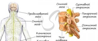
Oblong
This section extends from the spinal cord, its length is 25 mm. It is responsible for important respiratory and cardiovascular functions and metabolism. The medulla oblongata regulates:
- digestive reflexes: sucking, digesting food, swallowing;
- muscle reflexes: maintaining postures, walking, running;
- sensory reflexes: work of the vestibular apparatus, auditory, receptor, gustatory;
- receptors, processing of brain signals supplied by stimuli;
- defense reflex: blinking, sneezing, vomiting, coughing.
The medulla oblongata transmits signals to the brain from the spinal cord and back. Its structure is similar to the dorsal one, but has some differences. This section contains white matter, located on the outside, and gray matter, which collects in clusters, forming nuclei.
An important part of the central nervous system, which is called the bulbus in various medical descriptions. It is located between the cerebellum, pons, and spinal region. The bulbus, being part of the central nervous system trunk, is responsible for the functioning of the respiratory system and regulation of blood pressure, which is vital for humans.
In this regard, if this department is damaged in any way (mechanical damage, pathologies, strokes, etc.), then the probability of human death is high.
The most important functions of the oblongata are:
- Working together with the cerebellum to ensure balance and coordination of the human body.
- The department includes the vagus nerve with autonomic fibers, which helps ensure the functioning of the digestive and cardiovascular systems and blood circulation.
- Ensuring the swallowing of food and liquids.
- Presence of coughing and sneezing reflexes.
- Regulation of the respiratory system and blood supply to individual organs.
The medulla oblongata, the structure and functions of which differ from the spinal cord, has many common structures with it.
Structure functions
The main functions of the limbic system are:
- motivational processes. Development of certain needs, creation of functions for their implementation, assessment of the possibility of performing these actions, etc.;
- emotions. In this case, the structures in question are responsible for the formation of basic emotions in a person;
- memorization. This is one of the important points. This part of the brain is responsible for the so-called short-term memory function (it is created after a person’s full wakefulness cycle). further functioning is ensured by conservation of impulses;
- long-term memory;
- formation of higher centers of nervous regulation.
Among the emotions that originate in the olfactory brain are:
- feeling of fear. In this case, there is strong stimulation of the hypothalamus and tonsils;
- feeling of anger or complete calm;
- the feeling of pleasure or dependence on something is based on impulses passing through the brain's amygdala, nucleus accumbens and hippocampus.
Also, the olfactory brain, as a component of the limbic system, provides a person with functions responsible for sleep, appetite, sexual desire, memory, fear, etc.
If the functioning of one of its parts is disrupted, a person is diagnosed with serious pathologies.
Functions
Thanks to the ability to distinguish odors, a person receives information about the surrounding biochemical environment. Other functions of the olfactory brain are associated with the formation of instinctive reactions when interacting with the outside world. The sense of smell in the context of the formation of reflex behavioral patterns in response to external stimuli covers the nutritional, protective, sexual, and orientation spheres of the body’s life. The olfactory brain and limbic system are structures that are involved in the regulation of processes:
- Formation of emotions.
- Behavioral, motivational reactions.
- Sleep-wake mode.
- Learning, intuition and memory.
Odorants are volatile substances that have an odor. Once in the nasal passages, odorants reach the olfactory epithelium (Regio olfactorius), where they interact with chemoreceptors. The olfactory zone is located on the mucosal surface in the upper region of the nasal passages, covers an area of about 2.5 cm2, and contains about 50 million sensory neurons.
Axonal branches of nerve cells pass through the ethmoid bone towards the olfactory bulb within the brain. Here the processes form synaptic connections with the dendrites of second-level nerve cells and unite into glomeruli. Level 2 neurons include mitral (the largest cellular elements) and tufted cells. Their branches emerge from the bulb, which is a multilayer neural network.
The axons of nerve cells (mitral, fascicular) extending from the bulb transmit information to the primary zones of the cortex, where the stimulus is decoded, the information is interpreted and an adequate reflex response is formed. The signal is then transmitted to the center of smell, located in the cortical layer of the brain, which regulates the higher functions of the psyche, where a conscious sensation of aroma arises.
At the same time, the stimulus enters the limbic system, where emotions and conscious behavioral reactions are formed that are adequate to the perceived smell. Odorants are transmitted predominantly along unmyelinated (not covered with a protective layer of myelin) nerve fibers. Impaired smell function worsens a person's quality of life. For example, many people with such disorders have impaired digestive function, which is due to the close relationship between the smell and taste of food.
Pathologies
A neurologist treats problems associated with dysfunction of the olfactory brain. First of all, neurodegenerative processes should be attributed to pathological problems with this area. They cause serious disturbances in the hippothalamus, as a result of which the patient is diagnosed with Alzheimer's disease. Other diseases include:
- increased feeling of anxiety, which entails damage to the tonsils of the brain;
- epileptic seizures. They occur in both adult patients and children. In this case, partial sclerosis of the hippocampus occurs;
- depression;
- in case of disruption of the tonsils and some olfactory gyri, patients are diagnosed with autism or Asperger syndrome;
- the presence of malignant tumors that pinch the nerve endings in this area;
- problems with the functioning of the circulatory system, which are accompanied by insufficient nutrition of blood vessels in the area of the olfactory brain;
- schizophrenia;
- increased aggressiveness and excitability;
- congenital pathologies;
- impairment of olfactory functions, which may be accompanied by loss of taste, vision or smell.
In the latter case, the patient experiences a delay in the functions of transporting odors to the neuroepithelium in the olfactory area. They can occur against the background of chronic inflammatory or infectious processes, injuries, or exposure to chemical substances on the mucous membranes that can impair the function of the sense of smell.
To diagnose pathologies, MRI and CT examinations are used. They help to fully assess the state of all parts of the structure. Based on the research results, an effective treatment regimen is selected for the patient. For depression and increased excitability, tranquilizers and sedatives are used. Many disorders in the functioning of this area cannot be treated; the patient is given supportive therapy.
In older people, due to changes in this part of the brain, partial or complete memory loss occurs, tremors of the limbs occur, nerve cells die, etc. Unfortunately, such pathologies cannot be cured. In this case, the patient is constantly monitored by a specialized specialist.
Organ structure
The olfactory organ is the nose, which perceives appropriate stimuli dissolved in the air. The process of smell consists of:
- olfactory mucosa;
- olfactory filament;
- olfactory bulb;
- olfactory tract;
- cerebral cortex.
The olfactory nerve and receptor cells are responsible for the perception of odors. They are located on the olfactory epithelium, which is located on the mucous membrane of the upper posterior part of the nasal cavity, in the area of the nasal septum and the upper nasal passage. In humans, the olfactory epithelium covers an area of about 4 cm2.
All signals from the receptor cells of the nose (of which there are up to 10 million) enter the brain through nerve fibers. There the idea of the nature of the smell is formed or its recognition occurs.
In humans, there are olfactory and trigeminal nerves, to the endings of which odor receptors are attached. Nerve cells have two types of processes. The short ones, called dendrites, are rod-shaped, each containing 10-15 olfactory cilia. Others, the central processes (axons), are much thinner, forming thin nerves that resemble threads.
The visceral brain system, or limbic system, includes the cortical zones of the olfactory analyzer. These same systems are responsible for the regulation of innate activity - search, food, defensive, sexual, emotional. The visceral brain is also involved in maintaining homeostasis, regulating autonomic functions, forming motivational behavior and emotions, and organizing memory.
Front
Main functions:
- innate instincts;
- developed sense of smell;
- emotions, memory;
- reactions to stimuli.
The forebrain is one of the most extensive sections, consisting of the diencephalon and hemispheres (right and left), which are separated in the form of a gap, in the depth of which there are jumpers (corpus callosum).
The cerebral cortex is covered with nerve fibers - a white substance that forms the connection between neurons and parts of the brain. The hemispheres are covered with a cortex that contains gray matter. The cell bodies of neurons are components of the gray matter, arranged in columns in several layers. From the gray matter inside the hemispheres, connections are formed from nuclei located in the middle of the white matter, thereby forming the subcortical centers.
In the cerebral hemispheres, neurons are involved in processing nerve signals emanating from the sensory organs. This process occurs in the middle and posterior regions of the brain. Each lobe of the hemisphere is responsible for certain zones:
- the occipital lobe is responsible for visual functions;
- in the lobes of the temples there are neurons of the auditory zone;
- The parietal lobe controls muscle and skin sensation.
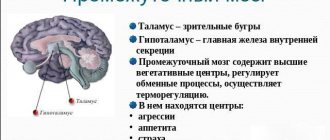
Intermediate
This section has a common edge with the midbrain and telencephalon, is located along the fibers of the optic thalamus to the real surface, and from the ventral tegmentum in front of the optic chiasm.
Based on its functions, the intermediate section is divided into types: thalamus and hypothalamus.
Thalamus
The thalamus is responsible for processing information transmitted from receptors to the cortex. Includes approximately 120 cores, which are divided into specific and non-specific. Signals passing through the thalamus: muscle, skin, visual, auditory. Impulses sent by the cerebellum and brainstem nuclei also pass through.
Hypothalamus
This department is responsible for the centers of smell, regulation of energy and metabolism, the constancy of hemeostasis (the internal environment of the body), for the center of vegetative work through the nervous system. The functional participation of other parts of the brain allows a person not only to move, but also to perform a cycle of actions - jumping, running, swimming.
Since the diencephalon contains many vegetative nuclei, pineal gland, pituitary gland, and visual thalamus, it is also responsible for the following aspects:
- Performing work related to metabolic processes (water-salt and fat balance, protein and carbohydrate metabolism) and heat regulation, since it is one of the centers of the nervous autonomic system.
- The body's sensitivity to various stimuli, as well as the processing and comparison of this information.
- Emotions, behavior, facial expressions, gestures associated with changes in the functioning of internal organs.
- Hormonal background, production and regulation of hormones produced by the pituitary gland and epiphysis.
The diencephalon performs the following main functions:
- control of endocrine glands;
- thermal control;
- regulation of falling asleep, awakening and wakefulness;
- water balance;
- responsible for the center of satiety and hunger;
- responsible for feelings of pleasure and pain.
The specific structure of the brain affects the structure of its main sections. For example, the diencephalon also consists of two main parts: ventral and dorsal. The dorsal section includes the epithalamus, thalamus, metathalamus, and the ventral section includes the hypothalamus. In the structure of the intermediate zone, it is customary to distinguish between the pineal gland and the epithalamus, which regulate the body’s adaptation to changes in biological rhythm.
The thalamus is one of the most important parts because it is necessary for humans to process and regulate various external stimuli and be able to adapt to changing environmental conditions. The main purpose is to collect and analyze various sensory perceptions (with the exception of smell), transmitting corresponding impulses to large hemispheres.
Considering the structure and function of the brain, it is worth noting the hypothalamus. This is a special separate subcortical center, completely focused on working with various autonomic functions of the human body. The impact of the department on internal organs and systems is carried out with the help of the central nervous system and endocrine glands. The hypothalamus also performs the following characteristic functions:
- creation and support of sleep and wakefulness patterns in everyday life.
- thermoregulation (maintaining normal body temperature);
- regulation of heart rate, breathing, blood pressure;
- control of the sweat glands;
- regulation of intestinal motility.
The hypothalamus also provides a person’s initial reaction to stress and is responsible for sexual behavior, so it can be described as one of the most important departments. When working together with the pituitary gland, the hypothalamus has a stimulating effect on the formation of hormones that help us adapt the body to a stressful situation. Closely related to the functioning of the endocrine system.
The pituitary gland is relatively small in size (about the size of a sunflower seed), but is responsible for the production of a huge amount of hormones, including the synthesis of sex hormones in men and women. It is located behind the nasal cavity, ensures normal metabolism, controls the functioning of the thyroid, gonads, and adrenal glands.
Hello student
finite brain
The telencephalon in mammals forms the largest part of the brain, which is so dominant morphologically that it covers all other parts of the brain up to the cerebellum, since even functionally it comes first in the hierarchical organization of the central nervous system.
Therefore, this part of the brain is called the cerebrum. The telencephalon is paired and consists of two hemispheres that lie above the brainstem with the mantle, pallium, and above the basal part, the ganglion hill (with the striatum, corpus striatum) connects to the diencephalon, and through it to all other parts of the brain. In phylogenesis, three phases of development of parts of the telencephalon are distinguished: paleopallium - the oldest section, embodies, together with the younger section, archipallium, the olfactory brain; between both passes the neopallium, which undergoes a significant change and pushes the more ancient parts of the brain basally and medially.
The olfactory brain, rhinencephalon, is separated from the neopallium by the lateral olfactory groove, sulcus rhinalis lateralis. It begins rostrally with the olfactory bulb, bulbus olfactorius, prominent, especially in dogs. Its surface is covered with olfactory filaments, fila olfactoria, which pass through the perforated plate, lamina cribrosa, ethmoid bone and go to the olfactory cells of the nasal mucosa (olfactory receptors).
Adjacent to the bulb caudally is the olfactory tract, tractus olfactorii, which in dogs runs in the form of two white cords (tractus olfactorius lateralis et medialis), and in cats only the tractus olfactorius lateralis is superficially noted. The olfactory tract is covered with gray matter, which continues laterally in the form of the lateral olfactory gyrus, gyrus olfactorius lateralis, and medially in the form of the medial olfactory gyrus, gyrus olfactorius medialis. Both gyri border a triangular field, the olfactory triangle, trigonum olfactoriurn, which in carnivores projects ventrally in the form of an olfactory tubercle, tuberculum olfactoriurn. Its caudal part forms a diagonal gyrus, gyrus diagonalis, running from the medial surface of the hemispheres.
Gyrus olfactorius lateralis goes caudally into the pyriform lobe, lobus piriformis. The stem part of the olfactory brain - the amygdala nucleus, corpus amygdaloideum, belongs to the ganglion hill. The nucleus connects to the hippocampus, and also through the terminal strip, stria terminalis, to the area septalis.
The connection between the halves of the olfactory brain occurs through the rostral commissure, commissura rostralis.
The piriformis lobe, lobus piriformis, is an important sensory area in the olfactory brain. The corpus amygdaloideum belongs to the limbic system.
During the process of phylogenesis, Archipallium is pushed aside by the medial wall of the hemispheres and characteristically coagulates when neopallium expands. At the same time, this area extends in an arcuate manner caudoventrally and, because of its shape, is called the hippocampus, hippocampus, or ammon's horn, cornu ammonis.
In domestic mammals, the surface of the hippocampus is smooth, lacking the characteristic structures that are present in humans. Carnivores especially have a ram's horn shape of the hippocampus, hence its name.
The superficial perihippocampal gyrus, gyrus parahippocampus, folds at the hippocampal sulcus, sulcus hippocampi, and at the end of a small curl is the dentate gyrus, gyrus dentatus. The cortex lies under the medulla, the hippocampal tray, alveus hyppocampi (-/4), which medially passes into the fimbria hippocampi (-/5). Fimbriae are collected in the legs of the vault, crura fomicis, which at first go separately, then protrude rostrally in the form of the body of the vault, corpus fomicis, into the third ventricle, plunge in the form of columns of the vault, columnae fomicis, into the lateral wall of the third ventricle and end in the mastoid body, corpus mamillare. .
The formation and increase in the volume of the neopallium commissure, the corpus callosum, corpus callosum, separates the hippocampus from the rostral area of the archi pallium, the paraterminal gyrus, gyrusparaterminalis, which with the adjacent subcallosal area, area subcallosa, belongs to the precommissural field, area praecommissuralis. It simultaneously forms the thin medial wall of the lateral ventricle. The corpus callosum is covered with a thin layer of cortex, archi pallium, indusium griseum.
The fornix, fornix, borders ventrally with the corpus callosum, but is separated from it at the transition to the arcuate columnae fornicis by a thin septum (area postcommissuralis, transparent septum, septum pellucidum), the volume of which depends on the size of the corpus callosum on the rostral side. Septum pellucidum most often lies in the midline. In carnivores, the growth of the septum occurs incompletely, resulting in the formation of a cavity, which in cats can be open on the rostral side (Thompson, 1932). The so-called cavity of the transparent septum, cavum septi pellucidi, has nothing in common with the ventricle of the brain.
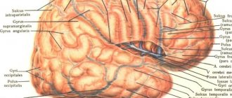
Depending on the structure of the neopallium, the large brain of dogs and cats has convolutions, while, for example, in rodents and insectivores it is smooth. The brain of all mammals has the following grooves: the longitudinal fissure of the cerebrum, fissura longitudinalis cerebri (between the hemispheres), sulcus hippocampi, lateral olfactory groove, sulcus rhinalis lateralis (between palaeopallium neopallium), as well as sulcus endorhinalis (between tuberculum olfactorium and tractus olfactorius lateralis) . Thus, the tortuous structure of the brain is due to the structure of the neopallium.
The number of convolutions of the brain is determined not only by the stage of development, but also by the size of the animal, which causes an increase in the cortex cerebri. In large animals at a high stage of development (mental hierarchy and brain performance are pronounced), neopallium has a larger number of convolutions.
On the convoluted brain there is always a lateral Sylvian fissure, fissura sylvia (lateralis cerebri)
It is located in the growth zone, from which the neopallium grows in an arched rostral and caudal manner and is folded, while the growth zone is preserved in the form of an island (insula, islet of Reil) and is buried in depth.
Unlike ungulates, in carnivores the islet is not folded or covered with grooves. That is why this groove in dogs and cats is called fissura pseudosylvia. In this book, the authors stick to the name fissura lateralis cerebri, or fissura sylvia. The difference between a simple (in carnivores) and a brain with a large number of convolutions is that in the latter case the immersion of the insular cortex occurs with the formation of a ventrolateral fossa, fossa lateralis (sylvii), while in the brain of carnivores the entire cortex lies superficially and separates the frontal and temporal lobes by the superficial groove, fissura lateralis. The remaining topographic names (neopallium growth zone, fence, ganglion hill) are similar.
Rice. 11 Appearance of the brain: on the left is the brain of a long-headed dog (German Shepherd), and on the right is the brain of a short-headed dog (French Bulldog) (after Seiferle, 1992)
In sulcus ectosylvius; With sulcus suprasylvius; D sulcus ectomarginalis; E sulcus marginalis; F sulcus endomarginalis; G sulcus coronalis; H sulcus ansatus; J sulcus cruciatus
1 bulbus olfactorius; 2 fissura longitudinalis; 3 vermis cerebelli; 4 hemisphaerium cerebelli
The insula is also a region of the cortex that provides connections between the neopallium and the diencephalon. This area develops into a multicellular region called the nucleus, nuclei, or telencephalon ganglion. Together with the directly attached nucleus of the diencephalon (globus pallidum), these areas form the so-called basal ganglia. These also include the corpus amygdaloideum, the nuclear region of the palaeoencephalon.
The cerebral hemispheres of dogs and cats are distinguished by a relatively simple pattern of convolutions and sulci. Arc-shaped and vertical convolutions predominate, the lateral convolutions are slightly pronounced.
In dogs, after birth, the brain elongates, especially the frontal lobes, and the bulbi olfactorii moves rostrally. In addition, the furrows increase. Sulci in newborns is only indicated. There is a significant difference between breeds in the formation of the brain in newborns and adult animals (Herge/Stephan, 1955).
Significant breed differences in dogs, which are especially evident in the shape of the head, lead to the formation of different brain shapes. Long-headed breeds have a rostrally elongated brain, while short-headed breeds have a more spherical brain. Bulbus olfactorius in the latter is located under the frontal lobe of the brain, while in the former it is prominent rostrally. Gyrus proreus, accordingly, is significantly developed in long-headed breeds. Breed characteristics of the brain develop postnatally and are expressed mainly in adult animals. For more details see Herre/Stephan (1955). Also in cats, after birth, the pattern of convolutions changes (Tilney, 1934). For more information on the pre- and postnatal development of the cerebral sulci in cats and dogs, see Morawski (1912).
In dogs, the lateral fissure of the cerebrum, fissura lateralis cerebri, is surrounded by two grooves, ectosylvian, sulcus ectosylvius (rostralis et caudalis) and suprasylvian, sulcus suprasylvius, on which 3 parts are distinguished (rostralis, medius et caudalis). The marginal groove, sulcus marginalis, runs higher, starting from the caudal surface of the hemispheres. Rostrally, it passes into the convex coronal groove, sulcus coronalis, from which the loop groove, sulcus ansatus, separates rostromedially in the area of transition. In the caudal part of the sulcus marginalis there are two short grooves on the sides, sulcus ectomarginalis and (not always pronounced) sulcus endomarginalis.
On the dorsal surface of the hemispheres there is a cruciate groove, sulcus cruciatus. It passes onto the medial surface and ends with the cingulum groove, sulcus cinguli, sulcus cruciatus is similar to the sulcus centralis in humans, sulcus proreus branches rostrally from sulcus praesylvius.
On the medial surface, the sulcus cinguli passes into the groove of the ridge, sulcus splenialis. The supravalicular groove, Sulcus suprasplenialis, is located dorsocaudally. Rostral to the corpus callosum is the genu groove, sulcus genualis, as well as sulcus ectogenualis.
According to Cohn/Papez (1933), 64 to 100 dogs studied have a posterior calcarine fissure, fissura calcarinaposterior (caudalis), which corresponds to the caudal horizontal branch of the sulcus splenialis.
Domestication, in general, leads to a decrease in brain volume compared to wild forms (Kruska, 1980). However, during the process of domestication in dogs, the number of convolutions in the frontal lobe increases, while the occipital lobes are similar to those in wild forms. Age and gender do not affect the number of brain convolutions (Klatt, 1921). For differences in the pattern of brain convolutions in dogs, see Filimonoff (1928), Marburg (1934) and Oboussier (1949).
The convolutions of cats are simpler. Sulcus ectosylvius is interrupted, the middle part is not expressed. Sulcus ectomarginalis is absent, as are sulcus proreus and sulcus ectogenualis. sulcus coronalis is not related to sulcus marginalis.
Cats always have fissura lateralis cerebri and sulcus cruciatus'; sulci postcruciatus, genualis et marginalis may be absent, and this can be observed mainly on one side (Kawamura, 1971). Sulcus diagonalis can go horizontally or vertically.
The structure of the grooves determines the structure of the convolutions. The gyri have the same names as the sulci or rest on them. The hemispheres are also divided into lobes, lobi. It should also be borne in mind that there are individual variations in the structure of the sulci and gyri.
According to nomina anatomica veterinaria gyrus suprasylvius is absent.
Gyrus suprasylvius rostralis corresponds to gyrus ectomarginalis rostralis;
Gyrus suprasylvius medius - gyrus ectomarginalis medius, pars lateralis;
Gyrus suprasylvius caudalis - gyrus ectomarginalis caudalis.
Gyrus ectomarginalis is called gyrus ectomarginalis medius, pars medialis.
The cerebral cortex, cortex cerebri, in dogs has a thickness of between 1.28 mm (occipital lobe) and 2.25 mm (temporal lobe).
Neopallium is usually characterized by the presence of a 6-layer cortex (neocortex, isocortex), the number of layers of palaeopallium and archipallium varies (palaeo-cortex, archicortex, allocortex).
The number and thickness of the layers, as well as the location and type of nerve cells, change. In the cerebral cortex there are fields called areae.
The division into fields is still carried out today according to Brodmann (1909), who distinguishes 52 fields in people and designates them with numbers. Thus, for domestic mammals, separate designations are used, for example, area 17 for vision.
The descriptive division of the cerebral cortex can be complemented by the functional division of the cortex. According to clinical and experimental studies, certain functions are localized in the cerebral cortex.
The perception of excitations from the senses is carried out in strictly defined areas of the cortex cerebri. Thus, the sense organs are projected through their receptors into the cerebral cortex. The correspondence in detail and location is widely known (eg, areas of the retina and hair cells of the inner ear). This also applies to the mechanoreceptors of the skin, which allows the entire body of the animal to be imaged in the cerebral cortex.
It follows that the surface of the hemispheres is related to the size of the animal's body (body surface). Therefore, large animals have brains rich in convolutions, unlike small animals. The structure of the cortex is also influenced by the level of organization. Thus, in large animals with the highest level of organization, the brain has more convolutions (for example, carnivores - primates).
Information about the projection onto the cerebral cortex is so accurate that the acoustic frequencies received by the hair cells of the cochlea, cochlea, are taken in kHz and are localized in area 47 in the hemispheres.
There is also a correlation of fields with certain muscle groups.
Although the fields of the cerebral cortex do not serve as exact landmarks of the boundaries of the functional areas of the cortex and do not correspond to them.
Rice. 12. Semi-schematic representation of a cross section of the left hippocampus of a dog (after Seiferle, 1992)
A sulcus hippocampi; In sulcus rhinalis lateralis; With sulcus suprasylvius
a gyrus parahippocampalis; b hippocampus; with gyrus dentatus
1 isocortex (new cortex); 2 allocortex (primitive cortex); 3 nucleus caudatus (caudate nucleus); 4 alveus (tray); 5 fimbria hippocampi (fringe); 6 plexus chorioideus ventriculi lateralis (choroid plexus of the lateral ventricle); 7 cornu temporale ventriculi lateralis (temporal horn of the lateral ventricle)
The sensitive field of the cortex, area sensoria, is located in the area gyrus postcrucia tusvi gyrus ectomarginalis rostralis. It receives afferent fibers from the opposite surface of the body, into a secondary field from both surfaces of the body.
The visual field, area optica, in dogs passes through the area of the gyrus splenialis and the caudal part of the guri marginalis et ectomargynalis, as well as the gyms occipitalis.
The auditory field, area acustica, in dogs is located in the gyrus ectosylvius and gyrus sylvius. Frequencies of 100–400 Hz are perceived in the gyrus ectosylvius rostralis, 400–8000 Hz in the gyrus ectosylvius medius, and 8000–16000 Hz in the gyrus ectosylvius caudalis.
The taste field, area gustatoria, with general visceral sensitivity is localized in the islet of Reil.
The motor field of the cortex, area motoria, in dogs and cats extends along the rostral half of the gyri postcruciatus et coronalis, as well as along the ventrolateral part of the gyrus praecruciarus to the ventral part of the sulcus praesylvius. According to Michaels/Davinson (1930), area motoria in cats is more than 3.7 cm2 (4.9% of the total surface of the hemispheres), in dogs it is higher than 14.3 cm2 (6.75%).
Rice. 13. Division of the cerebrum of dogs into lobes (according to Seiferle, 1992)
and the side view is on the left; b midplane incision; viewed from the dorsal side; d view from the ventral side
Projection fields are supplemented by associative fields that do not have a direct connection with the periphery.
Limbic system.
The hemispheres are surrounded in an arcuate manner by two gyri, gyri cinguli et parahippocampalis, the corpus callosum and form the area of separation between neoencephalon and palaeoencephalon. Because of this dividing zone, the arcuate gyrus is called the marginal gyrus, gyrus limbicus. This gyrus is functionally connected to other areas and contains the center of emotional manifestations. It controls the mental and emotional aspects of behavior and is therefore called the visceral or emotional brain.
The marginal system is functional, not morphological; gyri parahippocampalis, cinguli et paraterminalis are combined into the lobus limbicus. The marginal system includes the area septalis, corpus amygdaloideum and hippocampus. Meanwhile, this system is larger, as it also includes connections to named areas.
Papez first discovered in 1937 that part of the olfactory region of the brain is responsible for more than just smell. First of all, this is shown by behavioral disturbances when the structures of the olfactory brain are damaged during rabies.
The hypothalamus is central not only to the control of autonomic functions (temperature, heart rate, blood pressure, osmolarity, water intake and nutrition), but also related motivation and stimuli, and plays a large role in the expression of emotions. Parez points out the connection between the hypothalamus and cortex cerebri.
The medulla, corpus medullare, consists of interwoven fibers that can be partially isolated by certain methods.
Association fibers connect cortical areas with each other.
Rice. 14. Schematic representation of the motor, sensory and sensory areas of the dog’s cerebral cortex, according to experimental studies by Campbell (1905), Marquis (1934), Pinto Hamuy et al. (1956), Tunturi (1950) and Woolsey (1960) (no Seiferle, 1992)
a view from the lateral side, b view of the left hemisphere of the cerebrum from the medial side 1 fissura sylvia; 2 sulcus ectosylvius; 3 sulcus suprasylvius; 4 sulcus marginalis; 5 sulcus coronalis; 6 sulcus praesylvius; 7 sulcus cruciatus; 8 sulcus splenialis; 9 sulcus genualis; 10 caudomedial ending of sulcus rhinalis lateralis; 10' sulcus calcarinus; 11 sulcus hippocampi; 12 bulbus olfactorius; 13 lobus piriformis
Commissural fibers run transversely and connect identical areas of the brain. The rostral commissure, commissura rostralis, is a connection of the palaeopallium, also inside the olfactory brain. This commissure is very developed in carnivores. The commissure of the vault, commissura fornicis (or hippocampi), is located between the bodies of the vault, corpora fornicis (archicorfex) in the form of a plate. The transverse fibrous plate in the neopallium is the corpus callosum, corpus callosum, which covers the lateral ventricles, and rostrally with the knee, depi, and caudally with the splenium of the corpus callosum, splenium corporis callosi turns in an arcuate manner towards the hemispheres. Between the corpus callosum and the column of the fornix there is a transparent septum, septum pellucidum (area postcommissuralis of the septal region). The volume of the corpus callosum depends on the degree of development of the neopallium.
Rice. 15. Dog brain stem. Dorsal view Left: basal ganglia separated, hippocampus preserved, Right: deep thalamus visible
The roof of the third and fourth ventricles, as well as the areas of attachment of the plexus chorioideus (taeniae chorioideae) in the area of the lateral recesses of the fourth ventricle are not shown in the diagram
1 surface resolution neopallium; 2 capsula interna: 3 putamen; 4 capsula externa 5 claustrum; 6 capsules extrema; 7 nucleus caudatus, caput, 7′ cauda; 8 septum pellucidum (separated); 9 radiatio corporis callosi (separated 4); 10 hippocampus; 11 crus fornicis; 12 ventriculus lateralis, 13 III ventriculus; 14 stria medullaris thalami; 15 thalamus; lb stria tenninalis (at the base of the lateral ventricles, after removal of the plexus chorioideus); 17 sulcus terminalis; 18 cut surface to the telencephalon (basal ganglia); 19 glandula pinalis; 20 corpus geniculalum laterale; 21 corpus geniculatum mediale; 22 colliculus rostralis; 23 colliculus caudalis, 23′ commissura colliculi caudalis, 23″ brachium colliculi caudalis; 24 velum medullare rostrale (separated from the caudal side); 25 n. trochlearis, 25′ decussatio n. trochlearis; 26 pedunculus cerebellaris rostralis, 26′ pedunculus cerebellaris medius, 26″ pedunculus cerebellaris caudalis; 27 n. trigeminus; 28 n. facialis; 29 n. veslibulocochlearis, 29′ tubereulum acusticum; 30 IV ventriculus; 31 area postrema; 32obex; 33 fasciculus gracilis; 34 fasciculus cuneatus; 35 tractus spinalis n. trigemini; 36 fibrae arcuatae superficiales; 37 sulcus medianus dorsalis (spinal cord); 38 sulcus intermedius dorsalis (spinal cord)
“—apertura lateralis ventriculi quarti
Rice. 16. Dog brain stem. View from the lateral side on the left
1 cut surface of the corpus striatum; 2 capsules interna; 3 tractus opticus, radix lateralis, 3′ radix medialis; 4 corpus geniculatum laterale; 5 corpus geniculatum mediale. 6 thalamus; 7 colliculus rostralis; 8 colliculus caudalis; 7+8 tectum mesencephali; 9 crus cerebri; 10 tractus cruralis transversus; 11 no. oculomotorius; 12 no. trochlearis: 13 pons; 14 corpus trapezoideum; 15 no. trigeminus; 16 no. facialis; 17 no. vestibulo-cochlearis, 17′ tubereulum acuslicum; 18 no. abducens; 19 pedunculus cerebellaris rostralis, 19′ pedunculus cerebellaris medius, 19″ pedunculus cerebellaris caudalis; 20 pvramis; 21 no. glossopharyngeus; 22 no. vagus; 23 no. accessorius, 23′ radices spinales n. accessorii; 24 no. hyppglossus; 25 tractus spinalis n. trigemini; 26 fibrae arcuatae superficiales
Projection fibers are formed into ascending and descending pathways. They reach and leave the hemisphere through the internal capsule, capsula interna, in the area of the border of the telencephalon and diencephalon and continue into the cerebral peduncles, crus cerebri. This projection path is the tractus corticospinalis. The stem part of the telencephalon emerges from the connection with the diencephalon. The so-called basal ganglia lie deep in the insula. The neencephalon includes the caudate nucleus, nucleus caudatus, as well as the putamen, the olfactory brain in the broad sense of the word (palaeopallium and archipallium) - the amygdala nucleus, corpus amygdaloideum. The connections between the telencephalon and the diencephalon pass through the ganglion hill. Fibers and the internal capsule, capsula interna, give the surface of the cut through this area a striped structure, and this part of the brain stem is called corpus striatum. Also highlighted is a part of the thalamus, the pallid nucleus, pallidum (globus pallidum), approaching the putamen. The resulting lenticular nucleus, nucleus lentiformis, belongs to two different parts of the brain.
Outside the corpus striatum there is an external capsule, capsula externa, which is separated by a strip of gray matter, a fence, claustrum (-/5), from the terminal capsule located under the island, capsula extrema (-/6).
The caudate nucleus, nucleus caudatus, is located lateral to the sulcus terminalis, protrudes with its head, caput, into the lateral ventricle and accompanies its narrowed tail, cauda (-/7), in an arched lateral ventricle.
The lenticular nucleus, nucleus lentiformis, is located lateral to the nucleus caudatus and is delimited from the capsulae interna et externa. It consists of the putamen, the putamen of the neencephalon, and the pallidum, the pallidum of the diencephalon.
The fence, claustrum, runs in the form of a thin plate of gray matter lateral to the putamen under the insula and is separated from it by the capsula extrema (-/6).
The amygdala, corpus amygdaloideum, is located basally, protrudes in the form of pear-shaped lobes, lobus piriformis outward, and near the temporal horns, cornu temporale, inward. It relates not topographically, but functionally to the olfactory brain.
Nucleus caudatus and nucleus lentiformis are parts of the subcortical motor system, which includes the subthalamus, and especially the reticular formation (with the nucleus ruber) of the midbrain. These centers in domestic mammals are important motor centers with a corticospinal tract, tractus corticospinalis (pyramidal tract), extending from them, especially in humans, which contrasts with highly developed voluntary motor skills in the form of the extrapyramidal motor system.
