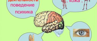
Like any other organ of the brain, the thalamus has an extremely important and irreplaceable function for the body. It’s hard to imagine, but this relatively small organ is responsible for all mental functions: perception and understanding, memory and thinking, because thanks to it we see, understand, feel the world and perceive everything that surrounds us. Thanks to its work, we navigate in space and time, feel pain, this “sensitivity collector” perceives and processes information received from all receptors, except the sense of smell, and transmits the necessary signal to the desired part of the cerebral cortex. As a result, the body gives the correct reaction, displays the correct behavior patterns to the corresponding stimulus or signal.
General information
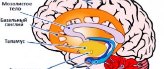
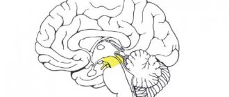
The diencephalon is located under the corpus callosum and consists of: the thalamus (thalamic brain) and the hypothalamus.
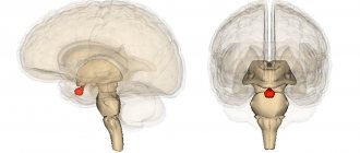
The thalamus (aka: visual thalamus, sensitivity collector, body informant) is a section of the diencephalon located in its upper part, above the brain stem. Sensory signals and impulses from various parts of the body and from all receptors (except smell) flow here. Here they are processed, the organ evaluates how important the incoming impulses are for a person and sends the information further to the central nervous system (central nervous system) or to the cerebral cortex. This painstaking and vital process occurs thanks to the components of the thalamus - 120 multifunctional nuclei that are responsible for receiving signals, impulses and sending processed information to the corresponding section of the cerebral cortex.
Thanks to its complex structure, the “visual thalamus” is capable of not only receiving and processing signals, but also analyzing them.
Ready information about the state of the body and its problems reaches the cerebral cortex, which, in turn, develops a strategy for solving and eliminating the problem, a strategy for further actions and behavior.
Structure
The thalamus is a paired ovoid formation consisting of nerve cells that are united into nuclei, thanks to which the perception and processing of signals and impulses coming from different sense organs occurs. The thalamus occupies the bulk of the diencephalon (approximately 80%). Consists of 120 multifunctional gray matter nuclei. It is shaped like a small chicken egg.
Based on the structure and location of individual parts, the thalamic brain can be divided into: metathalamus, epithalamus and subthalamus.
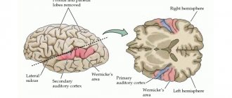
Metathalamus (subcortical auditory and visual center) - consists of medial and lateral geniculate bodies. The auditory lemniscus ends in the nucleus of the medial geniculate body, and the visual tracts end in the lateral geniculate nucleus.
The medial geniculate bodies constitute the auditory center. In the medial part of the metathalamus, from the subcortical auditory center, cell axons are directed to the cortical end of the auditory analyzer (superior temporal gyrus). Dysfunction of this part of the metathalamus can lead to hearing loss or deafness.
The lateral geniculate bodies constitute the subcortical visual center. This is where the optic tracts end. The axons of the cells form the optic radiation, along which visual impulses reach the cortical end of the visual analyzer (occipital lobe). Dysfunction of this center can lead to vision problems, and severe damage can lead to blindness.
Epithalamus (suprathalamus) - the upper posterior part of the thalamus, which rises above it: includes the pineal gland, which is the supracerebral endocrine gland (pineal gland). The pineal gland is in a suspended state, as it is located on leashes. It is responsible for the production of hormones: during the day it produces the hormone serotonin (the hormone of joy), and at night it produces melatonin (a regulator of the daily routine and the hormone responsible for the color of the skin and eyes). The epithalamus plays a role in the regulation of life cycles, regulates the onset of puberty, sleep and wakefulness patterns, and inhibits the aging process.
Lesions of the epithalamus lead to disruption of life cycles, including insomnia, as well as sexual dysfunction.
The subthalamus (subthalamus) or prethalamus is a small-volume brain substance. It consists mainly of the subthalamic nucleus and has connections with the globus pallidus. The subthalamus controls muscle responses and is responsible for action selection. Damage to the subthalamus leads to motor disturbances, tremors, and paralysis.
In addition to all of the above, the thalamus has connections with the spinal cord, with the hypothalamus, subcortical nuclei and, naturally, with the cerebral cortex.
Each department of this unique organ has a specific function and is responsible for vital processes, without which the normal functioning of the body is impossible.
Cores
Some scientists consider the term “nuclei” not entirely correct and within the structures that are so called they distinguish “subnuclei”. You can do this, or you can get by with larger structures. Therefore, we will talk about some clusters as “nuclei,” although perhaps it would be more correct to divide them into an even larger number. However, this article is not a textbook, and our goal is to simply and clearly talk about nuclei and groups of nuclei in general.
Thalamic nuclei
The group of anterior thalamic nuclei is designated in the figure as AN. They are thought to play a role in the control of wakefulness and are involved in learning and episodic memory. And they are also part of the limbic system.
The group of middle nuclei of the thalamus, or dorsomedial nucleus (MD in the figure) , occupies a fairly large volume of the thalamus and is important in the process of memorization, attention, planning, and abstract thinking. It is also involved when a person simultaneously performs many actions (the so-called multitasking, which we do not recommend to anyone). If the functioning of this nucleus is disrupted, a condition called “Korsakov's syndrome” occurs. A person stops remembering events of the present, but can more or less well reproduce memories of the past.
The ventral group of nuclei (VNG in the figure) consists of three nuclei. The first is the anterior ventral nucleus (VA), which is involved in the process of movement. It is often affected in Parkinson's disease. The second, the anterior lateral nucleus (VL), is involved in the coordination of movements and in the process of planning to make some kind of movement. It also plays a role in learning to move. This is relevant not only for small children, as you might immediately think, but also for adults who are learning to dance or swim. The third nucleus is the ventral posterior (VPL and VPM), an important part of the somatosensory system. It perceives information from touch, body position in space (or the “muscle sense” of proprioception), pain, taste, arousal, and even the urge to scratch.
The pulvinar nucleus (PUL in the picture) is responsible for visual attention. In humans, it occupies up to 40% of the thalamus, that is, it is one of the largest nuclei. If something goes wrong in it, then one-sided spatial neglect may appear - when a person’s brain does not react to what is shown from the side opposite to the affected one. For example, if there is a problem with the left side of this core, a person either does not see what is happening on the right, or cannot concentrate on what is happening. Another manifestation of a problem with the pulvinar nucleus may be attention deficit hyperactivity disorder.
The metathalamus (MTh in the figure) is represented by two formations: the paired medial geniculate bodies (MG in the figure), which play the role of the subcortical center of the auditory analyzer, and the lateral geniculate bodies (LG in the figure). The latter are exactly the same subcortical center, but this time for the visual analyzer. Both of these analyzers are connected to the centers of the corresponding analyzers in the cerebral cortex. It is believed that MGs can determine the intensity and duration of sound.
Functions of the thalamus
The “sensitivity collector” receives, filters, processes, integrates and sends information to the brain that comes from all receptors (except smell). We can say that in its centers the formation of perception, sensation, and understanding occurs, after which the processed information or signal enters the cerebral cortex.
The main functions of the body are:
- processing of information received from all organs (receptors of vision, hearing, taste and touch) senses (except smell);
- managing emotional reactions;
- regulation of involuntary motor activity and muscle tone;
- maintaining a certain level of activity and excitability of the brain, which is necessary for the perception of information, signals, impulses and irritations coming from the outside, from the environment;
- responsible for the intensity and feeling of pain.
As we have already said, each lobe of the thalamus consists of 120 nuclei, which, based on functionality, can be divided into 4 main groups:
- reticular;
- lateral (lateral);
- medial (middle);
- associative.
Reticular group of nuclei (responsible for balance) – responsible for ensuring balance when walking and balance in the body.
The lateral group (vision center) is responsible for visual perception, receives and transmits impulses to the parietal, occipital part of the cerebral cortex - the visual zone.
The medial group (hearing center) is responsible for auditory perception, receives and transmits impulses to the temporal part of the cortex - the auditory zone.
Associative group (tactile sensations) - receives and transmits tactile information to the cerebral cortex, that is, signals emanating from receptors of the skin and mucous membranes: pain, itching, shock, touch, irritation, etc.
Also, from a functional point of view, nuclei can be divided into: specific and nonspecific.
Specific nuclei receive signals from all receptors (except smell). They provide a person’s emotional reaction and are responsible for the occurrence of pain.
Specific kernels, in turn, are:
- external - receive impulses from the corresponding receptors and send information to specific areas of the cortex. Through these impulses feelings and sensations arise;
- internal - do not have direct connections with receptors. They receive information already processed by the relay cores. From them, impulses go to the cerebral cortex to the associative zones. Thanks to these impulses, primitive sensations arise and the relationship between sensory areas and the cerebral cortex is ensured.
Nonspecific nuclei support the general activity of the cerebral cortex, sending nonspecific impulses and stimulating brain activity. Having no direct connection with the cortex, the nonspecific nuclei of the thalamus transmit their signals to subcortical structures.
Reflex connections of the thalamus. Nuclei of the thalamus (specific and nonspecific) and their projections
Diencephalon
(diencephalons) includes several formations located anterior to the midbrain.
The largest of them are the thalamus
(optic thalamus),
metathalamus
(geniculate body) and
hypothalamus
(subthalamic region).
These are complexly organized structures, consisting of a large number of cores and providing many different functions. Together with the cerebral hemispheres, the diencephalon is involved in the organization of all complex forms of behavior and the regulation of all body functions. However, the structure, neural organization and functions of the thalamus and hypothalamus are so different that they are considered separately, as independent entities.
Thalamus
or visual thalamus - a pair of relatively massive formations consisting predominantly of gray matter. The human thalamus is massive, containing about 120 nuclei, which are separated by white matter fibers.
It has close connections with the bark. The thalamus is the subcortical (intermediate) center of all types of sensitivity, except olfactory. The ascending (afferent) pathways, through which information from various receptors (skin, vision, etc.) are transmitted, are approached and switched. From the thalamus, nerve fibers go to the cerebral cortex, forming thalamocortical pathways, and partly to the basal ganglia.
It is believed that the thalamus holds the keys to the secrets of the cerebral cortex. The nature of connections between the thalamic nuclei and the cortex is based on their structural and functional differences. Based on morphological differences and the nature of projections into the cortex, the thalamic nuclei are divided into:
Specific
:
o relay
(external). They receive impulses from afferent systems directly and transmit them to the primary projection zones of the cortex (strictly specific). Impulses also go to the associative nuclei. Destruction of relay nuclei leads to complete and irreversible loss of corresponding sensitivity or movement disorders
o associative
nuclei (internal) that do not have direct contacts with afferent systems. Receive impulses from relay cores. From them, impulses go to the secondary projection and associative (tertiary projection) zones of the cortex, provide communication between sensory systems in the cerebral cortex and create primitive sensations.
Nonspecific
. Nonspecific nuclei of the thalamus do not belong to any one projection zone. They are morphologically and functionally associated with many systems and participate, together with the reticular formation of the brain stem, in the implementation of nonspecific functions. From the nonspecific nuclei of the thalamus, impulses are projected diffusely onto the cerebral cortex and sent to neurons of all layers of the cortex. The nonspecific system enhances the specific one, increases the excitability of cortical neurons, and has a modulating effect on them. The activity of the nonspecific nuclei of the thalamus is closely related to the mechanisms of sleep development and integrative mechanisms of the brain.
The middle part of the thalamus faces the third ventricle, the upper surface faces the lateral ventricle, and the lateral and lower surfaces face adjacent brain structures.
Metathalamus or
geniculate bodies - are small elevations and consist of gray matter: the nuclei of the geniculate bodies.
Lateral geniculate bodies
– subcortical center of vision. Nerve fibers of the optic tract (from the retina) approach the neurons of its nuclei, and the axons go to the visual area of the cortex
Medial geniculate bodies
– subcortical hearing center. The nuclei receive the nerve fibers of the auditory pathway, and the axons of its neurons follow into the auditory zone of the cerebral cortex.
Separately about the visual thalamus
Previously, it was believed that the thalamus processed only visual impulses, and then the organ received the name - visual thalamus.
Now this name is considered obsolete, since the organ processes almost the entire range of afferent systems (except for smell). The system that provides visual perception is one of the most interesting. The main external organ of vision is the eye, a receptor that has a retina and is equipped with special cells (cones, rods) that transform the light beam and electrical signal. The electrical signal, in turn, passing through the nerve cells enters the lateral center of the thalamus, which sends the processed signal to the central part of the cerebral cortex. Here the final analysis of the signal occurs, thanks to which what is seen is formed, that is, the picture.