The cerebellum is the part of the brain that controls the musculoskeletal system. The human cerebellum is not directly connected to the receptors that convert the influence of external and internal factors into nerve impulses. This part of the brain interacts with all other parts of the central nervous system. Impulses coming from receptors of tendons, muscles, joint capsules, ligaments, cerebral cortex, subcortical nuclei and medulla oblongata are directed to it.
In turn, impulses depart from the cerebellar medulla in the direction of other parts of the central nervous system. In this way, two-way interaction is carried out. The functions of the cerebellum, located in the human brain, are associated with maintaining balance and coordinating movements of the limbs and other parts of the body.
General information about the organ
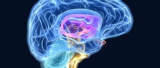
The cerebellum, also called the small brain, is located above the pons and the medulla oblongata. Outwardly it looks like other brain structures with characteristic convolutions, depressions, and folds on the surface. The condition of the organ and the presence of disturbances in its functioning can be clearly seen when performing fast movements - playing a musical keyboard instrument, typing using a typewriter or laptop keyboard, running.
The organ controls the consistency, clarity, and measuredness of actions performed by different parts of the body. It does not cause muscle contractions, but determines the sequence of movements and the time frame for their execution. Information about the desired sequence of actions enters the cerebellum from other parts of the brain and parts of the body.
Feedback information allows you to evaluate the consistency of planned and executed movements. The cerebellum adjusts the motor activity program taking into account deviations. Correction occurs by increasing or decreasing the activation of individual muscles. The organ, in cooperation with the cortical structures of the cerebrum, plans a new movement in advance. Irritation of this part of the brain provokes dilation of the pupils and an increase in blood pressure.
Serious damage to the cerebellar fibers leads to malfunctions in the body:
- Astasia. Impaired ability to hold the body in a standing position.
- Atony. Abnormally decreased tone of skeletal muscles and muscle fibers of internal organs.
- Asthenia. A state of powerlessness, general weakness.
- Ataxia. Impaired fine motor functions, lack of coordination of movements.
Autonomic functions are impaired when the structures that make up the cerebellum are damaged. The mechanisms of natural regulation of breathing, blood circulation, and maintaining normal body temperature are slowed down. Damage to all or part of the cerebellum does not provoke the development of paralysis and has virtually no effect on cognitive abilities.
A person experiences difficulties when it comes to learning and remembering new movements. To understand the questions of what the cerebellum is needed for and what it is responsible for, it is worth remembering that the organ is the control center for gross and fine motor skills. It coordinates complex and simple movements on a subconscious level without involving thought processes and involving the work of consciousness.
Yu.V. Zueva, N.K. Korsakova, L.A. Kalashnikov
Moscow, Russia
For many years, the cerebellum was considered as a structure responsible solely for the coordination and static movement of movements. And although clinical studies have long accumulated material on the connection between cerebellar pathology and diseases such as autism, schizophrenia, senile dementia, due to the lack of instrumental and technical base, these data remained at the phenomenological level (Sehmahmann, 1991; Snider, 1982; Weinberger et al ., 1980; Cole et al.., 1989). With the development of neuroimaging research methods, such as positron emission tomography, single-photon emission tomography and high-resolution magnetic resonance imaging, as well as the accumulation of a huge amount of data in the framework of anatomical, laboratory, and clinical studies, the role of the cerebellum in the organization of mental functions was shown (Akshoomoff & Courchersne , 1992; Botez-Marquard & Botez, 1993; Berquin et al., 1998; Kim et al., 1994; Leiner et al., 1986; Orioli & Stick, 1989; Paradiso et al., 1997; Paulesu et al., 1995; Petersen & Fiez, 1993; Sehmahmann, 1996, 1997, 1998; Sehmahmann & Handya, 1987; Silvery et al., 1994). The concept of the influence of the cerebellum on cognitive processes was formed relatively recently, but already in 1997 in the USA, under the editorship of Sehmahmann, the world's first monograph “Cerebellum and Cognition” was published, summarizing the currently available clinical, anatomical, physiological and neuroimaging data (Sehmahmann, 1997 ). The anatomical basis for the participation of the cerebellum in mental functions is its bilateral connections with the associative zones of the cortex of the predominantly contralateral cerebral hemispheres and the limbicoreticular complex. At the same time, the isolated existence of pathways going to the motor cortex and prefrontal parts of the brain was noted, which in turn determines the possibility of isolated occurrence of cognitive and motor disorders with damage to the cerebellum (Hook, 1997; Kim et al., 1994; Leiner et al. 1986 , 1993; Middleton & Stick, 1994; Orioli & Strick, 1989; Sehmahmann, 1997; Sehmahmann & Handya, 1987). It has also been shown that cerebellar pathology of various origins (tumors, degenerations, hypoplasia, vascular changes) leads to a wide range of mental dysfunctions in the form of disorders of planning, abstract thinking, working memory, deficits of spatial functions, speech, emotional and personal changes (Botez-Marquard & Botez, 1993; Berquin et al., 1998; Canavan et al., 1994; Cole, 1994; Grafman et al., 1992; Malm et al., 1998; Sehmahmann, 1998; Silvery et al., 1994).
However, most studies are limited to the consideration of either individual mental functions and their components, or the study of clinical syndromes, which prevents the possibility of finding general mechanisms for the formation of very diverse and numerous cognitive symptoms. It seems adequate to solve this problem by applying complex syndromic analysis to impaired mental functions. At the school of A.R. Luria for more than 20 years, T.V. Melnikova (1974) conducted a study of patients with subtentorial tumors (including a cerebellar tumor) and obtained data on the presence of neuropsychological syndromes of the frontal and temporoscopic type. parieto-occipital. At the same time, a relationship was shown between neuropsychological disorders and dysfunction of these particular parts of the cerebral cortex using EEG. However, the identified changes were explained by transient vascular disorders associated with a specific tumor location. The role of the cerebellum in cognitive deficits has not been identified and has not been developed in further studies.
Based on the experience of Western researchers and applying the methodology of syndromic analysis developed at the school of A.R. Luria (2000), we formulated the task of conducting a comprehensive study of disorders of higher mental functions (HMF) in cerebellar infarctions and identifying clinical parameters that influence the degree of cognitive decline in this form of pathology. Isolated infarctions, compared with other types of diseases (cerebellar degenerations, tumors, congenital anomalies), are the most adequate and correct clinical model, since they develop in patients without concomitant signs of damage to the cerebral hemispheres, which cannot be completely excluded in the case of degenerative diseases or tumors of the cerebellum, often accompanied hydrocephalus. However, isolated infarctions are a very rare clinical model (Malm et al., 1998: Schmahmann. 1998; Schmalimann & Handya. 1987) and the present study of 25 patients with this pathology is the first to be conducted on such a large sample.
switch – previous | next – neuropsychological study
A. R. Luria and psychology of the 21st century. Content
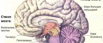
Structure of the cerebellum
The structure of the cerebellum is complex. The organ consists of many parts - lobes of the hemispheres, vermis, cortex, nuclei and legs, which perform certain functions and connect the cerebellum with parts of the brain. The anatomy of the cerebellum is represented by two hemispheres, lying on either side of the vermis. The cerebellar cortex is formed from three areas:
- Archicerebellum. Otherwise called the flocculonodular lobe.
- Paleocerebellum. Bilaterally interacts with the receptors of the spinal cord and the sensorimotor area of the cortex covering the cerebral hemispheres.
- Neocerebellum. Bilaterally interacts with cortical neurons that make up the cerebral hemispheres, receptors of the visual and auditory apparatus.
The cortex consists of an inner (granular), outer (molecular) and middle layer formed from Purkinje cells. The layered structure of the organ indicates integration processes that reflect the active interaction of different parts of the brain. The layers of the cerebellum provide its connection with different parts of the central nervous system. The thickness of the cerebellum contains 3 pairs of nuclei - lateral dentate, median tent nuclei, rounded and cork-like in the space between the first two pairs.
The cerebellum is connected to the parts of the brain through the peduncles. The fiber composition of the peduncles located in the cerebellum includes the nuclei of the vestibular nerve, as well as nerve bundles - the gentle and sphenoid. The lower legs connect the organ with a portion of the medulla oblongata. The middle legs in the form of a spacious, protruding part of the cerebellum are located laterally and pass into the pons. The cortical structures of the hemispheres located in the cerebrum control the functioning of the cerebellum using the middle peduncles. The flattened tufts of the upper legs are directed in both directions.
Worm
The structure and functions of the cerebellum cannot be imagined without the vermis, which connects both hemispheres of the organ together. The vermiform structure is composed of white matter nerve fibers. Pathological changes in the tissues of the worm cause ataxia.
Slices
The lobes of the hemisphere located in the cerebellum are delimited by grooves. The worm and hemispheres contain 8 lobules. The anterior, as well as the flocculonodular lobes, located in the cerebellum, are connected by working functions with the brain of the spinal column and the vestibular apparatus. The cerebellar hemispheres receive impulses from muscle and joint tissue receptors. On the front side, a small formation is adjacent to the hemisphere - a shred.
Cores
The nuclei located in the cerebellum control the axial and proximal muscles of the limbs. The idea of movement appears in the associative cortical structures of the brain, then is transmitted through afferent pathways towards the elements of the musculoskeletal system. The program of planned movement is formed in the dentate nucleus and simultaneously in the cerebellar hemispheres, then redirected to the motor centers of the cortex using the thalamic nuclei. The reproduced movement cannot be controlled using the somatosensory feedback system. Damage to the nuclei causes convulsive contraction of the muscles that control the limbs.
Functions of the small brain
The physiology of the cerebellum is mainly related to motor activity and regulation of the autonomic system. Increased heart rate and breathing during sports activities are triggered by the cerebellum’s reaction to increased physical activity. As a result, blood supply to tissues, their saturation with oxygen and nutrients improves. The organ plays a crucial role in maintaining balance. Functions of the cerebellum include:
- consistency, synchronization of movements;
- maintaining balance and a given posture;
- ensuring the accuracy of targeted movements with timing and taking into account clearly marked coordinates;
- interaction of agonist and antagonist muscles;
- regulation of the activity of the autonomic system.
The organ takes part in memorizing motor information. Its activities are associated with the correct execution of physical techniques, poses and exercises in sports.
Functions of the organ and what it is responsible for
The small brain plays the role of evaluator when making decisions. The functions of the cerebellum of the human brain are the reception and storage of information about the results of this action and possible consequences.
The cerebellum performs the following functions:
- maintaining the tone of muscle fibers and control over body posture;
- adjustment of slow movements and their coordination in relation to body position;
- control of the process of performing movements that require quick reactions.
To study as best as possible the characteristics of this organ and why it is needed, scientists conducted a number of studies on animals. The cerebellums were removed, and as a result, the following development was observed:
- astasia - the animal felt unsure in space, it spread its limbs wide and swayed;
- atony - disruption of the ability of normal functioning of muscle fibers, loss of the ability to flex and straighten limbs;
- asthenia - loss of the ability to exercise control over one’s movements;
- ataxia - sudden movements.
Symptoms of cerebellar damage
Pathological processes are often accompanied by dysmetria. Limbs cannot move smoothly to a given position due to incorrect force and incorrect direction of movement. Another sign of clinical damage is dysdiadochokinesis, which is expressed in awkward, angular execution of movements that alternate rapidly. To identify the disorder, the patient is asked to tap the fingertips of one hand on the back of the other hand or quickly turn the hand with the palm up and down.
An attempt to reproduce a complex movement leads to fragmentation of the action into separate stages. As a result, the movement is performed intermittently. If the patient tries to touch an object, an intention tremor of the limb occurs. The amplitude of the jitter increases as the distance to the target decreases. Patients with cerebellar dysfunction when walking move their legs too far apart, lose balance and often fall. Words and sentences are spoken at a slow pace, indistinctly. The speech takes on a chanting character.
Hypotonia is a decrease in muscle tone, manifested in combination with a decrease in muscle strength. Muscle resistance to passive movements decreases. Palpation reveals increased muscle softness. A pendulum-shaped knee reflex appears. When the doctor hits the knee tendon below the cap with a special hammer, the leg swings for a while due to weakened muscle tone. Normally, long-term vibrations of the limb do not occur. Other symptoms:
- Dizziness, nausea, headache. The conditions develop due to a breakdown in the functional connection between the vestibular apparatus and the cerebellum.
- Increased fatigue due to physical and mental stress.
- Malfunctions in the functioning of the cardiovascular system.
- Dysfunction of the gland that produces gastric juice.
- Neurological abnormalities.
The location of the cerebellum in the posterior cranial fossa determines the characteristics of symptoms in some diseases. For example, when a tumor forms, the disease in the first stages manifests itself as symptoms of damage to the brain stem or signs of blocking the drainage of cerebrospinal fluid.
Treatment and prognosis
For any type of lesion, therapy is divided into symptomatic and etiological. The chances of restoring dead cells are very small. Therefore, doctors only treat the symptoms of the disease. Medicines and surgical techniques are used. The prognosis for the disease is unfavorable.
How to develop the cerebellum?
Children with cerebellar insufficiency may lag behind their peers in development. And in older people, frequent falls are a source of dangerous complications. How to develop a “small” brain?
- The most ancient method is rocking a baby in a cradle. It’s not for nothing that our ancestors made cradles for the birth of their first child. This is not only caring for the mother, but also the development of the child’s vestibular apparatus;
- Next, a swing is suitable for training a two-year-old. They must be modern and safe. Just play with your baby and develop not only vestibular skills, but also conversational speech;
- After 5-7 years of age, children are taught on Dr. Bilgow’s balancing board. You can make it yourself or buy it in a store. It’s better to attend courses with an experienced exercise therapy instructor;
- There's more to the board than just balancing. Exercises are used with throwing and catching bags of sand, a ball and other devices.
Important Barbiturates in urine: drugs, elimination times, permissible limit, what they threaten
It is important to exercise regularly (3-4 times a week). Lesson duration: at least 30 minutes
If you feel well, such training is also available for older people.
Diseases
Diseases that affect the cerebellum - cyst, malignant tumor, malformations and anomalies, abscess, olivopontocerebellar degenerative processes, cerebellar ataxia caused by hereditary factors, known as Pierre Marie's ataxia. Pathologies of the cerebellum cause disorders in the functioning of the musculoskeletal system. Violations affect:
- coordination of movements (dynamic ataxia);
- maintaining balance (static ataxia);
- muscle tone.
The defeat of a specific lobe is reflected in symptoms. For example, damage to the tissue of the flocculo-nodular area leads to atony of the muscles running along the spinal column.
The influence of the cerebellum on autonomic functions
The small brain is endowed with the ability to influence the functioning of all body systems. Damage to the cerebellum leads to a decrease in blood pressure and the development of bradypnea. The tone of the respiratory muscles on the affected side will be decreased, while on the opposite side there will be an increase.
The tone of the intestinal muscle fibers changes, it will be reduced, and this causes a disruption in the removal of the contents of the intestines and stomach. There is a suppression of the absorption of nutrients. Metabolic processes accelerate, the level of glucose in the blood increases, which persists for a long period of time compared to the norm. Appetite worsens, the patient loses weight, and the process of tissue regeneration is suppressed.
In drawing a conclusion, I would like to focus on the fact that the cerebellum, despite its small size, plays a very important role in the functioning of the entire organism. Its defeat can cause severe disorders; in the absence of adequate therapy, this is fraught with irreversible consequences.
Diagnosis of pathologies
Instrumental diagnostics are carried out using ultrasound, MRI and CT methods. Other research methods are cerebrospinal fluid puncture and vascular Dopplerography. To identify disturbances in the functioning of the organ, vestibulometry is performed - a series of neurological tests:
- Romberg's test. The patient is in a standing position, the feet are moved together, the eyes are closed, the upper limbs are extended forward, then spread to the sides.
- Romberg test with complication. The body position is the same as in the usual Romberg test. The only difference is that the feet are on the same line. In this case, the right foot is located in front of the left foot.
- Walking along a straight line. The test is performed with eyes open, then with eyes closed.
- Unterberger's test. The patient walks in one place with his eyes closed, raising his knees high. The angle of deviation from the initial position after taking 50 steps normally does not exceed 30°.
- Finger test. An attempt to touch your own nose with your index finger with your eyes closed ends in a miss or trembling of the finger.
A blood test will show the presence of specific markers of inflammation or stroke. A lumbar puncture is performed if the neurologist suspects an infectious disease occurring in the cerebrospinal fluid.
The cerebellum is an organ that regulates the functions of the musculoskeletal system. Failures in the functioning of this part of the brain are accompanied by multiple disorders of motor activity. Organ dysfunction impairs quality of life. With significant damage to the cerebellar tissue, a person is unable to hold the body in the desired position, walk smoothly, or make precise movements with the arms and legs.
150