| Striatum | |
| lat. corpus striatum | |
| Striatum. | |
| Catalogs | |
| |
| Media files on Wikimedia Commons |
Striped body
(lat. corpus striatum) - anatomical structure of the telencephalon, belonging to the basal ganglia of the cerebral hemispheres. In horizontal and frontal sections of the brain, the striatum appears as alternating stripes of gray matter and white matter. The striatum includes the caudate nucleus and the lentiform nucleus.
Functions[ | ]
The striatum regulates muscle tone, reducing it; participates in the regulation of the work of internal organs; in the implementation of various behavioral reactions (food-procuring behavior); participates in the formation of conditioned reflexes. When the striatum is destroyed, the following occurs:
- hypertonicity of skeletal muscles,
- disturbance of complex motor reactions and food-procuring behavior;
- the formation of conditioned reflexes is inhibited.
The ventral striatum and nucleus accumbens regulate reward and reinforcement in the brain. The dorsal part of the striatum is more involved in regulating motor functions. The dorsal part is also associated with the presence of impulsive behavior.[2][3][4][5][6]
Afferent connections of the basal ganglia.
Most of the afferent signals coming to the basal ganglia enter the striatum.
These signals come almost exclusively from three sources: - from all areas of the cerebral cortex;
- from the intralamellar nuclei of the thalamus;
- Basal ganglia structure, development, functions
- Grooved Features
- Basal ganglia of the brain
- Associated disorders
- Histology
- Circulation
- Shells
- Does this mean that psychopaths are not to blame?
- Functionality of the basal ganglia
- from the substantia nigra (along the dopaminergic pathway).
Efferent fibers from the striatum go to the globus pallidus and substantia nigra. From the latter, not only the dopaminergic path to the striatum begins, but also the paths going to the thalamus.
The most important of all efferent tracts of the basal ganglia originates from the internal part of the globus pallidus, ending in the thalamus, as well as in the roof of the midbrain. Through the stem formations with which the basal ganglia are connected, centrifugal impulses follow to the segmental motor apparatus and muscles along descending conductors.
- from the red nuclei - along the rubrospinal tract;
- from the Darkshevich nucleus - along the posterior longitudinal fasciculus to the nuclei of nerves 3, 4,6 and through it to the nucleus of the vestibular nerve;
- from the nucleus of the vestibular nerve - along the vestibulospinal tract;
— from the quadrigeminal region — along the tectospinal tract;
- from the reticular formation - along the reticulospinal tract.
Thus, the basal ganglia play mainly the role of an intermediate link in the chain connecting the motor areas of the cortex with all its other areas.
Notes[ | ]
- ↑ 1234
Striatum // Fundamental Model of Anatomy - L. M. Yager, A. F. Garcia, A. M. Wunsch, S. M. Ferguson.
The ins and outs of the striatum: Role in drug addiction (English) // Neuroscience. — 2015-08. - Vol. 301. - P. 529–541. - doi:10.1016/j.neuroscience.2015.06.033. - M. Foster Olive, Taylor, Lewis.
The neurocircuitry of illicit psychostimulant addiction: acute and chronic effects in humans (English) // Substance Abuse and Rehabilitation. — 2013-02. - P. 29. - ISSN 1179-8467. - doi:10.2147/SAR.S39684. - Sergi Ferré, Carme Lluís, Zuzana Justinova, César Quiroz, Marco Orru.
Adenosine-cannabinoid receptor interactions. Implications for striatal function: Adenosine-cannabinoid receptor interactions // British Journal of Pharmacology. — 2010-06. - Vol. 160, iss. 3. - P. 443–453. - doi:10.1111/j.1476-5381.2010.00723.x. - Nestler, Eric J. (Eric Jonathan), 1954-.
Molecular neuropharmacology: a foundation for clinical neuroscience. — 2nd ed. — New York: McGraw-Hill Medical, 2009. — 1 online resource p. — ISBN 978-0-07-164119-7, 0-07-164119-X, 0-07-148127-3, 978-0-07-148127-4, 978-1-281-79174-0, 1- 281-79174-1. - BaekSun Kim, Heh‐In Im.
The role of the dorsal striatum in choice impulsivity // Annals of the New York Academy of Sciences. — 2019-09. - Vol. 1451, iss. 1. - P. 92–111. — ISSN 1749-6632 0077-8923, 1749-6632. - doi:10.1111/nyas.13961.
Basal ganglia structure, development, functions
In the article we will talk about the basal ganglia
What is it and what role does this structure play in human health? All questions will be discussed in detail in the article, after which you will understand the importance of absolutely every detail in your body and head
Basal ganglia: physiology
These nuclei are located near the cerebral hemispheres. They have many long processes called axons. Thanks to them, information, that is, nerve impulses, is transmitted to different structures of the brain.
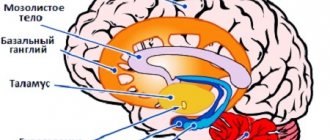
The basal nuclei can be considered the red and caudate nuclei, globus pallidus, putamen, substantia nigra and reticular formation.
The structure of the basal ganglia is varied. Basically, according to this classification, they are divided into those that belong to the extrapyramidal and limbic system. Both of these systems have a huge impact on the functioning of the brain and are in close interaction with it. They affect the thalamus, parietal and frontal lobes. The extrapyramidal network consists of the basal ganglia. It completely permeates the subcortical parts of the brain, and it has a major influence on the functioning of all functions of the human body. These modest formations very often remain underestimated, and yet their work has not yet been fully studied.
The functions of the basal ganglia are not many, but they are significant. As we already know, they are strongly connected to all other brain structures. Actually, from the understanding of this statement the main functions of the kernels follow:
- Control over the implementation of integration processes in higher nervous activity.
- Influence on the functioning of the autonomic nervous system.
- Regulation of human motor processes.
What are they involved in?
There are a number of processes in which nuclei are directly involved. The basal ganglia, the structure, development and functions of which we are considering, are involved in the following actions:
- affect a person’s dexterity when using scissors;
- accuracy of driving nails;
- reaction speed, dribbling the ball, accuracy of hitting the basket and dexterity of hitting the ball when playing basketball, football, volleyball;
- control of the voice while singing;
- coordination of actions while digging the earth.
New research has shown that the basal ganglia can also influence the type of movement:
- controllable or sudden;
- repeated many times or new, completely unknown;
- simple monosyllabic or sequential and even simultaneous.
In ordinary life, the basal ganglia simply transmit impulses that come from the frontal lobes to other brain structures. The goal is to purposefully perform known actions without stressing the central nervous system. However, in dangerous situations, the ganglia switch and allow a person to automatically make the most optimal decision.
Lesions of the basal ganglia can be very different. Let's look at some of them. These are degenerative lesions of the human brain (for example, Parkinson's disease or Huntington's chorea). These may be hereditary genetic diseases that are associated with metabolic disorders. Pathologies characterized by malfunctions of enzyme systems. Diseases of the thyroid gland can also occur due to disturbances in the functioning of the nuclei. Possible pathologies resulting from manganese poisoning. Brain tumors can affect the functioning of the basal ganglia, and this is perhaps the most unpleasant situation.
Important Ladasten: instructions for use and what it is needed for, price, reviews, analogues. drug for the treatment of asthenic disorders Ladasten: instructions, reviews Ladasten instructions for use analogues
Basal ganglia of the brain
These structures (ganglia) are located directly under the cortical part of the telencephalon. They significantly affect the motor functionality of the human body. Their violation mainly affects muscle tone.
What are the basal ganglia?
The subcortical ganglia of the brain are dense anatomical structures localized in the white matter of the cerebral hemispheres.
The ganglion structures are connected to:
- Lenticular and caudate nuclei of the brain
- Fence
- Amygdala
The subcortical nuclei of the ganglion have membranes, which include white matter. The caudate nucleus, together with the lentiform nucleus, is anatomically represented by the striatum.
Three large subcortical nuclei form the extrapyramidal system, which is involved in controlling movements and maintaining muscle tone.
The main function of the ganglia is to slow down or accelerate the transmission of impulses from the thalamus to the cortical areas that are responsible for motor functions.
The caudate nucleus, the terminal ganglion, makes up the striopalidal system and is responsible for muscle contraction.
The purpose of the basal ganglia is closely related to the activity of the hypothalamus and pituitary gland. Most often, a number of disorders in the structure and functions of the ganglia are accompanied by a decrease in the functions of the pituitary gland.
Additional structures
The fence appears to be a thin layer of gray matter, which is localized between the shell and the insula. The entire fence is literally enveloped by a white substance that forms two capsules:
- External, which is localized between the fence and the shell
- The outermost one, located next to the island
The limbic system, or visceral brain, stands out for its structural complexity. The functions of the limbic system are multifaceted, as is the specificity of its structure.
Limbics are responsible for:
- Autonomic reactions
- Active activities aimed at acquiring and developing skills
- Psychological and emotional processes
Pathological conditions of the ganglia
Experts have found that characteristic pathological changes lead to a number of other diseases:
- Functional deficiency of the basal ganglia
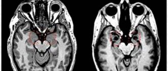
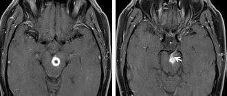
A pathological neoplasm in the brain occurs as a result of abnormal metabolism, atrophy or damage to soft tissue, as well as infectious processes. The most unfavorable complication caused by the pathology of the basal ganglia is hemorrhage. If in this case the patient is not provided with timely medical care, then a possible rupture of the cavity will result in death.
A benign neoplasm or cyst that does not increase in size causes virtually no inconvenience to the patient. If the doctor notes the progression of the evolution of the ganglia, the patient is assigned a disability.
Associated disorders
Most striatum-related disorders and diseases affect movement, both voluntary and automatic. Parkinson's disease and Huntington's disease are two major examples of basal ganglia dysfunction.
However, some psychological changes appear to depend on the functioning of this structure, mainly due to its role in the brain's reward system.
Parkinson's disease
Parkinson's disease causes lesions in the brain, mainly in the basal ganglia. The death of dopaminergic neurons in the substantia nigra prevents the release of dopamine in the striatum, causing motor symptoms such as slowness, rigidity, tremors and postural instability. Depressive symptoms are also produced.
Important Nimotop
Huntington's disease
At the initial stage, Huntington's disease mainly affects the striatum; This explains why early symptoms are related to motor control, emotions and executive functions. In this case, the basal ganglia are unable to restrain unnecessary movements and hyperkinesia occurs.
Bipolar disorder
Research shows that in some cases of bipolar disorder, there are changes in genes that regulate striatal function. Evidence in this regard has been found for both bipolar disorder type I and type II.
Related article: "Bipolar Disorder: 10 Features and Curiosities You Didn't Know"
Obsessive-compulsive disorder and depression
Obsessive-compulsive disorder and depression, which have a similar biological basis, have been associated with dysfunctions in the striatum. This explains the decrease in mood that occurs in both disorders; Difficulty inhibiting movements is also a factor in OCD.
You may be interested: "Are there several types of depression?"
Dependencies
Dopamine is a neurotransmitter involved in the brain's reward system; the pleasurable sensations we experience when dopamine is released in the basal ganglia explain our motivation to return to seeking experiences that we know are pleasurable. This explains cravings from a physiological point of view.