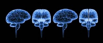
General information about the parietal lobe
The parietal lobe is a part of the cerebral hemisphere that is located behind the central sulcus.
The posterior border runs along the parieto-occipital sulcus and borders the occipital lobe. On the sides, the parietal lobe is limited by the Sylvian fissure. This structure of the cerebral hemisphere has the following main grooves:
- postcentral, which in turn is divided into upper and lower;
- interparietal.
The intersection of these grooves is called the helix, or star.
In the anterior part is the postcentral gyrus. The other two gyri are the superior parietal and inferior parietal, which are located horizontally.
Structure of the parietal lobe
The parietal lobe occupies the superior lateral surfaces of the hemisphere. From the frontal lobe, the parietal lobe is limited in front and to the side by the central sulcus, from the temporal lobe below - by the lateral sulcus, from the occipital - by an imaginary line running from the upper edge of the parieto-occipital sulcus to the lower edge of the hemisphere.
On the superolateral surface of the parietal lobe there are three gyri: one vertical - posterior central and two horizontal - superior parietal and inferior parietal. The part of the inferior parietal gyrus, which encircles the posterior part of the lateral sulcus, is called the supramarginal (supramarginal) region, the part surrounding the superior temporal gyrus is the nodal (angular) region.
The parietal lobe, like the frontal lobe, makes up a significant part of the cerebral hemispheres. In phylogenetic terms, it is divided into an old section - the posterior central gyrus, a new one - the superior parietal gyrus and a newer one - the inferior parietal gyrus.
The function of the parietal lobe is associated with the perception and analysis of sensory stimuli and spatial orientation. Several functional centers are concentrated in the gyri of the parietal lobe.
In the posterior central gyrus, sensitivity centers are projected with a body projection similar to that in the anterior central gyrus. The face is projected in the lower third of the gyrus, the arm and torso are projected in the middle third, and the leg is projected in the upper third (see Fig. 2 A). In the superior parietal gyrus there are centers in charge of complex types of deep sensitivity: muscular-articular, two-dimensional spatial sense, a sense of weight and range of motion, a sense of recognizing objects by touch.
Posterior to the upper parts of the posterior central gyrus, a center is located that provides the ability to recognize one’s own body, its parts, their proportions and relative positions (field 7).
Fields 1, 2, 3 of the postcentral region constitute the main cortical nucleus of the skin analyzer. Together with field 1, field 3 is the primary, and field 2 is the secondary projection zone of the skin. analyzer. The postcentral region is connected by efferent fibers to the subcortical and stem formations, to the precentral and other areas of the cerebral cortex. Thus, the cortical section of the sensitive analyzer is localized in the parietal lobe.
Primary sensory areas –
these are areas of the sensory cortex, irritation or destruction of which causes clear and permanent changes in the sensitivity of the body (analyzer nuclei, according to I.P. Pavlov). They consist mainly of unimodal neurons and form sensations of the same quality. In the primary sensory zones there is usually a clear spatial (topographic) representation of body parts and their receptor fields.
Around the primary sensory areas are less localized secondary sensory areas,
whose neurons respond to the action of several stimuli, i.e. they are multimodal.
The most important sensory area is the parietal cortex of the postcentral gyrus and the corresponding part of the paracentral lobule on the medial surface of the hemispheres, which is designated as somatosensory area I. Here there is a projection of the skin sensitivity of the opposite side of the body from tactile, pain, temperature receptors, interoceptive sensitivity and musculoskeletal sensitivity. musculoskeletal system - from muscle, joint, tendon receptors (see Fig. 2A).
In addition to somatosensory area I, a smaller somatosensory area II is distinguished, located at the border of the intersection of the central sulcus with the upper edge of the temporal lobe, in the depth of the lateral sulcus. The degree of localization of body parts is less pronounced here.
are located in the inferior parietal lobe .
Praxis refers to purposeful movements that have become automated in the process of repetition and exercise, which are developed in the process of learning and constant practice throughout an individual’s life. Walking, eating, dressing, the mechanical element of writing, various types of work activities (for example, the movements of a driver while driving a car, mowing, etc.) are praxis. Praxis is the highest manifestation of the motor function inherent in humans. It is carried out as a result of the combined activity of various areas of the cerebral cortex.
The center of the analyzer of interoceptive impulses is located in the lower parts of the anterior and posterior central gyri
internal organs and blood vessels. The center has close connections with subcortical vegetative formations.
What fields are included?
In total, the parietal lobe of the brain has nine fields.
The first three fields, which are 1, 2 and 3, are primary sensorimotor fields. They are located in the vertical gyrus of the parietal lobe (postcentral). The fourth field is the primary motor cortex. Areas 5 and 6 are secondary somatosensory and motor areas, respectively. The seventh field, which is located in the upper part of the parietal lobe, is the tertiary motor cortex. Field 39, in turn, is responsible for analyzing written speech. The main function of field 40 is reading comprehension, as well as ensuring the reading process. This part of the brain is characterized by the presence of the following centers:
- - responsible for recognizing the position of the body and individual organs in space;
- sensitivity center – analyzes information about any changes in the environment;
- center of praxia – responsible for performing complex actions;
- vocabulary center – helps in recognizing letters and other signs;
- calculus center - responsible for carrying out numerical calculations in the mind.
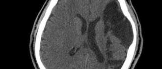
Classification
Depending on the type of brain cells that degenerated and gave rise to pathogenesis, the following types of tumors are distinguished:
- astrocytomas are the most common type, their share in the general population is about 50%;
- oligodendrogliomas – the share in the general population of tumors of this type is up to 10%;
- Ependymomas are the rarest forms (occurrence less than 7%).
According to the WHO classification (which is generally accepted), neoplasia is ranked depending on the degree of malignancy.
Benign gliomas
These are tumors of the first degree of malignancy, for example, astrocytomas: giant cell, pilocytic, juvenile subependymal. They are benign because they grow slowly, have no signs of cancer, and are easy to treat. The prognosis for life with benign glioma is favorable. After tumor removal, patients live 10 years or longer.
Low grade glioma
Second degree of malignancy. It is also classified as a benign neoplasm, but with a borderline degree of malignancy. The growth of pathological tissue is slow (low-grade), they are well differentiated, as a rule, there is only one sign of cancer (cell atypia). Tumors of this type can degenerate into cancer and easily progress to the third and fourth degree of malignancy. These include diffuse and fibrillary astrocytomas. Treatment is complex: surgery to remove atypical tissues, additionally radio and chemotherapy.
Malignant gliomas
These include gliomas of grade 3 and 4:
- Third degree of malignancy . There are all signs of malignancy, except for tissue necrosis. Tissues lose clear differentiation, tumor growth accelerates (high-grade), the boundaries are unclear, and growth into nearby tissues is typical. The most striking example is anaplastic astrocytoma, which most often develops in middle-aged and older people. Treatment is hampered by the lack of clear boundaries of the tumor; life expectancy depends on how much atypical tissue is removed, as well as the stage of carcinogenesis at the beginning of treatment.
- The most dangerous is the fourth degree of malignancy (glioblastoma). It develops between the ages of 40 and 70 years. In this case, all the signs of malignancy are present, including necrosis. The tumor grows quickly, penetrates other tissues and has no clear boundaries. This significantly complicates therapy. The prognosis is unfavorable.
Neoplasia is divided into two types, depending on growth characteristics:
- Tumors of nodular growth . As a rule, these are benign neoplasms with clear contours. They are characterized by formation anywhere in the brain and the presence of cysts. Examples: pleomorphic xanthoastrocytoma and piloid astrocytoma.
- Diffuse type formations . There are no obvious boundaries, the size can reach significant sizes, neoplasia grows into adjacent brain tissue, which complicates the removal of dysplasia. These are often malignant tumors, such as glioblastoma or anaplastic astrocytoma, or those that can quickly degenerate.
What is he responsible for?
The main function of this brain structure is to analyze information about space.
It is thanks to the parietal lobe that a person can determine the position of body parts and other objects, sizes, and proportions.
In addition, it is responsible for the analysis of oral and written speech, the perception of texts, verbal memory, as well as gaze control.
There are centers where analyzers are located that are responsible for the sensitivity of the skin, limbs and head. Thanks to this, a person can feel pressure on these organs, any changes in the temperature of the external environment, a feeling of goosebumps.
At the bottom of this brain structure are the praxis centers. Their main role is for a person to perform certain actions that are purposeful. This may include eating, running, dressing, and other activities.
Symptoms
Clinical manifestations are divided into general and focal. The latter manifest themselves depending on which part of the brain the pathological formation is formed.
General symptoms
General signs of brain cancer (or a benign tumor) arise due to increased pressure inside the skull, compression of neighboring tissues by the tumor body and the negative influence of its metabolites. Common symptoms include:
- dizziness, headache, discomfort in the head;
- dyspeptic manifestations not related to food intake or poisoning;
- loss of appetite, weight loss;
- blurred vision;
- changing the psycho-emotional behavior habitual for a person;
- memory problems, weakening of mental activity;
- signs of epilepsy.
It's important to understand. That general symptoms are not specific. If they bother you regularly, you need to urgently visit a doctor.
The pathological process in the early stages, as a rule, occurs latently or its signs are weak. Often neoplasms are found by chance, when examining a patient for another disease or for preventive purposes.
Focal symptoms
These are specific signs that arise as a result of damage to the cerebral structures of the central nervous system. The clinical picture depends on the location of the pathological focus:
- Glioma of the optic nerve is formed from cells of its trunk. Tissue growth is slow, accompanied by infiltration, but does not grow into the dura mater. The neoplasm is considered benign; it can form along the entire length of the nerve, but is more often located in the orbital part. Symptoms of optic nerve glioma: first, visual acuity decreases (as the nerve and elements of the eye are damaged), then bulging eyes develop.
- Diffuse glioma of the brainstem is highly malignant. Since diffuse dysplasia is formed in the area where the nerves responsible for the functioning of the limbs are located, when they are damaged, the coordination of the arms and legs is disrupted, up to paresis and paralysis. Clinical signs of diffuse pontine glioma are significant even with small sizes of dysplasia. There are many nerve cords passing through the bridge, so the clinic can be more diverse.
- Glioma of the frontal lobe of the brain is manifested by apathy, mental changes, the person becomes nervous, hot-tempered, and sudden mood changes are characteristic. The later stages of the disease are characterized by the onset of paralysis.
- Glioma of the temporal region causes impaired coordination of movements, memory deteriorates, and diction suffers. In addition, temporal lobe glioma negatively affects the sense of smell.
- Glioma of the cerebellum and cerebellar vermis leads to movement abnormalities. The gait is disturbed, it seems that the person is drunk. This is because it becomes difficult for the patient to maintain balance, his arms and legs do not obey, and maintaining the body in space becomes problematic.
- Glioma of the parietal lobe is accompanied by disturbances in fine motor skills, so a person’s handwriting changes, and over time it becomes difficult to write. Tactile sensations become dull.
- Glioma of the quadrigeminal plate or quadrigeminal plate causes visual disturbances, abnormalities in eye movement, and hearing impairment.
- Glioma of the corpus callosum is accompanied by visual hallucinations (the same ones occur in the presence of a tumor in the occipital lobe), violations of logic, as well as many other signs: changes in sensitivity, muscle weakness, and so on.
- Pontine glioma (diffuse or common) leads to disruption of conduction functions, since the nuclei of the cranial nerves are localized in this area. Therefore, the clinical picture will be quite broad. A violation of sensory perception, movements, and so on is recorded.
- Glioma of the cerebral ventricle leads to an increase in intracranial pressure due to disruption of the outflow of cerebrospinal fluid. Hydrocephalus may develop.
Consequences of damage
The occipital lobes are connected to the parietal lobes and help them through the work of the visual centers. Interaction with frontal neurons contributes to logical thinking, learning languages, mathematics, perception of topographic objects and navigation among them. Damage to one area can affect the course of the entire process.
The parietal lobes of the brain may lose their functions due to injury, ischemic or hemorrhagic stroke, tumor growth, or metastases from other organs (breast).
Since this most important and extensive part of the central nervous system is responsible for a large number of functions (sensitivity, skills, coordination of movements), and also actively interacts with other areas, when it is damaged, serious, often irreversible disorders develop.
The consequences of the lesion depend on its area and location. There are three main clinical syndromes:
- Gerstmann's syndrome. Occurs with injuries, tumor development, hemorrhage in the left parietal lobe. As a result, the patient loses the ability to perform mathematical calculations, perceive oral and written speech, and logical thinking. The following symptoms appear: acalculia, alexia, agraphia, agnosia (impaired recognition) of fingers.
- Balint's syndrome. Occurs when both parietal lobes are damaged - left and right. This leads to loss of motor skills and visual attention. A person becomes incapable of holistic visual perception, and voluntary eye movements are weakened. The ability to evaluate the parameters of an object by touch or perform any actions with it is lost.
- Right-sided lesion. The patient becomes unable to take full care of himself, as he does not notice half of his body (anosognosia). Drawing skills deteriorate significantly, and constructive apraxia develops.
Important! When the inferior parietal lobule of the dominant hemisphere is damaged (for left-handers - right, for right-handers - left), bilateral (bilateral) apraxia or loss of motor skills develops.
Kinesthetic apraxia is a violation of practical skills associated with an incorrect assessment of the efforts required to move objects and other manipulations with them. Man is unable to calculate force. Movements become rough and clumsy.
Damage to the lower parts leads to ideomotor and ideator apraxia - loss of the ability to perform actions on command. When the non-dominant hemisphere is damaged, anosognosia develops - ignoring the half of the body that has undergone paralysis (hemiplegia) and loss of sensitivity (hemianesthesia).
Everything about the pons: structure, functions, symptoms in pathological conditions.
Find out what the pituitary gland is responsible for: diseases associated with dysfunction of the pituitary gland.
Read what the left hemisphere of the brain is responsible for: structure and functions.
The angular gyrus is responsible for reading, writing, arithmetic skills, and distinguishing between the left and right halves of the body. When it is damaged, these functions suffer.
Symptoms of the lesion include homonymous and inferior quadrant hemianopsia. This is loss of the visual field, disappearance of natural nystagmus, ataxia, loss of topographic memory, spontaneous pain, hallucinations, apraktoagnosia (ignoring the loss of skills on one side of the body), tactile agnosia.