Anatomy of the human midbrain - information:
The midbrain, mesencephalon , develops in the process of phylogenesis under the predominant influence of the visual receptor, therefore its most important formations are related to the innervation of the eye. Hearing centers were also formed here, which, together with the vision centers, later grew in the form of four mounds of the roof of the midbrain.
With the appearance in higher animals and humans of the cortical end of the auditory and visual analyzers in the forebrain cortex, the auditory and visual centers of the midbrain themselves fell into a subordinate position and became intermediate, subcortical. With the development of the forebrain in higher mammals and humans, pathways began to pass through the midbrain, connecting the telencephalon cortex with the spinal cord (cerebral peduncles). As a result, the human midbrain contains:
- subcortical centers of vision and nuclei of nerves innervating the muscles of the eye;
- subcortical auditory centers;
- all ascending and descending pathways connecting the cerebral cortex with the spinal cord and passing through the midbrain;
- bundles of white matter connecting the midbrain with other parts of the central nervous system.
Accordingly, the midbrain, which is the smallest and most simply structured part of the brain in humans, has two main parts: the roof, where the subcortical centers of hearing and vision are located, and the cerebral peduncles, where the pathways predominantly pass.
- Dorsal part, roof of the midbrain, tectum mesencephali. It is hidden under the posterior end of the corpus callosum and is divided by two criss-crossing grooves - longitudinal and transverse - into four colliculi, arranged in pairs. The upper two hillocks, colliculi superiores, are subcortical centers of vision, the two lower ones, colliculi inferiores, are subcortical centers of hearing. The pineal body lies in a flat groove between the superior tubercles. Each hillock passes into the so-called handle of the hillock, brachium colliculi, heading laterally, anteriorly and upward, to the diencephalon. The handle of the superior colliculus, brachium colliculi superioris, goes under the pillow, pulvinar, of the thalamus to the lateral geniculate body, corpus geniculatum laterale. The handle of the lower colliculus, brachium colliculi inferioris, passing along the upper edge of the trigonum lemnisci to the sulcus lateralis mesencephali, disappears under the medial geniculate body, corpus geniculatum mediale. The named geniculate bodies already belong to the diencephalon.
- The ventral part, the cerebral peduncles, pedunculi cerebri, contains all the pathways to the forebrain. The cerebral peduncles look like two thick semi-cylindrical white cords that diverge from the edge of the pons at an angle and plunge into the thickness of the cerebral hemispheres.
- The midbrain cavity, which is a remnant of the primary cavity of the midbrain bladder, has the appearance of a narrow canal and is called the cerebral aqueduct, aqueductus cerebri. It is a narrow, ependymal-lined canal 1.5-2.0 cm long, connecting the IV ventricle with the III. Dorsally, the aqueduct is limited by the roof of the midbrain, ventrally by the tegmentum of the cerebral peduncles. Internal structure of the midbrain.
In a cross section of the midbrain, three main parts are distinguished:
- roof plate, lamina tecti;
- tire, tegmentum, representing the upper section of the pedunculi cerebri;
- ventral section of the pedunculi cerebri, or the base of the cerebral peduncle, basis pedunculi cerebralis.
According to the development of the midbrain under the influence of the visual receptor, it contains various nuclei related to the innervation of the eye. In lower vertebrates, the superior colliculus serves as the main termination of the optic nerve and is the main visual center. In mammals and in humans, with the transfer of visual centers to the forebrain, the remaining connection of the optic nerve with the superior colliculus is important only for reflexes. The fibers of the auditory loop (lemniscus lateralis) end in the nucleus of the inferior colliculus, as well as in the medial geniculate body. The roof of the midbrain has a bilateral connection with the spinal cord - tractus spinotectalis and tractus tectobulbaris et tectospinalis. The latter, after crossing in the tegmentum, go to the muscle nuclei in the medulla oblongata and spinal cord. This is the so-called visual-sound reflex pathway, which was mentioned when describing the spinal cord.
Thus, the plate of the roof of the midbrain can be considered as a reflex center for various types of movements that arise mainly under the influence of visual and auditory stimuli.
The cerebral aqueduct is surrounded by central gray matter, which in its function is related to the autonomic system. In it, under the ventral wall of the aqueduct, in the tegmentum of the cerebral peduncle, the nuclei of two motor cranial nerves are located - n. oculomotorius (III pair) at the level of the superior colliculus and n. trochlearis (IV pair) at the level of the inferior colliculus.
The nucleus of the oculomotor nerve consists of several sections, corresponding to the innervation of several muscles of the eyeball. Medial and posterior to it there is also a small, also paired, vegetative accessory nucleus , nucleus accessories, and an unpaired median nucleus .
The accessory nucleus and the unpaired median nucleus innervate the involuntary muscles of the eye, m. ciliaris and m. sphincter pupillae. This part of the oculomotor nerve belongs to the parasympathetic system. Above (rostral) the nucleus of the oculomotor nerve in the tegmentum of the cerebral peduncle is the nucleus of the medial longitudinal fasciculus. Lateral to the cerebral aqueduct is the nucleus of the midbrain tract of the trigeminal nerve , nucleus mesencephalicus n. trigemini.
The cerebral peduncles are divided into the ventral part, or the base of the cerebral peduncle, basis pedunculi cerebralis, and the tegmentum, tegmentum. The boundary between them is the black substance, substantia nigra, which owes its color to the black pigment contained in its constituent nerve cells - melanin. The tegmentum mesencephali is a part of the midbrain located between its roof and the black substance (substantia nigra) of the cerebral peduncles. The tractus tegmentalis centralis - the central tegmental tract - is a projection descending nerve pathway located in the central part of the midbrain tegmentum. It contains fibers coming from the thalamus, globus pallidus, red nucleus and reticular formation of the midbrain to the reticular formation and olive of the medulla oblongata; belongs to the extrapyramidal system.
Substantia nigra extends along the entire length of the cerebral peduncle from the pons to the diencephalon; according to its function it belongs to the extrapyramidal system. Located ventral to the substantia nigra, the base of the cerebral peduncle contains longitudinal nerve fibers descending from the cerebral cortex to all underlying parts of the central nervous system (tractus corticopontinus, corticonuclearis, corticospinalis, etc.). The tegmentum, located dorsal to the substantia nigra, contains predominantly ascending fibers, including the medial and lateral lemniscus. As part of these loops, all sensory pathways ascend to the cerebrum, with the exception of the visual and olfactory ones.
Among the nuclei of gray matter, the most significant is the red nucleus, nucleus ruber. This elongated, sausage-shaped formation extends in the tegmentum of the cerebral peduncle from the hypothalamus of the diencephalon to the inferior colliculus, where it begins an important descending tract, the tractus rubrospinal, connecting the red nucleus with the anterior horns of the spinal cord. This bundle, after leaving the red nucleus, intersects with a similar bundle of the opposite side in the ventral part of the median suture - the ventral decussation of the tegmentum.
Nucleus ruber is a very important coordination center of the extrapyramidal system, connected with its other parts. Fibers pass to it from the cerebellum as part of the upper peduncles of the latter after their decussation under the roof of the midbrain, ventrally from the aqueductus cerebri, as well as from the pallidum - the lowest and most ancient of the subcortical nodes of the brain that are part of the extrapyramidal system. Thanks to these connections, the cerebellum and the extrapyramidal system, through the red nucleus and the tractus rubrospinal extending from it, influence the entire skeletal muscle in the sense of regulating unconscious automatic movements. The reticular formation, formatio reticularis, and fasciculus longitudindlis medialis also continue into the tegmentum of the midbrain. The latter originates in various places. One of its parts begins from the vestibular nuclei, passes on both sides along the sides of the midline, directly under the gray matter of the bottom of the aqueduct and the IV ventricle, and consists of ascending and descending fibers going to the nuclei of the III, IV, VI and XI cranial nerves . The medial longitudinal fasciculus is an important associative pathway that connects the various nuclei of the nerves of the eye muscles with each other, which determines the combined movements of the eyes when they are deviated in one direction or another. Its function is also associated with eye and head movements that occur when the balance apparatus is stimulated.
Types of substantia nigra
The substantia nigra differs from the white matter and gray matter in its appearance, location, structure and function. However, within the substantia nigra you can also identify two specific areas.
This differentiation mainly corresponds to the types of neurons that the substantia nigra includes. In some regions, a certain cell type predominates, while in others, different neurons are connected.
Likewise, the two areas of the substantia nigra are associated with different functions, as well as different types of pathologies.
The two parts of the substantia nigra are the compact part and the reticulate part. The pars compacta includes the adjacent dopaminergic groups, and the pars reticularis also touches the lateral part of the substantia nigra.
Compact black substance
The substantia nigra pars compacta is characterized by nigral neurons stained with neuromelanin pigment. This pigment increases with age, so the neurons in this area become darker as we age.
This portion of the substantia nigra may be divided between the ventral and dorsal floors. Pars compacta neurons receive inhibitory signals from collateral axons of substantia nigra pars reticularis neurons.
Dopaminergic cells in this area also innervate other structures of the basal ganglia system, such as the medial pallidum, the substantia nigra pars reticularis, and the subthalamic nucleus.
Important Cerebral cortex
His activities are mainly related to educational processes. However, the workings of this region are complex and little studied at present.
Some studies suggest that degeneration of pigmented neurons in the substantia nigra compacta is a core feature of Parkinson's disease, so this region is expected to be involved in the development of the pathology.
Regarding electrophysiological studies, some authors indicate that neurons in this area are characterized by the presence of action potentials with a triphasic signal, with a first positive phase and with an average duration of more than 2.5 milliseconds.
Reticulated black substance
The cross-linked substantia nigra differs from the compact substantia nigra in that it has a significantly lower neuron density. In fact, what results is a somewhat diffuse area, and the dendrites of the neurons are preferentially perpendicular to the grooved afferents.
It consists of a heterogeneous population of GABAergic neurons, mainly large and medium-sized projection neurons, as well as small star-shaped interneurons.
The low neuronal density of the substantia nigra reticularis is anatomically very similar to that of the balloon pallidum and entopeduncular nucleus. In fact, due to its cytology, connections, neurochemistry and physiology, the substantia nigra reticularis can be considered an extension of these brain structures.
Medium neurons have a neural body of variable shape. It may be triangular, fusiform, ovoid or polygonal, usually containing 3 to 5 primary dendrites that originate in the neuron body.
The main dendrites of the substantia nigra reticularis are formed at the poles of fusiform neurons, dichotomously dividing at a short distance from the body. Tertiary dendrites usually appear at a great distance, near the terminal dendrites.
Neuronal axons millenize and originate from the body or primary dendrites of the cell. Most of them end up in the substantia nigra reticularis or substantia nigra compacta.
Regarding its functions, the substantia nigra reticulata appears to be associated with the processes of orientation and oculomination. Additionally, this brain structure has been linked to Parkinson's disease and epilepsy.
Difference from white matter
This section is intended to calibrate concepts and answer the question of what gray and white matter of the brain are.
Gray matter
- Created by the nuclei of nerve cells and its relatives.
- It is located mainly in the central parts of the nervous system.
- Makes up no more than 40% of the total mass of the brain.
- Consumes about 3-5 ml of oxygen per minute.
- A structure that has a regulatory function.
White matter
- Formed by long myelinated axons.
- It is located primarily in the peripheral nervous system.
- Makes up more than 60% of the weight of the human brain.
- Consumes less than 1 ml of oxygen per minute.
- Responsible for conducting nerve impulses through the nervous system
It should be remembered that unlike the structure of the cerebral cortex, where the gray matter is a shell and covers the white substance, in the spinal cord the gray matter is surrounded by the white matter of the brain.
Claude syndrome
In 1912, when the famous transatlantic liner Titanic sank and the first metro line was opened in Hamburg, Henri Claude first described the syndrome, which was named after its discoverer. The essence of Claude's syndrome is that when the lower part of the red nuclei is damaged, the fibers from the cerebellum to the thalamus, as well as the oculomotor nerve, are damaged.
After the lesion, the patient’s eyelid muscles stop working, causing them to droop or one eyelid to droop on the side where the disorder occurred. Pupil dilation is also observed, and divergent strabismus appears. Body weakness and hand tremors are observed.
Claude's syndrome is caused by damage to the lower part of the red nucleus, through which the third nerve root passes. In addition, there are dento-rubral connections passing through the superior cerebellar peduncle. When these important connections are disrupted, a person begins to experience intentional tremors, hemiataxia, and muscle hypotonia.
External structure
The midbrain is located between the pons and the diencephalon. Of all the structures we have analyzed, the midbrain is the shortest - its size barely reaches two centimeters. In this illustration, the myelencephalon is tinted blue, the pons is yellow, and our mesencephalon, that is, the midbrain, is red.
The midbrain has two surfaces - ventral and dorsal. The ventral surface is directed towards the abdomen, and the dorsal surface is directed towards the back. If we look at the ventral surface of the midbrain, we see two massive projections that are located horizontally. These projections are called the cerebral peduncles (pedunculi cerebri). You've probably heard that brains can leak, according to the term "brain drain"? So, they may rather not flow away, but run away - after all, they have brain stems.
I would like to immediately warn you against a popular misconception. By association with the name, the cerebral peduncles are often mistakenly classified as part of the cerebral hemispheres. Of course, the cerebral peduncles are connected to the cerebral hemispheres by many pathways, however, they are a direct part of the midbrain, and not the cerebral hemispheres. Also, do not confuse the cerebral peduncles (1 pair) and the cerebellar peduncles (like the cockroach, 3 pairs) - these are completely different anatomical formations.
On our sagittal section we can see not only the cerebral peduncle, but also a fairly large trunk of the oculomotor nerve (nervus oculomotorius). To avoid confusion, I have outlined the oculomotor nerve in green and the cerebral peduncle itself in black.
The space located between the cerebral peduncles is called the interpeduncular fossa (fossa interpeduncularis mesencephali). The interpeduncular fossa is an area of white matter through which many vessels pass. If these vessels are removed, we will see a white substance with many holes. That is why the bottom of the interpeduncular fossa is called the posterior perforated substance (substantia perforata posterior).
We can only see these structures in a horizontal section of the bridge, so I'll have to jump ahead a little and use a horizontal section. The arrow points to the interpeduncular fossa, and the space with small circles is the perforated substance:
The dorsal surface of the midbrain looks like four small elevations, two of which are located above and are called the superior colliculi (colliculi superioris), and two below (colliculi inferioris). If we remove the cerebellum and the thin plate called the superior medullary velum (velum medullare superius), and then expand it to the frontal plane, we will see the lamina quadrigemina in all its glory:
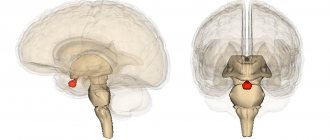
Just above the superior colliculi hangs a small, round structure called the epiphysis. This is a very important endocrine gland, which belongs to the diencephalon (diencephalon). I talked a little about the pineal gland here, but undoubtedly, this gland’s finest hour on my blog is yet to come. In this illustration (unfortunately, I don’t know who the author is - it’s very coolly done) we see the epiphysis (which the arrow points to) and the quadrigeminal.