Structure and functions of a neuron[edit | edit code]
A. Structure and functions of a nerve cell
Excitable cells respond to stimuli by changing the state of their membranes. There are two types of excitable cells: nerve cells, which conduct and transduce impulses in the nervous system, and muscle cells, which contract either in response to nerve impulses or autonomously.
Nervous system
The human body consists of more than 1010 nerve cells, or neurons.
A neuron
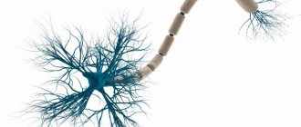
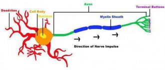
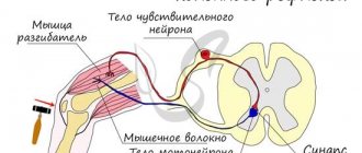
is a structural and functional unit of the nervous system. A typical neuron (motoneuron, A1) consists of a soma, or cell body, and two types of processes - axon and dendrites. In addition to the usual cellular organelles, such as the nucleus and mitochondria (A2), the neuron contains neurofibrils and neurotubules. A neuron receives afferent signals (excitatory and inhibitory) from several and sometimes several thousand neighboring neurons through dendrites (usually tree-like), and the signals are summed along the neuron body on the cell membrane (summation).The axon starts from the axon hillock of the neuron body: it carries out transmission of efferent nerve signals to nearby or distant effectors (muscle and secretory cells) and nearby neurons. Axons often have branches (collaterals), which branch further and end in swellings - synaptic vesicles or synaptic endings. If the total potential at the axon hillock exceeds a certain threshold, an action potential is generated and transmitted down the axon, where it reaches the next synapse through the synaptic terminal (A1, 3), described below.
Vesicles containing various substances (proteins, lipids, sugars and neurotransmitter molecules) are transported from the Golgi complex in the soma to the synaptic terminal and to the tips of dendrites by rapid axonal transport (40 cm/day). This type of anterograde (forward-directed) transport along neurotubules is carried out by kinesin (myosin-like protein), and the energy required for this is supplied by ATP. Endogenous and exogenous substances, such as nerve growth factor (NGF), herpes virus, polio virus, and tetanus toxin, are transported retrogradely from peripheral sites to the soma at a rate of ~25 cm/day. Slow axonal transport (~1 mm/day) plays an important role in the treatment of severe neuritis.
The plasma membrane of the soma continues along the axon and is called the axolemma (A1, 2).
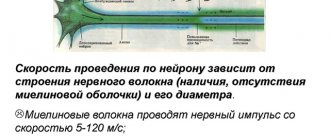
In the central nervous system (CNS), the axolemma is surrounded by oligodendrocytes, and in the peripheral nervous system, by Schwann cells (A1, 2). A nerve fiber consists of an axon and its sheath. In some neurons, Schwann cells form a multilayered myelin sheath of double phospholipid layers (A1, 2) around the axon, which insulates the axon from ionic currents. The myelin sheath is interrupted approximately every 1.5 mm at the nodes of Ranvier (A1). The conductivity of myelinated nerve fibers is much higher than that of unmyelinated nerve fibers and increases with the diameter of the nerve fiber.
A synapse (A3) is the site where a neuron's axon interacts with effectors or other neurons. Synaptic transmission in almost all mammals is carried out by chemical compounds rather than by electrical signals. In response to an electrical signal in the axon, neurotransmitters are released from vesicles on the presynaptic membrane by exocytosis. The transmitter diffuses through the synaptic cleft (10-40 nm) to the postsynaptic membrane, where it connects with receptors that create new electrical signals (AZ). Depending on the type of neurotransmitter and receptor involved in the process, the neurotransmitter has either an excitatory (for example, acetylcholine in skeletal muscle) or an inhibitory effect (for example, glycine in the central nervous system) on the postsynaptic membrane. Because the postsynaptic membrane does not normally release neurotransmitters (there are only a few exceptions), nerve impulses can only pass through the synapse in one direction. Thus, the synapse acts as a valve that ensures orderly signal transmission. Synapses are also sites at which the transmission of a nerve impulse can be converted by other (excitatory or inhibitory) neurons.
Neurokinesiology
Neurons
Admin
09.01.2019
Uncategorized
A neuron (nerve cell) is a structural and functional unit of the nervous system. This cell has a complex structure, is highly specialized and in structure contains a nucleus, a cell body and processes. There are more than one hundred billion neurons in the human body.
Review
The complexity and diversity of the nervous system depends on the interaction between neurons, which, in turn, represent a set of different signals transmitted through the interaction of neurons with other neurons or muscles and glands. Signals are emitted and propagated by ions that generate an electrical charge that travels along the neuron.
Structure
Cell body
A neuron consists of a body with a diameter of 3 to 100 μm, containing a nucleus (with a large number of nuclear pores) and other organelles (including a highly developed rough ER with active ribosomes, the Golgi apparatus), and processes. There are two types of processes: dendrites and axons. The neuron has a developed cytoskeleton that penetrates its processes. The cytoskeleton maintains the shape of the cell; its threads serve as “rails” for the transport of organelles and substances packaged in membrane vesicles (for example, neurotransmitters). A developed synthetic apparatus is revealed in the teleneuron; the granular ER of the neuron is stained basophilically and is known as “tigroid”. The tigroid penetrates the initial sections of the dendrites, but is located at a noticeable distance from the beginning of the axon, which serves as a histological sign of the axon.
There is a distinction between anterograde (away from the body) and retrograde (toward the body) axon transport.
Dendrites and axon
Neuron structure diagram
An axon is usually a long process adapted to conduct excitation from the neuron body. Dendrites are, as a rule, short and highly branched processes that serve as the main site of formation of excitatory and inhibitory synapses influencing the neuron (different neurons have different ratios of axon and dendrites lengths). A neuron may have several dendrites and usually only one axon. One neuron can have connections with many (up to 20 thousand) other neurons.
Dendrites divide dichotomously, while axons give off collaterals. Mitochondria are usually concentrated at branching nodes.
Dendrites do not have a myelin sheath, but axons may have one. The place of generation of excitation of most neurons is the axon hillock - a formation at the point where the axon departs from the body. In all neurons this zone is called the trigger zone.
Synapse
A synapse is a point of contact between two neurons or between a neuron and an effector cell receiving a signal. It serves to transmit a nerve impulse between two cells, and during synaptic transmission the amplitude and frequency of the signal can be adjusted. Some synapses cause depolarization of the neuron, others cause hyperpolarization; the former are excitatory, the latter are inhibitory. Typically, stimulation from several excitatory synapses is necessary to excite a neuron.
Classification
Structural classification
Based on the number and arrangement of dendrites and axons, neurons are divided into axonal neurons, unipolar neurons, pseudounipolar neurons, bipolar neurons, and multipolar (many dendritic arbors, usually efferent) neurons.
Axonless neurons are small cells grouped near the spinal cord in intervertebral ganglia that do not have anatomical signs of division of processes into dendrites and axons. All processes of the uklet are very similar. The functional purpose of axonless neurons is poorly understood.
Unipolar neurons are neurons with one process, present, for example, in the sensory nucleus of the trigeminal nerve in the midbrain.
Bipolar neurons are neurons with one axon and one dendrite, located in specialized sensory organs - the retina, olfactory epithelium and bulb, auditory and vestibular ganglia;
Multipolar neurons - Neurons with one axon and several dendrites. This type of nerve cells predominates in the central nervous system
Pseudounipolar neurons are unique in their kind. One tip extends from the body, which immediately divides in a T-shape. This entire single tract is covered with a myelin sheath and structurally represents an axon, although along one of the branches the excitation goes not from, but to the body of the neuron. Structural dendrites are branches at the end of this (peripheral) process. The trigger zone is the beginning of this branching (i.e., it is located outside the cell body).
Functional classification
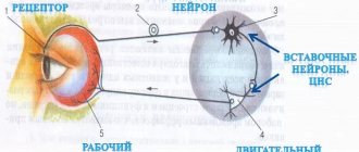
According to their position in the reflex arc, afferent neurons (sensitive neurons), efferent neurons (some of them are called motor neurons, sometimes this name does not apply to the entire group of efferents) and interneurons (interneurons) are distinguished.
Afferent neurons (sensory, sensory or receptor). Neurons of this type include primary cells of the sensory organs and pseudounipolar cells, whose dendrites have free endings.
Efferent neurons (effector, motor or motor). Neurons of this type include the final neurons - ultimatum and penultimate - non-ultimatum.
Associative neurons (interneurons or interneurons) - this group of neurons communicates between efferent and afferent, they are divided into comisural and projection (brain).
Morphological classification
Nerve cells are stellate and spindle-shaped, pyramidal, granular, pear-shaped, etc.
Neuron development and growth
Growth cone
A neuron develops from a small precursor cell that stops dividing even before it produces its processes. (However, the issue of neuronal division currently remains controversial. [1] (Russian)) As a rule, the axon begins to grow first, and dendrites form later. At the end of the developing process of the nerve cell, an irregularly shaped thickening appears, which, apparently, makes its way through the surrounding tissue. This thickening is called the growth cone of the nerve cell. It consists of a flattened part of the nerve cell process with many thin spines. The microspines have a thickness of 0.1 to 0.2 µm and can reach 50 µm in length; the wide and flat region of the growth cone is about 5 µm wide and long, although its shape can vary. The spaces between the microspines of the growth cone are covered with a folded membrane. Microspikes are in constant motion - some are retracted into the growth cone, others elongate, deviate in different directions, touch the substrate and can stick to it.
The growth cone is filled with small, sometimes connected to each other, membrane vesicles of irregular shape. Directly below the folded areas of the membrane and in the spines there is a dense mass of entangled actin filaments. The growth cone also contains mitochondria, microtubules and neurofilaments found in the body of the neuron.
It is likely that microtubules and neurofilaments elongate mainly due to the addition of newly synthesized subunits at the base of the neuron process. They move at a rate of about a millimeter per day, which corresponds to the speed of slow axonal transport in a mature neuron. Since the average speed of advancement of the growth cone is approximately the same, it is possible that during the growth of the neuron process, neither assembly nor destruction of microtubules and neurofilaments occurs at its far end. New membrane material is added, apparently, at the end. The growth cone is an area of rapid exocytosis and endocytosis, as evidenced by the many vesicles present there. Small membrane vesicles are transported along the neuron process from the cell body to the growth cone with a stream of fast axonal transport. The membrane material is apparently synthesized in the body of the neuron, transported to the growth cone in the form of vesicles and incorporated here into the plasma membrane by exocytosis, thus lengthening the process of the nerve cell.
The growth of axons and dendrites is usually preceded by a phase of neuronal migration, when immature neurons disperse and find a permanent home.
Source: https://kineziolog.su/content/kholinergicheskaya-sistema-regulyatsii
You can sign up online or get the schedule of the nearest master classes at the link: https://qviz.matomba.ru/1c06710015fbab389df6
Artificial stimulation of a nerve cell[edit | edit code]
When an electrical impulse from an external source is applied to a nerve cell, current flows from the positively charged electrode (anode) and exits to the negatively charged electrode [cathode]. The nerve fiber below the cathode depolarizes and, provided the threshold potential is reached, an action potential is generated.
The speed of conduction of an impulse along a nerve can be measured by placing two electrodes on the skin along the nerve at a known distance from each other, then stimulating that nerve (containing numerous neurons) and recording the time it takes for the total action potential to travel the distance between the electrodes. The speed of signal transmission in humans is usually from 40 to 70 m/s. Values below 40 m/s are considered pathological.
- Accidental exposure to electricity. High voltage, especially low-frequency alternating current (for example, from an electrical outlet), and also under conditions of reduced resistance (bare feet, accident in the bathroom), mainly affects the conduction of signals in the heart, which can cause ventricular fibrillation.
Direct current usually acts as a stimulus only when switched on and off: high-frequency alternating current (> 15 kHz), in contrast, is not capable of causing depolarization, but damages body tissues. Diathermy is based on this principle.