The human spinal cord is a complex mechanism that consists of many different “parts”. And each of them is important for our full life. It is thanks to the fact that we have a spine that we move. The reflex function of the spinal cord is one of the main ones it performs. In addition to it, there is also a conductor. It is worth considering these functions in more detail and finding out what each of them is responsible for.
The formation of the spinal cord in the fetus occurs in the mother's womb, and at a time when she is not yet aware of her pregnancy. By the end of the first month, the first foundation of the spinal column is laid. However, it will take some more time for its complete formation after the birth of the child. Some parts of the brain are fully formed when the baby is 2 years old.
What does the spinal cord look like?
Not everyone knows what the spinal cord looks like. Moreover, not all people have an idea of what its role is in the life of every person. It is therefore worthwhile to fill this knowledge gap. In addition, many people mistakenly believe that the brain and spinal cord are separate parts.
To find out what the reflex function of the spinal cord is for, let's try to determine what it looks like. It is impossible to clearly understand where the spinal cord begins and ends. It starts from the first vertebra just below the skull, smoothly connecting to the brain in this area. The division into the spinal cord and the brain is formal, but in reality the spinal cord smoothly passes into the brain. Thus, we can conclude that these two parts are a single whole.
Segments
A segment of the spinal cord is a separate part of the organ that is responsible for certain parts of the body, as well as for the functioning of all organs. There are 31 segments in total. To make it easier to understand the functions of each of the segments, which together make up the departments, you need to create a simple table.
Sections of the spinal cord and their functions: table
| Departments | What are they responsible for? |
| Cervical | Movement of the diaphragm and elbow joints |
| Breasts | Skin sensitivity, muscle function, lungs, heart, digestive system |
| Lumbar | Work of the prostate, kidneys, bladder, adrenal glands, uterus, ureters |
| Sacral | Centers for defecation, urination and erection |
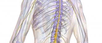
Location of the spinal cord and its membrane
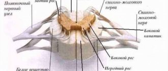
The brain is protected by the cranium, and the spinal cord is hidden in the spine and surrounded by three membranes. The first of them is the most delicate, thin and soft. It contains blood vessels that deliver nutrients to the brain. In other words, the spinal cord is a kind of “courier” for delivering food.
Continuing to talk about how the reflex function of the spinal cord works, we cannot ignore the analysis of the structure of the second arachnoid membrane. There is a special space here called the subarachnoid space. Along the entire length of the spine it is filled with cerebrospinal fluid (CSF). It is this that is taken during puncturing for analysis in order to determine the health status of the spinal cord.
The last shell is located on the outside and has a harder surface, which allows it to provide protective functions against various types of external damage.
Thickenings and grooves
Only two parts of the spinal canal have significant thickening - the cervical and lumbosacral vertebrae.
It is there that the greatest concentration of nerve endings is observed, which are responsible for the proper functioning of the upper and lower extremities. Therefore, a spinal cord contusion can negatively affect a person’s coordination and movement.
Since the spinal canal has symmetrical halves, specific division boundaries run through them - the anterior median fissure and the posterior groove.
The anterior lateral groove runs from the median fissure on both sides. The motor root originates in it.
Thus, the groove serves to separate the lateral and anterior cords of the spinal cord. In addition, there is also a lateral groove at the back, which also serves as a dividing border.
Characteristics of the spinal cord
In adults, the spinal cord reaches 45 cm in length and 1.5 cm in thickness. Its weight, by the most conservative standards, is no more than 35 grams. The entire brain is divided into several sections, from which various roots extend:
- cervical;
- chest;
- lumbar;
- cross;
- coccygeal
Since the reflex function of the spinal cord is carried out, the cervical and lumbosacral regions are the most important parts of the spine. In this regard, they are best protected - nature itself took care of this, making them significantly thicker and denser. It is in these places that important nerve endings are located, the defeat of which can lead to serious consequences. In the cervical region there is a cluster of roots responsible for the movement of the arms. The roots of the lower section are responsible for the movement of the lower extremities.
The human spinal cord controls the activity of all internal organs. Each of them is associated with a specific department. In addition, the entire spinal canal is divided into segments and each of the listed sections has its own number. There are 8 of them in the cervical, 12 in the thoracic, 5 in the lumbar and sacral, and one or two in the coccygeal.
Segmental structure of the spinal cord - what is it?
How is the spinal cord connected to the organs? In the transverse direction, the spinal cord is divided into special sections, or segments. From each segment there are roots, a pair of anterior ones and a pair of posterior ones, which communicate the nervous system with other organs. The roots emerge from the spinal canal and form nerves that are directed to various structures of the body. The anterior roots transmit information primarily about movements (stimulate muscle contraction), therefore they are called motor roots. The dorsal roots carry information from receptors to the spinal cord, that is, they send information about sensations, which is why they are called sensitive.
The number of segments is the same for all people: 8 cervical segments, 12 thoracic, 5 lumbar, 5 sacral and 1-3 coccygeal (usually 1). The roots from each segment rush into the intervertebral foramen. Since the length of the spinal cord is shorter than the length of the spinal canal, the roots change their direction. In the cervical region they are directed horizontally, in the thoracic region - obliquely, in the lumbar and sacral regions - almost vertically downwards. Due to the difference in the length of the spinal cord and spine, the distance from the exit of the roots from the spinal cord to the intervertebral foramen also changes: in the cervical region the roots are the shortest, and in the lumbosacral region they are the longest. The roots of the four lower lumbar, five sacral and coccygeal segments form the so-called cauda equina. It is this that is located in the spinal canal below the second lumbar vertebra, and not the spinal cord itself.
Each segment of the spinal cord is assigned a strictly defined zone of innervation on the periphery. This zone includes an area of skin, certain muscles, bones, and part of the internal organs. These zones are almost the same for all people. This structural feature of the spinal cord allows one to diagnose the location of the pathological process in the disease. For example, knowing that the sensitivity of the skin in the navel area is regulated by the 10th thoracic segment, if the sensation of touching the skin below this area is lost, we can assume that the pathological process in the spinal cord is located below the 10th thoracic segment. This principle works only taking into account the comparison of zones of innervation of all structures (skin, muscles, and internal organs).
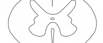
If you cut the spinal cord in a transverse direction, it will not look the same in color. On the cut you can see two colors: gray and white. Gray color is the location of the cell bodies of neurons, and white color is the peripheral and central processes of neurons (nerve fibers). In total, there are more than 13 million nerve cells in the spinal cord.
The bodies of gray neurons are arranged in such a way that they have a bizarre butterfly shape. This butterfly has clearly visible convexities - the front horns (massive, thick) and the rear horns (much thinner and smaller). Some segments also have lateral horns. The area of the anterior horns contains the bodies of neurons responsible for movement, the area of the posterior horns contains neurons that receive sensory impulses, and the lateral horns contain neurons of the autonomic nervous system. In some parts of the spinal cord, the bodies of nerve cells responsible for the functions of individual organs are concentrated. The locations of these neurons have been studied and clearly defined. Thus, in the 8th cervical and 1st thoracic segments there are neurons responsible for the innervation of the pupil of the eye, in the 3rd - 4th cervical segments - for the innervation of the main respiratory muscle (diaphragm), in the 1st - 5th thoracic segments - for regulation of cardiac activity. Why do you need to know this? It is used in clinical diagnosis. For example, it is known that the lateral horns of the 2nd - 5th sacral segments of the spinal cord regulate the activity of the pelvic organs (bladder and rectum). If there is a pathological process in this area (hemorrhage, tumor, destruction due to injury, etc.), a person develops urinary and fecal incontinence.
The processes of neuron bodies form connections with each other, with different parts of the spinal cord and brain, and tend upward and downward, respectively. These nerve fibers, which are white in color, constitute the white matter in cross section. They also form the cords. In the cords, the fibers are distributed in a special pattern. In the posterior cords there are conductors from the receptors of muscles and joints (articular-muscular feeling), from the skin (recognition of an object by touch with closed eyes, sensation of touch), that is, the information goes in an upward direction. In the lateral cords there pass fibers that carry information about touch, pain, temperature sensitivity to the brain, to the cerebellum about the position of the body in space, muscle tone (ascending conductors). In addition, the lateral cords also contain descending fibers that provide body movements programmed in the brain. In the anterior cords there are both descending (motor) and ascending (sensation of pressure on the skin, touch) pathways.
The fibers can be short, in which case they connect the segments of the spinal cord with each other, and long, in which case they communicate with the brain. In some places, the fibers may cross or simply move to the opposite side. The crossing of different conductors occurs at different levels (for example, the fibers responsible for the feeling of pain and temperature sensitivity cross 2-3 segments above the level of entry into the spinal cord, and the fibers of the joint-muscular sense go uncrossed to the very upper parts of the spinal cord). The result of this is the following fact: in the left half of the spinal cord there are conductors from the right parts of the body. This does not apply to all nerve fibers, but is especially true for sensory processes. Studying the course of nerve fibers is also necessary to diagnose the location of the lesion in the disease.
Gray matter
Gray matter or substantia grisea is represented by several columns connected to each other by two plates (anterior and inferior), called commissures. On a cross-section of one of these pillars, you can see that the gray matter in its shape resembles a butterfly with spread wings or the Latin letter H.
In addition, you can also notice that there are projections extending from the substance, which are otherwise called horns. They can be either front, located on the front wall, or rear, running along the back wall. Both the first and second pairs, and have a narrow and wide shape. But in addition to the rear and anterior ones, there are also lateral horns, which contain the centers of the autonomic nervous system.
What is the reflex function of the spinal cord? The fact is that in the anterior horns there is a special type of motor neurons, the processes of which form nerve roots.
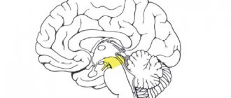
In the middle of the gray matter there is a central canal, which is also filled with cerebrospinal fluid. In the upper part, the canal is connected to the ventricles of the brain. In this case, all sections: the ventricles, the central canal and the subarachnoid space take an active part in the circulation of cerebrospinal fluid.
What happens when tracks become damaged?
To understand the neurophysiology of motor and sensory pathways, one must understand the anatomy of the spine. In its structure, the brain of the back resembles a cylinder surrounded by muscles. Paths pass through its gray matter, thanks to which movements and control of the functioning of organs are carried out. Associative pathways make tactile sensations and pain possible, and movements provide reflex functions of the body.
Due to injury, illness or pathology of brain development, impulse conductivity may decrease or even disappear. This phenomenon occurs due to the death of nerve branches. As a rule, conduction disturbances are accompanied by paralysis and lack of sensation in the limbs. In addition, there are disturbances in the functions of organs that were controlled by damaged nerve fibers. Thus, with damage to the lower back brain, urinary incontinence and spontaneous defecation are possible.
The reflex, conductive function of the brain undergoes changes immediately after the appearance of degenerative changes. There is a death of nerves, which are very difficult to recover in the future. The disease progresses rapidly, causing conduction disturbances to become noticeable. That is why treatment in this case should be started without delay.
White matter
White matter - substantia alba, envelops the gray matter, is formed by a collection of nerve fibers, which also come in three types:
- front;
- rear;
- lateral.
Moreover, all the roots have a different direction, and some of them are connected directly to the brain and central nervous system (hereinafter simply the CNS). And if the reflex function of the spinal cord is to transmit signals from the motor neurons of the gray matter, then the task of the white matter neurons is the prompt delivery of impulses from the muscles and joints to the medulla oblongata. Thus, the transmission of all commands along the entire spinal cord is realized.
Here are the paths through which all information regarding sensitivity and pain is transmitted. Only before entering the cerebral cortex, the information first reaches the diencephalon, and only then rushes further to its destination.
Localization of pathways
The entire set of nerve tissue is located in the white, gray matter, uniting the spinal horns and the cerebral cortex. Pathways are often understood as a set of threads and tissues of nerves located in certain areas of the gray, white matter of the brain. Impulses are transmitted through neural communication.
The morphofunctional characteristics of the descending pathways make it likely that impulses are transmitted in strictly one direction. Irritation of synapses occurs from the presynaptic to the postsynaptic membrane. Such possibilities and the location of ascending and descending pathways can be compared with the conductive function of both brains.
- Associative paths. They are called “bridges”, which unite the zones between the cortex and the nuclei of gray matter. They include long and short fibers. Thus, short fibers are located within one half or lobe of the hemisphere, but long fibers can transmit impulses through 2-3 segments of gray matter. Spinal neurons form intersegmental bundles.
- Commissural fibers. They make up the corpus callosum and unite the newly formed sections of the two brains. As a rule, they diverge in rays and are located in the white matter of the brain tissue.
- Projection fibers. They, located in the spinal cord, allow signals to reach the brain at maximum speed. By their nature and specific functions, fibers are divided into ascending (called afferent pathways) and descending. In turn, they are divided into interoceptive (provide communication with organs), proprioceptive (responsible for movements), exteroceptive (vision, hearing). Such receptors are located between the ridge and the hypothalamus.
The descending pathways of the brain of the back, as a rule, include the pyramidal and central motor neurons, as well as the spino-cerebellar filaments of nerves. The pyramidal neuron originates in the cerebral cortex and descends through the entire trunk. Each of its bundles ends on the horn of the spinal substance. The central motor neuron connects the cerebral cortex with the anterior horns of the brain through axons - nerve roots.
If we talk about the spinocerebellar filaments of nerves, then they cover a thin and wedge-shaped path that connects the legs and the spinal cord of a person. The specifics of such paths are very difficult for a person who does not have a medical education. However, you need to realize: neural signaling is what turns the human body into one whole.
How our brain works
Ascending and descending pathways are responsible for the fast and correct functioning of our body. The latter flows are formed using the red nuclear and lateral pathways. It is thanks to these pathways that the reflex and conduction functions of the spinal cord are carried out. Thanks to the red nuclear spinal tract, involuntary motor impulses are produced. While the lateral corticospinal tracts are responsible for voluntary impulses.
All roots are supplied by personal veins and arteries, which as a result form neurovascular bundles. Each such beam is responsible only for its own segment and works autonomously, analyzing incoming information and transmitting the necessary impulses.
Damage to these bundles leads to serious pathological and sometimes irreversible changes in the human body. And so that specialists can determine which particular bundle is damaged and localize the pain, it is necessary to conduct a whole range of studies.
What are the pathways formed by?
The main pathways of the spinal cord are formed by bundles of cells - neurons. It is this structure that guarantees the required speed of impulse transmission. The division of the functions of the paths is associated with the peculiarities of their purpose. Ascending pathways perceive and transmit signals from the skin, mucous membranes, and human organs. In addition, they are responsible for the functioning of the musculoskeletal system.
The descending pathways of the spinal cord transmit signals to human organs - tissues, glands. They connect to the cortical gray matter. The transmission of signals to organs is carried out due to the spinal neural connection.
The spinal cord is characterized by a double direction of such pathways, due to which rapid impulse transmission of information from systems under control occurs. The conductive spinal function becomes possible only due to the transmission of signals through nerve tissue. The following designations are often used for these pathways in medicine.
- Corticospinal tract. This is a collection of nerve threads that is responsible for movement functions. Based on its purpose and direction, it is divided into several parts: lateral, cortical-nuclear, anterior cortical-cerebral system.
- Tectospinal tract. It is represented by the descending projection NS, which originates in the midbrain cortex, passes through the trunk, cord of the hemispheres, and ends on the anterior horns of the ridge. Otherwise it is called the tectospinal tract.
- vestibulospinal tract. Manages the work of the vestibular apparatus. It begins from the lateral nucleus of the prespinal nerve.
- Reticular-spinal tract. Provides muscle tone, and the task of its fibers can be called the transmission of nerve impulses to the gray matter of the cerebral hemispheres.
- Pyramid path. Its components include the lateral and direct bundle of nerve fibers.
Reflex function
Everything in our body is thought out to the smallest detail, and our body reacts differently to every external stimulus. The defense mechanism is based on reflexes. We sneeze, cough, get burns, flinch from a sharp sound, or react in our own way to gusts of wind. These are all examples of the reflex function of the spinal cord and such actions occur outside of our control.
So that we can respond in a timely manner to any irritant, including critical situations, pain receptors are located throughout the surface of our skin. As a striking example: after touching a hot kettle or any surface, we almost instantly withdraw our hand. The reaction speed is so fast that it is impossible to understand the time frame. In a split second, a reflex ring is formed, which causes the muscles to contract.
Another common case can be cited. If you accidentally swallow a portion of smoke or sniff dust particles with your nose, you will start sneezing or coughing. Thus, it became clear that in such a short time the information was received, processed, and our “defenders” received instructions to free the body from the presence of foreign bodies.
Conductor function
So, it is now clear what the reflex function of the spinal cord is expressed in, we can move on to another, also significant task - conduction. It involves transmitting signals along ascending pathways to the main brain. From it, depending on the situation, the impulse is directed along descending pathways to some organ.
The conductor function allows us to perform meaningful actions:
- take or throw;
- stand up or sit down;
- walk slowly or run;
- draw;
- cut off.
We perform all these actions in everyday life: at home or at work, and usually we simply do not notice.
This whole connection of the brain, spinal cord, the entire central nervous system, internal organs and all limbs makes the human body unique in nature. Even the most modern robot cannot boast of the number of movements that any biological organism can perform.
Introduction
The pathways of the nervous system and the complex reflex arcs consisting of them are the most important and complex section of neurology. It is important because it affirms the cellular nature of the nervous system (neural doctrine) and shows the ordered nature of the arrangement and connections of neurons (in the form of reflex arcs), which underlies its regulatory function.
There is a significant difference from the method of descriptive anatomy. The latter makes it possible to demonstrate the shape, size and localization of a particular formation of the nervous system, as well as its belonging to gray or white matter, but does not reveal at all the structural organization of the nervous system and the mechanisms of its functioning.
This “separation” of structure from function, which is dangerous for the worldview, eliminates the systematic approach to the nervous system in the form of the study of reflex arcs. Here the emphasis is placed precisely on the presence of connections between neurons, on their interaction, leading to the functioning of both the nervous system itself and the entire organism. However, at the same time, the number of mental operations among students increases (synthesis is added to analysis), which increases the complexity of mastering the material and its subjective complexity. Nevertheless, only the study of the nervous system as a set of reflex arcs allows us to understand its organization and functional significance. Finally, only knowledge of neural connections and interactions allows for topical diagnosis of damage to the nervous system, i.e. take a meaningful approach to the diagnosis and treatment of nervous and many other diseases and injuries.
How do the concepts of “conducting path” and “reflex arc” relate to each other? Here it should be clearly understood that any conductive path is part of one or another reflex arc. Since there are two main links in the reflex arc: afferent and efferent, the pathways are classified into afferent and efferent. Taking into account the hierarchical principle of construction of the central nervous system (the presence of higher and lower nerve centers subordinate to them) and the possibility of closing reflex arcs at the level of higher nerve centers, it is clear that both afferent and efferent pathways should be localized in both the peripheral and central parts nervous system. Since the closure of somatic reflex arcs (the connection of afferent and efferent links through interneurons) always occurs in the central nervous system, the latter also contain an associative link and corresponding associative pathways, localized only within the central nervous system.
Afferent nerve pathways conduct impulses from the receptor to the nerve center and are sensitive. Afferent nerve pathways ending in the projection centers of the cerebral cortex are classified as pathways of conscious sensitivity. The same afferent pathways that end in the subcortical sensory nerve centers are classified as pathways of unconscious sensitivity.
Efferent nerve pathways conduct impulses from nerve centers to the working organ. Since we are talking here only about the somatic nervous system, the working organ is the skeletal muscle, therefore the efferent nerve pathways are called motor. Depending on which nerve centers the efferent pathways are connected to, the latter are responsible for performing both conscious and unconscious movements.
Any conducting pathway (afferent, associative or efferent), depending on the level of closure and complexity of the reflex arc, can be single-neuron or multi-neuron (several neurons connected in series in a chain). If we consider a multineuron pathway as a chain, then within its boundaries we can distinguish links represented by the corresponding neurons. Compactly located neuron bodies form nerve centers (nodal, nuclear or screen type), and axons collected in bundles form nerve tracts. Thus, a multineural pathway consists of nerve centers and tracts. In this case, the nerve centers and tracts of the same pathway are localized in certain but different parts of the nervous system. Each tract within the central nervous system conducts nerve impulses usually in one direction and in most cases - of the same functional content. It should be clearly understood how the tracts within the central nervous system differ from the bundles of fibers that form the cranial or spinal nerves. Nerves contain both afferent and efferent fibers, and different afferent fibers can carry different sensory impulses.
In the future, material will be presented that concerns primarily the somatic part of the nervous system.