The cerebral cortex is a multi-level brain structure in humans and many mammals, consisting of gray matter and located in the peripheral space of the hemispheres (the gray matter of the cortex covers them). The structure controls important functions and processes occurring in the brain and other internal organs.
The hemispheres (hemispheres) of the brain in the cranium occupy about 4/5 of the total space. Their component is white matter, which includes the long myelinated axons of nerve cells. On the outer side, the hemisphere is covered with the cerebral cortex, which also consists of neurons, as well as glial cells and unmyelinated fibers.
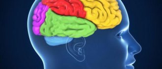
It is customary to divide the surface of the hemispheres into certain zones, each of which is responsible for performing certain functions in the body (for the most part these are reflexive and instinctive activities and reactions).
There is such a thing as “ancient bark”. This is the evolutionarily most ancient structure of the telencephalon of the cerebral cortex in all mammals. They also distinguish the “new cortex,” which in lower mammals is only outlined, but in humans forms the majority of the cerebral cortex (there is also the “old cortex,” which is newer than the “ancient” one, but older than the “new one”).
Functions of the cortex
The human cerebral cortex is responsible for controlling many functions that are used in different aspects of the human body. Its thickness is about 3-4 mm, and its volume is quite impressive due to the presence of channels connecting the central nervous system. How perception, information processing, and decision-making occur through an electrical network using nerve cells with processes.
Various electrical signals are produced within the cerebral cortex (the type of which depends on the current state of the person). The activity of these electrical signals depends on the person’s well-being. Technically, electrical signals of this type are described in terms of frequency and amplitude. A larger number of connections and neurons are localized in places that are responsible for ensuring the most complex processes. At the same time, the cerebral cortex continues to actively develop throughout a person’s life (at least until his intellect develops).
In the process of processing information entering the brain, reactions (mental, behavioral, physiological, etc.) are formed in the cortex.
The most important functions of the cerebral cortex are:
- The interaction of internal organs and systems with the environment, as well as with each other, the correct course of metabolic processes within the body.
- High-quality reception and processing of information received from the outside, awareness of the information received due to the flow of thinking processes. High sensitivity to any information received is achieved due to a large number of nerve cells with processes.
- Supporting a continuous relationship between various organs, tissues, structures and systems of the body.
- Formation and proper functioning of human consciousness, the flow of creative and intellectual thinking.
- Exercising control over the activity of the speech center and processes associated with various mental and emotional situations.
- Interaction with the spinal cord and other systems and organs of the human body.
The cerebral cortex in its structure has the anterior (frontal) sections of the hemispheres, which are currently least studied by modern science. These areas are known to be virtually impervious to external influences. For example, if these sections are influenced by external electrical impulses, they will not give any reaction.
Some scientists are confident that the anterior sections of the cerebral hemispheres are responsible for a person’s self-awareness and his specific character traits. It is a known fact that people whose anterior sections are affected to one degree or another experience certain difficulties with socialization, they pay practically no attention to their appearance, they are not interested in work activity, and are not interested in the opinions of others.
From a physiological point of view, the importance of each section of the cerebral hemispheres is difficult to overestimate. Even those that have not yet been fully studied.
Cytoarchitecture and myeloarchitecture of the cerebral cortex
The cerebral cortex is represented by a layer of gray matter with an average thickness of about 3 mm (1.3-4.5 mm), the grooves and convolutions significantly increase the area of the gray matter of the brain. The cortex contains about 10-14 billion nerve cells. Its various sections, which differ from each other in certain features of the location and structure of cells (cytoarchitectonics), the arrangement of fibers (myeloarchitectonics) and functional significance, are called Brodmann's fields; there are no sharply defined boundaries between them.
1. Histological types of cortex:
- new cortex (lat. neocortex) - 6 layers, most of the cerebral cortex:
1) agranular type of cortex - in the motor centers of the cortex (for example, in the anterior central gyrus), layers III, V and VI are highly developed and layers II and IV are poorly expressed.
2) granular type of cortex - in the sensitive centers of the cortex (for example, visual), layers III, V and VI are poorly developed, granular layers (II and IV) reach their maximum development.
- old cortex (lat. archipallium) - 3 layers, the hippocampus, located in the depths of the hippocampal sulcus, and the dentate gyrus;
- ancient cortex (lat. paleopallium) - 2 layers, lower-inner surface of the temporal lobe (olfactory tubercle and the surrounding cortex, including a section of the anterior perforated substance);
- intermediate cortex (lat. mesocortex) - mixed structure, divided into two zones: one separates the new cortex from the old (peri-archicortical zone), the other from the ancient (peripaleocortical zone). These areas occupy the inferior insula, the parahippocampal gyrus, and the inferior limbic region.
2. The structure of the layers of the new cortex:
- 1st layer - molecular (lat. lamina molecularis) - small associative cells of a spindle shape, axons - parallel to the surface of the brain as part of the tangential plexus of the molecular layer (branching of the dendrites of the neurons of the underlying layers).
- layer 2 - external granular (lat. lamina granularis externa) - small neurons (10 µm), having a round, angular and pyramidal shape, and stellate neurons; dendrites - into the molecular layer; axons - in layers 3, 5 and 6.
- layer 3 - pyramidal neurons (lat. lamina pyramidalis) - the widest layer, pyramidal cells; the main dendrite is in the molecular layer, other dendrites are synapses with the cells of this layer; axon in small cells - within the cortex; large cell axon - forms myelin association or commissural fiber.
- layer 4 - internal granular (lat. lamina granularis interna) - small sensory stellate neurons and tangential plexus of the internal granular layer , developed very strongly in the visual zone of the cortex, practically absent in the precentral gyrus, dendrites - as part of the projection and commissural tracts, axons - in 3, 5 and 6 layers.
- layer 5 - ganglion (layer of Betz cells) (lat. lamina ganglionaris) - large pyramidal cells (in the precentral gyrus - giant pyramids of Betz), the main dendrite is from the molecular layer, the remaining dendrites are within the layer, forming a tangential plexus of the ganglion layer , the axon forms commissural and projection pathways.
- 6th layer - multiform (polymorphic) cells (lat. lamina multiformis) - neurons of various, mainly spindle-shaped, dendrites - from the molecular layer, axons - as part of commissural and projection pathways
3. General principles of the functioning of the cortex:
- afferent information along thalamo-cortical fibers -> cells of layer IV -> pyramidal cells of layers III and V,
— cells of layer III form fibers (associative and commissural) that connect different parts of the cortex.
— cells of layers V and VI form projection fibers to other parts of the central nervous system.
— in all layers of the cortex there are inhibitory neurons that play the role of a filter by blocking pyramidal neurons.
4. General principles of the brain center structure:
— “Nucleus” – a morphologically homogeneous group of cells with an accurate projection of receptor fields;
— “Scattered elements” – cells and groups of cells located outside the “nucleus” and carrying out elementary analysis and synthesis.
5. Areas of the cerebral cortex:
— Primary – projection zones (sensitive and motor), responsible for elementary acts,
— Secondary – projection-associative zones responsible for the operations of gnosis and praxis,
— Tertiary – associative-integrative zones, overlap of cortical representations of various analyzers.
6. Functional blocks of the cortex (according to A.R. Luria):
– energetic – regulation of cortical tone (limbic-reticular complex),
- receiving, processing and storing information (occipital, parietal and temporal cortex),
- programming, regulation and control (frontal lobes).
7. Integrative levels of the nervous system:
— The first signaling system is a system of conditioned reflex connections formed in the cerebral cortex of animals and humans when exposed to specific stimuli (light, sound, pain, etc.), is a form of direct reflection of reality in the form of sensations and perceptions.
— The second signaling system is a system of conditioned reflex connections formed in the cerebral cortex, in which an abstract conventional sign (word) produces a specific effect of the objects or actions it denotes. The key link is speech, which is achieved through the formation of a conditioned reflex connection between the word and the first-signal (specific) reaction.
Layers of the cerebral cortex
The cerebral cortex is formed by several layers, each of which has a unique structure and is responsible for performing specific functions. They all interact with each other, doing a common job. It is customary to distinguish several main layers of the cortex:
- Molecular. In this layer, a huge number of dendritic formations are formed, which are woven together in a chaotic manner. The neurites are parallel oriented and form a layer of fibers. There are relatively few nerve cells here. It is believed that the main function of this layer is associative perception.
- External. Many nerve cells with processes are concentrated here. Neurons vary in shape. Nothing is known yet about the exact functions of this layer.
- The outer one is pyramidal. Contains many nerve cells with processes that vary in size. Neurons are predominantly conical in shape. The dendrite is large.
- Internal grainy. It includes a small number of small neurons that are located at some distance. Between the nerve cells there are fibrous grouped structures.
- Internal pyramidal. Nerve cells with processes that enter into it are large and medium in size. The upper part of the dendrites may be in contact with the molecular layer.
- Cover. Includes spindle-shaped nerve cells. It is characteristic of neurons in this structure that the lower part of the nerve cells with processes reaches all the way to the white matter.
The cerebral cortex includes various layers that differ in shape, location, and functional components of their elements. The layers contain pyramidal, spindle, stellate, and branched neurons. Together they create more than fifty fields. Despite the fact that the fields do not have clearly defined boundaries, their interaction with each other makes it possible to regulate a huge number of processes associated with receiving and processing impulses (that is, incoming information), creating a response to the influence of stimuli.
The structure of the cortex is extremely complex and not fully understood, so scientists cannot say exactly how some elements of the brain work.
The level of a child’s intellectual abilities is related to the size of the brain and the quality of blood circulation in the brain structures. Many children who have had hidden birth injuries in the spinal area have a noticeably smaller cerebral cortex than their healthy peers.
Prefrontal cortex
A large section of the cerebral cortex, which is represented in the form of the anterior sections of the frontal lobes. With its help, control, management, and focusing of any actions that a person performs are carried out. This department allows us to properly distribute our time. The famous psychiatrist T. Galtieri described this area as a tool with the help of which people set goals and develop plans. He was confident that a properly functioning and well-developed prefrontal cortex was the most important factor in a person’s effectiveness.
The main functions of the prefrontal cortex also include:
- Concentration, focusing on obtaining only the information a person needs, ignoring other thoughts and feelings.
- The ability to “reboot” consciousness, directing it in the right thinking direction.
- Perseverance in the process of performing certain tasks, the desire to achieve the intended result, despite the emerging circumstances.
- Analysis of the current situation.
- Critical thinking, which allows you to create a set of actions to search for verified and reliable data (checking the information received before using it).
- Planning, development of certain measures and actions to achieve set goals.
- Forecasting events.
The ability of this department to control human emotions is especially noted. Here, the processes occurring in the limbic system are perceived and translated into specific emotions and feelings (joy, love, desire, grief, hatred, etc.).
Regions
Different functions are attributed to different structures of the cerebral cortex. There is still no consensus on this issue. The international medical community now comes to the conclusion that the cortex can be divided into several large zones, including cortical fields. Therefore, taking into account the functions of these zones, it is customary to distinguish three main sections.
Area responsible for processing pulses
Impulses entering through the receptors of the tactile, olfactory, and visual centers go precisely to this zone. Almost all reflexes associated with motor skills are provided by pyramidal neurons.
This is also where the department is located, which is responsible for receiving impulses and information from the muscular system and actively interacts with different layers of the cortex. It receives and processes all impulses that come from the muscles.
If for some reason the scalp cortex is damaged in this area, then the person will experience problems with the functioning of the sensory system, problems with motor skills and the functioning of other systems that are associated with sensory centers. Externally, such disorders will manifest themselves in the form of constant involuntary movements, convulsions (of varying degrees of severity), partial or complete paralysis (in severe cases).
Sensory zone
This area is responsible for processing electrical signals entering the brain. There are several departments located here that ensure the human brain’s sensitivity to impulses coming from other organs and systems.
- Occipital (processes impulses coming from the visual center).
- Temporal (processes information coming from the speech-hearing center).
- Hippocampus (analyzes impulses coming from the olfactory center).
- Parietal (processes data received from taste buds).
In the sensory perception zone there are departments that also receive and process tactile signals. The more neural connections there are in each department, the higher its sensory ability to receive and process information will be.
The sections noted above occupy about 20-25% of the entire cerebral cortex. If the sensory perception area is damaged in some way, a person may have problems with hearing, vision, smell, and the sensation of touch. The received impulses will either not arrive or will be processed incorrectly.
Not always violations of the sensory zone will lead to the loss of some sense. For example, if the auditory center is damaged, this will not always lead to complete deafness. However, a person will almost certainly have some difficulties with the correct perception of the sound information received.
Association zone
The structure of the cerebral cortex also contains an associative zone, which ensures contact between the signals of neurons in the sensory zone and the motor center, and also provides the necessary feedback signals to these centers. The associative zone forms behavioral reflexes and takes part in the processes of their actual implementation. It occupies a significant (comparatively) part of the cerebral cortex, covering sections included in both the frontal and posterior parts of the cerebral hemispheres (occipital, parietal, temporal).
The human brain is designed in such a way that in terms of associative perception, the posterior parts of the cerebral hemispheres are especially well developed (development occurs throughout life). They control speech (its understanding and reproduction).
If the anterior or posterior parts of the association zone are damaged, this can lead to certain problems. For example, if the departments listed above are damaged, a person will lose the ability to competently analyze the information received, will not be able to make simple forecasts for the future, will not be able to build on facts in the thinking process, or will not be able to use previously acquired experience stored in memory. There may also be problems with spatial orientation and abstract thinking.
The cerebral cortex acts as a higher integrator of impulses, while emotions are concentrated in the subcortical zone (hypothalamus and other departments).
Paul Brodmann
Different areas of the cerebral cortex are responsible for performing specific functions. You can examine and determine the difference using several methods: neuroimaging, comparison of electrical activity patterns, study of cellular structure, etc.
At the beginning of the 20th century, K. Brodmann (a German researcher of human brain anatomy) created a special classification, dividing the cortex into 51 sections, basing his work on the cytoarchitecture of nerve cells. Throughout the 20th century, the fields described by Brodmann were discussed, refined, and renamed, but they are still used to describe the cerebral cortex in humans and large mammals.
Many Brodmann fields were initially defined based on the organization of neurons within them, but later their boundaries were refined in accordance with correlations with various functions of the cerebral cortex. For example, the first, second and third fields are defined as the primary somatosensory cortex, the fourth field is the primary motor cortex, and the seventeenth field is the primary visual cortex.
However, some Brodmann fields (for example, area 25 of the brain, as well as fields 12-16, 26, 27, 29-31 and many others) have not been fully studied.
Speech motor area
A well-studied area of the cerebral cortex, which is also commonly called the speech center. The zone is conventionally divided into three large sections:
- Broca's speech motor center. Forms a person's ability to speak. Located in the posterior gyrus of the anterior part of the cerebral hemispheres. Broca's center and the motor center of the speech motor muscles are different structures. For example, if the motor center is damaged in some way, then a person will not lose the ability to speak, the semantic component of his speech will not suffer, but speech will cease to be clear, and the voice will become poorly modulated (in other words, the quality of pronunciation of sounds will be lost). If Broca's center is damaged, the person will not be able to speak (just like a baby in the first months of life). Such disorders are commonly called motor aphasia.
- Wernicke's sensory center. Located in the temporal region, it is responsible for the functions of receiving and processing oral speech. If Wernicke's center is damaged, sensory aphasia will form - the patient will not be able to understand speech addressed to him (and not only from another person, but also his own). What the patient says will be a collection of incoherent sounds. If simultaneous damage to Wernicke's and Broca's centers occurs (usually this occurs during a stroke), then in these cases the development of motor and sensory aphasia is observed simultaneously.
- Center for Comprehension of Written Speech. Located in the visual part of the cerebral cortex (field No. 18 according to Brodmann). If it turns out to be damaged, then the person experiences agraphia - loss of the ability to write.
8.7.2. Sensory cortical areas
8.7. NEW CORTEX
The new cortex (neocortex) is a layer of gray matter with a total area of 1500-2200 cm2, covering the cerebral hemispheres; it makes up about 72% of the total area of the cortex and about 40% of the mass of the brain. There are about 14 billion neurons in the cortex, the number of glial cells is about 10 times more. The cerebral cortex is phylogenetically the youngest neural structure; in humans, it carries out the highest regulation of body functions and psychophysiological processes that provide various forms of behavior.
8.7.1. STRUCTURAL AND FUNCTIONAL CHARACTERISTICS
A. Neural organization of the neocortex.
In the direction from the surface to the depths of the crust, 6 horizontal layers are distinguished.
I - the molecular layer has very few cells, but a large number of branching dendrites of pyramidal cells, forming a plexus located parallel to the surface. Afferent fibers coming from the associative and nonspecific nuclei of the thalamus form synapses on these dendrites.
II - the outer granular layer is composed mainly of stellate cells and partially small pyramidal cells. The fibers of the cells of the second layer are located mainly along the surface of the cortex, forming cortico-cortical connections.
III - the outer pyramidal layer consists mainly of medium-sized pyramidal cells. The axons of these cells, like granule cells of layer II, form cortico-cortical associative connections.
IV - the inner granular layer is similar in the nature of the cells (stellate cells) and the arrangement of their fibers to the outer granular layer. In this layer, afferent fibers have synaptic endings coming from neurons of specific nuclei of the thalamus; The highest capillarization density is noted here.
V - the inner pyramidal layer is formed by medium and large pyramidal neurons, and Betz's giant pyramidal cells are located in the motor cortex. The axons of these cells form the efferent corticospinal and corticobulbar motor tracts.
VI - layer of polymorphic cells is formed mainly by spindle-shaped cells, the axons of which form corticothalamic tracts.
Assessing the afferent and efferent connections of the cerebral cortex in general, it can be noted that in layers I-IV the perception and processing of signals entering the cortex occurs. On the contrary, efferent pathways leaving the cortex are formed predominantly in layers V-VI. A more detailed division of the cortex into various fields was carried out on the basis of cytoarchitectonic features (shape and arrangement of neurons) by K. Brodmann (1909), who identified 52 fields; many of them are characterized by functional and neurochemical features.
Histological evidence shows that the elementary neural circuits involved in information processing are located perpendicular to the surface of the cortex. Electrophysiological studies
[Mountcastle V., 1957] with immersion of microelectrodes perpendicular to the surface of the somatosensory cortex showed that all neurons encountered along the way respond to a stimulus of only one quality (type) (for example, tactile). On the contrary, when the electrode was immersed at an angle, neurons of different modalities came across its path. It was concluded that in the cerebral cortex there are functional associations of neurons located in a cylinder with a diameter of 0.5-1.0 mm. These associations were called neural columns.
They are found in the motor cortex and in various areas of the sensory cortex. Adjacent neural columns can interact with each other.
B. Corticolization of functions -
an increase in phylogenesis of the role of the cerebral cortex in the analysis and regulation of body functions and subordination to the cortex of the underlying parts of the central nervous system. For example, the regulation of motor functions such as locomotion (jumping, walking, running) and righting reflexes in lower vertebrates (amphibians) is completely provided by the brain stem; removal of the cerebral hemispheres practically does not change them. In cats, transection of the trunk between the midbrain and diencephalon does not significantly affect the righting reflexes, but disrupts locomotion in the acute period, which is subsequently partially restored. Switching off the cerebral cortex in monkeys and humans leads to the loss of not only locomotion, but also righting reflexes.
B. Localization of functions in the cerebral cortex
intensively studied in clinical and experimental medicine since the mid-19th century. When developing this problem, two opposite concepts were formulated: narrow localizationism and functional equivalence (equipotentiality) of various cortical structures. The modern concept of localization of functions is based on the principle of multifunctionality (but not equivalence) of cortical fields. The property of multi-functionality allows one or another cortical structure to be involved in supporting various forms of activity, while realizing the main, genetically inherent function of it [Adrianov O.S., 1976]. The degree of multifunctionality of different cortical structures is not the same. In the fields of the associative cortex it is higher than in the primary sensory fields. Multifunctionality is based on the multichannel entry of afferent excitation into the cerebral cortex, the overlap of afferent excitations, especially at the thalamic and cortical levels, the modulating influence of various structures (for example, nonspecific thalamus, basal ganglia) on cortical functions, the interaction of cortical-subcortical and intercortical paths of excitation. The results of studying the localization of functions allowed scientists in the middle of the 20th century. divide the cerebral cortex into different functional areas (cortical mapping). Detailed functional maps (for example, K. Kleist, 1959) are used in neurological practice, but one must keep in mind the simplified nature of these diagrams. One of the most well-known options for the functional division of the cerebral cortex is the separation of sensory, associative and motor areas in it.
These are the zones into which sensory stimuli are projected (syn.: projection cortex, cortical sections of the analyzers). They are located predominantly in the parietal (fields 1-3), temporal (fields 21, 22, 41, 42) and occipital (fields 17-19) lobes. Afferent pathways to the sensory cortex come predominantly from the relay sensory nuclei of the thalamus - the ventral posterior lateral (VPL) and medial (VPM).
Areas of the sensory cortex, irritation or destruction of which causes clear and
permanent changes in the body's sensitivity are called primary sensory areas
(nuclear parts of analyzers, according to I.P. Pavlov).
They consist predominantly of unimodal neurons and form sensations of the same quality. In the primary sensory zones there is usually a clear spatial (topographic) representation of body parts and their receptor fields. Surrounding the primary sensory areas are less localized secondary sensory areas, whose multimodal
The most important sensory areas are the parietal cortex of the postcentral gyrus and the corresponding part of the paracentral lobule on the medial surface of the hemispheres (fields 1-3). This
area is designated somatosensory area
I. Here there is a projection of skin sensitivity on the opposite side of the body from tactile, pain, temperature receptors, interoceptive sensitivity and sensitivity of the musculoskeletal system from muscle, joint, and tendon receptors. The projection of the head and upper parts of the torso is located in the inferolateral areas of the postcentral gyrus, the projection of the lower half of the torso and legs is in the superomedial zones of the gyrus, the projection of the lower part of the lower leg and feet is located in the cortex of the paracentral lobule on the medial surface of the hemispheres (Fig. 8.7), with the projections of the most sensitive areas (tongue, lips, larynx, fingers) have relatively large areas compared to other parts of the body. It is assumed that in somatosensory area I, in the zone of tactile sensitivity of the tongue, there is a projection of taste sensitivity.
In addition to somatosensory area I, a smaller somatosensory area II
,
located The degree of localization of body parts is less pronounced here; the projection of the face is in front and below, the arms are central, the legs are behind and above. The functions of area II are poorly understood. It is known that signals to this area come from both sides of the body and from other sensory areas of the brain, such as visual and auditory. Stimulation of area II leads to complex body movements; suggest its role in sensory control of movement.
A well-studied primary projection zone is the auditory cortex (fields 41, 42), which is located deep in the lateral sulcus (cortex of Heschl's transverse temporal gyri). In this zone, in response to irritation of the auditory receptors of the organ of Corti, sound sensations are formed that differ in volume, tone and other characteristics. There is a clear topical projection here: different parts of the organ of Corti are represented in different parts of the cortex. The projection cortex of the temporal lobe also includes the center of the vestibular analyzer in the superior and middle temporal gyri (fields 20 and 21). The processed sensory information is used to form a “body schema” and regulate the functions of the cerebellum (temporo-pontine tract).
The most important primary projection area of the neocortex is located in the occipital
The cortex is the primary visual area (cortex of part of the sphenoid gyrus and lingual lobule, area 17). Here there is a topical representation of retinal receptors. Each point of the retina corresponds to its own section of the visual cortex, while the macula zone has a relatively large area of representation. Due to the incomplete decussation of the visual pathways, the same halves of the retina are projected into the visual area of each hemisphere. The presence of a retinal projection in both eyes in each hemisphere is the basis of binocular vision. Irritation of the cortex of area 17 leads to the appearance of light sensations. Near field 17 is the cortex of the secondary visual area (fields 18 and 19). The neurons of these zones are multimodal and respond not only to light, but also to tactile and auditory stimuli.
In this visual area, a synthesis of different types of sensitivity occurs, more complex visual images and their recognition arise. Irritation of these fields causes visual hallucinations, aura (obsessive sensations), and eye movements.
The main part of the information about the environment and internal environment of the body, received in the sensory cortex, is transferred for further processing to the associative cortex.
Thickness
All mammals that have relatively large brains (in a general sense, not in comparison with body size) have a fairly thick cerebral cortex. For example, in field mice its thickness is about 0.5 mm, and in humans it is about 2.5 mm. Scientists also highlight a certain dependence of the thickness of the bark on the weight of the animal.
With modern examinations (especially MRI), it is possible to accurately measure the thickness of the cerebral cortex in any mammal. However, it will vary significantly in different areas of the head. It is noted that in the sensory areas the cortex is much thinner than in the motor (motor) areas.
Research shows that the thickness of the cerebral cortex largely depends on the level of human intelligence. The smarter the individual, the thicker the cortex. Also, a thick cortex is recorded in people who constantly and for a long time suffer from migraine pain.
Sensory areas of the cerebral cortex
Afferent impulses sent to the cerebral cortex are switched on the cells of the thalamic nuclei and from there are projected into certain cortical fields. In each hemisphere, primary zones of representation of somatic (skin and muscle-articular) and visceral sensitivity are distinguished. These zones are designated as somatosensory zones I and II of the cortex. The first somatosensory zone is located in the posterior central gyrus and has a significantly larger area than the second. It receives fibers from the posterior ventral nucleus of the thalamus. A particularly large surface area is occupied by the representation of receptors in the hands, vocal apparatus and face, and a much smaller surface area is occupied by the torso, thighs and lower legs. These differences reflect differences in the number of receptor formations present in the skin of the torso and the most sensitive areas of the body - lips, tongue, fingertips.
Ventral to the first somatosensory zone, in the lateral (Sylvian) fissure, a somatosensory zone was found, which also receives fibers from the cells of the posterior ventral nucleus of the thalamus. The postcentral cortex has often been subjected to direct electrical stimulation in patients during surgical procedures. The result of this stimulation was sensations of pressure, touch or heat felt on the opposite side of the body.
Removal of areas of the somatosensory area leads to the loss of a fine gradation of sensitivity of that part of the body that is represented in the removed area of the cortex. In addition, there is a certain awkwardness and sloppiness when moving this part of the body. Thus, the main function of the somatosensory area is to integrate and critically evaluate the information that comes from specific nuclei of the thalamus. Here, the relative intensity of sensations is assessed, the spatial relationships of the irritated areas of the body are determined, and the similarities and differences of felt irritations are identified.
Another clear primary projection area of the cortex is the inner surface of the occipital cortex in the region of the calcarine sulcus. The axons of the cells of the lateral geniculate body enter this area, delivering visual information to the cortex. In visual area I (field 17), a topographically organized representation of the retina is found. In mammals, in connection with binocular vision, the primary visual area of each hemisphere receives projections from the retinas of both eyes.
In this case, the same halves of the retinas are projected into each hemisphere (both right halves are projected to the left, and both left halves are projected to the right).
With electrical stimulation of the 17th field, a person experiences light sensations. Fields 18 and 19 are associated with the association of visual and other types of sensitivity. Here visual, tactile and partly auditory influences undergo synthesis, providing a more complete visual sensation. Damage to fields 18 and 19 leads to disruption of visual evaluation, so that, for example, written or printed words are not perceived. Irritation of the 19th field causes visual hallucinations and eye movements.
The auditory cortex (fields 41 and 42) is mainly located in the lateral sulcus. Only a small part of this zone is visible on the superior edge of the temporal lobe. In this area of the cortex, sound signals entering the cochlea are perceived as sounds varying in tone, volume and quality.
The sensory auditory area is distinguished by a rich cellular and fiber composition and has a well-defined layer of large pyramidal neurons. A well-defined layer of stellate cells is also typical of the auditory cortex. In addition to the auditory pathways, vestibular afferents are also projected into the auditory area.
Electrical stimulation of the auditory cortex in people with preserved consciousness causes subjective sensations of noise in both ears. Due to bilateral representation, damage to the temporal lobe on one side, although it causes hearing impairment, does not lead to deafness.
In the auditory area of the cortex there is a topical representation of various parts of the cochlea. The zone lying on the periphery of the primary auditory area forms an associative center associated primarily with assessing the significance of sounds. Irritation of this area causes auditory hallucinations and head movements in the opposite direction. Special zones of the superior and middle temporal protrusions (convolutions) in the left hemisphere are associated with speech understanding. When they are damaged, the ability to pronounce or understand spoken words is lost.
Furrows, convolutions, fissures
Among the structural features and functions of the cerebral cortex, it is customary to distinguish also fissures, grooves and convolutions. These elements form a large surface area of the brain in mammals and humans. If you look at the human brain in section, you can see that more than 2/3 of the surface is hidden in the grooves. Fissures and grooves are depressions in the bark that differ only in size:
- The fissure is a large groove that divides the mammalian brain into parts, into two hemispheres (longitudinal medial fissure).
- A sulcus is a shallow depression surrounding the gyri.
However, many scientists consider this division into grooves and fissures to be very arbitrary. This is largely due to the fact that, for example, the lateral sulcus is often called the “lateral fissure” and the central sulcus the “central fissure.”
The blood supply to the parts of the cerebral cortex is carried out using two arterial basins at once, which form the vertebral and internal carotid arteries.
The most sensitive area of the cerebral hemispheres is considered to be the central posterior gyrus, which is associated with the innervation of different parts of the body.