Publisher:
Bustard
Authors:
Sapin M.R., Sonin N.I.
At first glance, biology is a fairly simple subject. But many choose it to take the first final tests - the OGE and the Unified State Exam. To prepare for this important event, you need a lot of time and effort. And the student may have difficulties because of this. To prevent problems, it is worth turning to methodological literature. One of the best of its kind is rightfully considered the “GDZ in biology for grade 9 Textbook Sapin, Sonin, Bustard.”
Textbook structure
One of the main criteria by which reference books with ready-made homework assignments are selected is the most complete coverage of the school course. The workbook published by the Drofa publishing house includes the following main topics:
- molecular level (proteins, fats and carbohydrates);
- cell theory, its composition;
- photosynthesis and chemosytnesis;
- sexual and asexual reproduction of organisms;
- Morgan's law, Vavilov's work;
- development of the evolutionary theory of Charles Darwin.
Thus, “GDZ in biology for grade 9 Textbook M.R. Sapin, N.I. Sonin, Bustard” is compiled in accordance with all the norms and requirements of the Federal State Educational Standard.
How to use the GDZ collection
Anyone who believes that GDZ can only be considered as cheat sheets for cheating, and they are good for nothing else, is mistaken. This is a wrong point of view. You just have to find the right approach to working with them, and the result will not take long to arrive. We offer the following option:
- First of all, you should solve everything assigned to the house yourself.
- Only after this is it possible to check your answers with the correct keys given at the end of the paragraph.
- It is recommended to correct existing errors and try not to make them again.
It is better that the solver lessons are regular and systematic.
Transmission and brakes
A transmission is a system of elements that collectively transmit force from a person’s legs to the wheels. It includes:
- Carriage.
Bearing unit that provides torque when pedaling. - Connecting rods.
The pedals are connected to the carriage and frame. - Pedals.
There’s probably no point in talking about this detail in the design of children’s and adult bicycles. Despite the apparent simplicity of the pedals, there are 5 types, and the stability and confidence of the cyclist when pedaling greatly depends on them. - Front and rear sprockets.
Provides switching of bicycle speeds and chain mobility. - Chain.
The connecting link between the front and rear wheels. Transmits torque to the front and is mounted on the sprockets. - Brakes.
There are rim brakes (clamp and V-brake), disc and drum-bushing.
Who will benefit from the GDZ collection in biology for grade 9 from Sapin
The manual can be used not only by students with problems in mastering the material, but also by many others:
- excellent students, for example, will use it to prepare for any test or test;
- Children who missed a lesson due to illness will catch up with their peers and go through this or that topic on their own;
- teachers will learn many new and interesting tasks, thereby diversifying the curriculum.
In conclusion, we can say that the problem book edited by Sapin will become a real assistant for a ninth-grader; it will come to the rescue in any difficult situation.
It is known that age-related changes manifest themselves in all organs and systems of the human body. In this case, the processes occurring during the aging period of the nervous system are of particular importance, which is the subject of this review, which summarizes the main facts in this area.
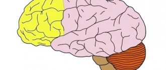
Aging of the central nervous system (CNS) is accompanied by atrophic processes expressed to varying degrees. The mass of the brain slowly but steadily decreases. The cerebral cortex, and subsequently the cerebellum, becomes thinner. According to V.V. Frolkis, the weight of the brain of a person aged 60 to 75 years decreases by 6%, and unevenly in different parts. The cerebral cortex decreases by 4%, the greatest changes (12-15%) occur in the frontal lobe. Gender differences in the degree of brain atrophy with aging have been noted. The weight of the brain of women is approximately 110-115 g less than that of men. Between 40 and 90 years, brain weight decreases in men by 2.85 g per year, and in women by 2.92 g [1]. The dura mater becomes thicker, becomes sclerotic, and fuses with the bones of the skull. The pia mater also noticeably thickens, the convolutions become thinner, the furrows widen and deepen. The arachnoid membrane progressively hyperplasias and sclerosis with age. According to H. Brody [2] and H. Chugani et al. [3], and brain weight decreases by 6-7% by the age of 80, the cerebellum loses up to 25% of Purkinje cells with age, the thalamic nuclei - up to 18%; changes most often occur in the prefrontal and medial temporal regions. According to E. Kensinger et al. [4], atrophy of both white and gray matter is observed in the prefrontal region. The decrease in gray matter volume is caused by a decrease in the number of neurons due to their degeneration. Axonal pathological changes and delayed neurotransmission are noted in the white matter. At the same time, R. Cabeza et al. [5] found that the decrease in interhemispheric asymmetry observed in older adults is most pronounced in the prefrontal cortex. It is still unclear whether bilateral alignment is a reflection of compensatory activation of one of the hemispheres or whether it is the result of pathological changes.
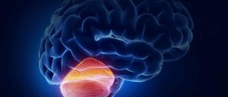
Another area of the brain where age-related changes are most pronounced is the hippocampus [6]. Considering the role of the hippocampus in memory formation, it becomes clear that functional and structural changes in this area during aging cause difficulties in remembering the context in which information was received [7, 8].
Thus, most human brain researchers point to predominant neuronal loss in the cortex, hippocampus, and cerebellum. In most subcortical formations, the cellular composition remains unchanged until old age [1]. At the same time, phylogenetically “newer” brain structures associated with cognitive function are more susceptible to age-related neuronal loss than phylogenetically “older” ones (brain stem).
During the aging process, the neurons themselves and their processes decrease in size, and lipofuscin and fat vacuoles accumulate in them. Myelin fibers become thinner. Electron microscopic examination reveals senile chromatolysis of neurons, their sclerotic changes and transformation into “shadow cells”. Not only signs of damage and degeneration of neurons (homogenization of the cytoplasm, displacement and pyknosis of nuclei, cytolysis, tigrolysis) of varying severity are revealed, but also signs of hypertrophy of intracellular structures, which indicates adaptive processes in conditions of age-related neuronal degeneration [9, 10]. Due to the death of neurons, one of the typical morphological signs of the aging brain occurs - cell rarefaction. The voids in areas of complete disappearance of neurons contain granular basophilic material and vacuoles, and they are also replaced by glial elements [11, 12].
With aging of the brain in the cerebral cortex, mainly in the frontal lobes, as well as in the hippocampus and subcortical ganglia, the number of glial elements increases and senile (senile) plaques are detected. They are located next to the vessels of the microvasculature of the cortex and represent accumulations of argyrophilic structureless material containing amyloid and surrounded by interweavings of thickened axons and macro- and microglial cells with little cytoplasm [13, 14].
With aging, synaptic density decreases. However, synapse loss does not occur equally in all parts of the central nervous system. Thus, in the frontal lobe, a decrease in the number of synapses with age has been reliably proven, while in the temporal lobe, age-related changes are not observed. Changes in the state of synapses are observed not only in the cortex, but also in subcortical structures. For example, age-related impairments in spatial memory have been attributed to decreased specificity, efficiency, and plasticity of synaptic transmission in the hippocampus. With aging, the ability to form new synapses decreases. Reduction of synaptic plasticity in old age can contribute to memory loss, deterioration of motor activity and the development of other disorders. At the same time, interneuron contacts in various areas of the central nervous system deteriorate, neurons seem to undergo deafferentation, and therefore their response to environmental signals, nervous and hormonal stimuli is disrupted, i.e., the synaptic mechanisms of brain activity are damaged [1].
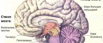
As age increases, the state of the brain's neurotransmitter systems changes significantly. One of the most characteristic phenomena of aging is the degeneration of the dopaminergic system of the brain, which is directly related to the development of diseases such as parkinsonism in old age. Disturbances in the activity of the cholinergic neurotransmitter system of the brain play a major role in disorders of memory, perception and other cognitive processes that occur in Alzheimer's disease [15].
Aging is also accompanied by changes in the activity and content in human brain tissue of enzymes related to the synthesis and destruction of tyrosine hydroxylase, DOPA decarboxylase in the substantia nigra, caudate nucleus and putamen; choline acetylase and acetylcholinesterase - in the cortex, striatum, hippocampus and cerebellum, and therefore, the synthesis of acetylcholine in these structures is reduced. On the contrary, the content of monoamine oxidase increases in the midbrain. Disturbances in the exchange of neurotransmitters in dopaminergic neurons of the brain entail a decrease in it in the basal ganglia, caudate nucleus and putamen, which causes a disturbance in motor activity. A decrease in the content of serotonin and norepinephrine, a decrease in the content and rate of dopamine metabolism in the hypothalamus is associated with the development of depression in the elderly [16].
Age-related deterioration of blood supply to the brain through extra- and intracranial vessels is accompanied by changes in small vessels: sclerosis and hyalinosis of the walls, narrowing of the lumen. With aging, cerebral blood flow decreases, the functions of the blood-brain barrier are disrupted, the coupling between cerebral blood flow and glucose metabolism decreases due to the use of ketone bodies as an energy substrate, the levels of tissue respiration and oxidative phosphorylation decrease, as well as intracellular pH in the brain, which characterizes changes in cerebral energy exchange at all levels. Such changes are relatively weakly expressed during normal aging; nevertheless, they increase the sensitivity of the brain to oxidative stress and other damaging factors [17–19].
Age-related changes in the choroid plexus are manifested by sclerosis, the formation of cysts, calcification, and the appearance of psammotic bodies. Calcification processes in them progress with age: computed tomography reveals them in 1 out of 3 50-year-old patients, 3 out of 4 60-year-old patients, and 5 out of 6 80-year-old patients [11].
In the process of aging, there is a gradual decline in higher mental functions - perception, attention, memory, thinking. The speed of information processing, the amount of RAM, the ability to learn and remember new information decrease [20, 21].
Elderly people are characterized by emotional instability, decreased mental performance, increased threshold of unconditioned reflex reactions, difficulties in developing conditioned reflexes, as well as their slower decline compared to young people [22, 23].
As a person gets older, the process of perceiving new information and encoding it for subsequent storage requires more and more time, which is associated with reduced efficiency of neural transmission and sensory deficits that limit a person's ability to quickly and accurately perceive information presented for memorization [24]. , 25]. Older people also have a reduced ability to retrieve information stored in memory. This is partly due to the fact that it is more difficult for them to differentiate the necessary piece of information from the abundant stores of information and knowledge accumulated over many years of life. This process of delimitation (differentiation) can be especially difficult in the case when new information is similar in content to long-learned information. As a result, older adults perform much worse than younger adults on tests of free recall in which they are asked to recall learned information with minimal prompting. However, this difference is minimized when older subjects are given enough cues and cues to narrow the focus of their memory search for the desired information, or when they are asked to choose the correct answer from a small number of options [26–28].
It is believed that older people have better memory for events that happened in the distant past than for recent events. This is mainly due to the fact that long-standing events either have a special personal meaning for a person, or are so special in content that they could not be “erased” from memory by later events during life [29, 30]. Older people access these memories many times throughout their lives, which makes them more accessible for retrieval than everyday events, much of which are forgotten over time. It should be borne in mind that it is much more difficult to identify the errors of subjects in the reproduction of long-standing events compared to current events, when recall errors are obvious. Thus, short-term memory weakens significantly with age and is often impaired in older people. Age-related differences in long-term memory are much less pronounced and are thought to be a consequence of the use of ineffective encoding strategies or deficits in retrieval function [31, 32]. Semantic memory is not impaired at a later age. Progressive memory loss in some people is observed between 50 and 60 years of age, which is likely the result of degenerative changes in neurons, lipofuscin deposition, and the formation of senile plaques in brain tissue [27].
Speech is preserved relatively well during aging [3]. Older people aged 60–70 years use a more diverse range of grammatical forms in their speech compared to older age groups. Speech fluency in older people is no different from that of younger people. However, there are changes in the processes of understanding someone else's speech due to sensory deficits and a slowdown in the speed of information processing. In terms of written language, certain changes are also observed with age. Comprehension and perception slow down, making it more difficult for older people to grasp the meaning of what they read.
Another feature of neurodynamic disorders is a decrease in the ability to concentrate attention for a long time, so older people are often distracted by extraneous stimuli when performing certain tasks, this is especially pronounced when it is necessary to remember information against a background of “noise” [33]. They also have difficulty working with multiple sources of information. The latter may be associated with a decrease in the ability to switch attention, that is, with a certain intellectual rigidity.
Due to morphological changes in the brain, its bioelectrical activity also changes slowly and progressively. Starting from the age of 50 years, a restructuring of the spectrum of EEG rhythms is observed, expressed in a decrease in the amplitude and relative amount of the alpha rhythm and theta waves and an increase in the power of the beta rhythm [34, 35]. As for slow wave activity, the results obtained here are contradictory. According to some studies [36, 37], there is an increase in the power of slow rhythms and less often [38-40] no changes and a decrease in the power of slow rhythms were detected. A number of authors note that the dominant frequency after 60-70 years tends to decrease, and according to visual analysis of the EEG, theta and delta waves predominate. It is believed that the EEG slowdown is associated with ischemia, which leads to a progressive increase in the number of borderline normal and pathologically altered EEGs [41]. There is evidence that significant deviations in the background EEG in people over 70 years of age may be caused by dysfunctions in the normal regulation of sleep and wakefulness. By 90-100 years, the frequency of the dominant rhythm continues to decrease, the representation of slow activity increases, and its asymmetry appears in the temporal leads. Quantitative analysis shows a decrease in the power of the dominant rhythm and a decrease in its differences in different zones compared to 60-year-old healthy people. Theta rhythm is associated with memory and emotional regulation [42, 43]. Since memory impairment is one of the most significant manifestations of brain aging, this largely explains the decrease in the power of the theta rhythm [44, 45]. A decrease in alpha rhythm power was also noted. Alpha 1 rhythm is associated with attention and task difficulty, while alpha 2 is a neurophysiological correlate of complex semantic memory [46]. At the same time, the reactivity of the alpha rhythm to activating loads decreases, and the power of beta activity during functional loads increases [47]. N.V. Wolf and A.A. Glukhikh [34] found in their studies an increase in the power of high-frequency beta-2 and gamma rhythms in all leads compared to a young group, and these differences were most pronounced in the frontal parts of the hemispheres. In older people, there is also a decrease in the ability to assimilate imposed rhythms; the range of rhythm assimilation is narrowed and shifts towards low frequencies.
There is also a steady increase in the latency of the P300 wave of evoked potentials with age (P300 latency lengthens by 1.25 ms per year, and the amplitude decreases at a rate of 0.09 μV per year) and a decrease in the amplitude of visual evoked potentials. When studying auditory evoked potentials, an increase in the latency of N1 and P2 was noted, and their amplitudes change depending on the type of stimulus: the amplitude of N1 increases in response to a speech stimulus compared to a non-speech stimulus [48]. P2 amplitude, according to J. Lister et al. [49], less in older people compared to younger people due to a decrease in the activity of inhibition processes. However, K. Rufener et al. [48] in their study did not find any significant modulation of P2 in the elderly compared to the young. With age, the speed of propagation of excitation along the nerves decreases, and synaptic conduction slows down [50, 51].
According to V.F. Fokina et al. [52, 53], a qualitative analysis of the nature of changes during aging can be presented in relation to two parameters: the average level of constant potentials (ALP) and the interhemispheric difference in the temporal leads. At the same time, it is possible that the picture of cerebral energy exchange will change depending on biological age, social and psychological status, region of residence and other factors.
There is evidence that in the frontal areas, where age-related changes predominate - a decrease in blood flow and glucose hypometabolism, a secondary slight increase in SMR is recorded, reflecting a decrease in cerebral pH. In old age, there is a certain discrepancy between the dynamics of glucose metabolism and changes in ACR: glucose consumption decreases with aging, but the pH in the brain tissue increases, which may be due to a complex of reasons: a decrease in blood flow and energy metabolism, destructive processes [18].
Studies have been conducted on the distribution of SPP in elderly northerners, which have shown that characteristic changes during aging in them include: low values of SPP in the frontal leads, increased values in the central and parietal leads, as well as an increase in individual variability in indicators of interhemispheric differences. There was a smoothing of interhemispheric asymmetry in northern men in the frontal leads, and in northern women in the central leads, and right hemispheric dominance in the central leads in men [54].
Thus, during the aging process, the most significant changes are observed in the medial temporal and prefrontal areas of the brain, which in turn leads to a decrease in cognitive functions: a decrease in the speed of information processing, the amount of working memory, the ability to learn and remember new information. Morphological changes in the brain cause changes in its functional activity, which is reflected in the EEG, in the analysis of evoked potentials, as well as SCP.
The work was carried out within the framework of the project part of the state assignment in the field of scientific activity
of the Ministry of Education and Science of the Russian Federation for 2014-2016,
No. 2025 to the Northern (Arctic) Federal University named after. M.V. Lomonosov.
There is no conflict of interest.