Basal ganglia and their functions
basal ganglia,
called ganglia by histologists of the last century, they are nuclear-type structures that are located in the thickness of the white matter of the forebrain closer to its base.
In mammals, the basal nuclei include the strongly elongated and curved caudate nucleus
and
the lentiform nucleus embedded in the thickness of the white matter.
It is divided into three parts by two white plates: the largest, laterally lying
shell,
and
the globus pallidus,
consisting of internal and external sections (Fig. 3.29).
| Rice. 3.29 Afferent and efferent connections of the basal nuclei 1 - caudate nucleus, 2 - putamen, 3 - substantia nigra, 4 - thalamus, 5, 5* - internal and external parts of the globus pallidus, 6 - subthalamic nucleus, 7 - sensorimotor cortex, 8 - motor cortex, 9 - paths to the brainstem; arrows indicate afferent pathways, efferent pathways, internuclear interactions. |
These anatomical formations form the so-called striopallidal system
(From Latin striatus - striped and pallidus - pale.)
,
which, according to phylogenetic and functional criteria, is divided into the ancient part paleostriatum and the new part neostriatum.
The paleostriatum
is represented by the globus pallidus, and
the neostriatum,
which appears for the first time in reptiles, consists of the caudate nucleus and putamen, which are collectively called
the striatum,
or striatum.
The caudate nucleus and putamen are anatomically connected and are characterized by alternating white and gray matter, which justifies the term striatum.
The subthalamic nucleus is also often included in the striopallidal system.
(Lewis body) and
the substantia nigra
of the midbrain, which form a functional unity with the basal ganglia. The striatum consists mainly of small cells, the axons of which are directed to the globus pallidus and the substantia nigra of the midbrain.
The striatum is a kind of collector of afferent inputs going to the basal ganglia. The main sources of these inputs are the neocortex (mainly sensorimotor), nonspecific nuclei of the thalamus, and dopaminergic pathways from the substantia nigra.
In contrast to the striatum, the globus pallidus
consists of large neurons and is the concentration of the output, efferent pathways of the striopallidal system. The axons of neurons localized in the globus pallidus approach various nuclei of the diencephalon and mesencephalon, including the red nucleus, where the red nucleus-spinal tract of the extrapyramidal motor regulation system begins.
Another important efferent pathway runs from the internal part of the globus pallidus to the anteroventral and ventrolateral nuclei of the thalamus, and from there continues to the motor areas of the cerebral cortex. The presence of this pathway determines a multi-link loop-like connection between the sensorimotor and motor areas of the cortex, which occurs through the striatum and globus pallidus to the thalamus. It is noteworthy that, as part of this striopallidothalamocortical pathway, the basal ganglia act as an afferent link in relation to the motor areas of the cerebral cortex. Numerous connections of the striopallidal system with various parts of the brain indicate its participation in integration processes, however, to date, much remains unclear in the knowledge about the functions of the basal ganglia.
The basal ganglia play an important role in the regulation of movement
and
sensorimotor coordination.
It is known that when the striatum is damaged,
athetosis is observed -
slow worm-like movements of the hands and fingers.
Degeneration of cells of this structure also causes another disease - chorea,
expressed in convulsive twitching of the facial muscles and muscles of the limbs, which are observed at rest and when performing voluntary movements.
However, attempts to elucidate the etiology of these phenomena in animal experiments did not yield results. Destruction of the caudate nucleus in dogs and cats did not lead to the occurrence of hyperkinesis,
characteristic of the diseases described above.
Local electrical stimulation of certain areas of the striatum causes so-called circulatory motor reactions in animals,
characterized by turning the head and torso in the direction opposite to the irritation. Stimulation of other areas of the striatum, on the contrary, leads to inhibition of motor reactions caused by various sensory stimuli.
The presence of certain discrepancies between experimental and clinical data apparently indicates the occurrence of systemic disturbances in the mechanisms of movement regulation during pathological processes in the basal ganglia. Obviously, these disorders are associated with changes in the function of not only the striatum, but also other structures.
As an example, we can consider the possible pathophysiological mechanism of parkinsonism.
This syndrome is associated with damage to the basal ganglia and is characterized by a complex of symptoms such as
hypokinesia -
low mobility and difficulty in the transition from rest to movement;
waxy rigidity,
or
hypertonicity,
independent of the position of the joints and the phase of movement;
static tremor
(shaking), most pronounced in the distal limbs.
All these symptoms are caused by hyperactivity of the basal ganglia, which occurs when the dopaminergic (most likely inhibitory) pathway that runs from the substantia nigra to the striatum is damaged. Thus, the etiology of parkinsonism is due to dysfunction of the striatum and midbrain structures, which are functionally combined into the striopallidal system.
To clarify the role of the basal ganglia in the implementation of movements, data from microelectrode studies are successfully used. Experiments on monkeys have shown a correlation between the discharges of neurons in the striatum and slow, side-to-side worm-like movements of the paw. As a rule, the neuron discharge precedes the onset of slow movement, and during fast “ballistic” movements it is absent. These facts allow us to conclude that striatal neurons are involved in the generation of slow movements that are subject to correction by sensory feedback. The basal ganglia represent one of the levels of a movement regulation system built on a hierarchical principle.
Receiving information from the associative zones of the cortex, the basal ganglia are involved in creating a program of targeted movements, taking into account the dominant motivation. Next, the relevant information from the basal ganglia enters the anterior thalamus, where it is integrated with information coming from the cerebellum. From the thalamic nuclei, impulses reach the motor cortex, which is responsible for implementing the program of purposeful movement through the underlying brainstem and spinal motor centers. So, in general terms, we can imagine the place of the basal ganglia in the entire system of motor centers of the brain.
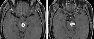
How does the midbrain develop?
Children in their mother's womb must go through many stages of development. During the embryonic stage, the midbrain grows from a small vesicle and remains intact throughout life. Throughout development, more and more new cells appear in this part, they compress the cerebral aqueduct. If there are disturbances at this stage, problems with the cerebral aqueduct may develop - partial or complete blockage. One of the most dangerous consequences is such a dangerous disease as hydrocephalus.
Helpful information.
Every time a person remembers information, neural connections are formed. This means that the structures of various parts, including the midbrain, are constantly changing; it does not freeze in a certain state.
Lenticular nucleus
The lentiform nucleus (nucl. lentiformis) is located lateral and anterior to the thalamus. It is wedge-shaped with the apex facing the midline. Between the posterior edge of the lenticular nucleus and the thalamus is the posterior leg of the internal capsule (crus posterius capsulae internae). The anterior face of the lentiform nucleus below and in front is fused with the head of the caudate nucleus.
Two strips of white matter divide the lenticular nucleus into three segments: the lateral segment - the putamen , which has a dark color, is located on the outside, and the two ancient parts of the globus pallidus (globus pallidus) of a conical shape are facing the middle.
Caudate nucleus
The caudate nucleus (nucl. caudatus) is club-shaped and curved backwards.
Its anterior part is expanded, called the head (caput) and is located above the lenticular nucleus, and its posterior part - the tail (cauda) passes above and lateral to the thalamus, separated from it by the medullary stripes (stria medullaris). The head of the caudate nucleus participates in the formation of the lateral wall of the anterior horn of the lateral ventricle (cornu anterius ventriculi lateralis). The caudate nucleus consists of small and large pyramidal cells. Between the lentiform and caudate nuclei there is an internal capsule (capsula interna).
The internal capsule (capsula interna) is located between the thalamus, lentiform and caudate nuclei and is a layer of white matter formed by projection fibers on the way to the cortex and from the cortex to the underlying parts of the central nervous system.
On a horizontal section of the cerebral hemisphere at the level of the middle of the thalamus, the internal capsule is white and resembles the shape of an angle open outward. The internal capsule is divided into three sections: the anterior leg (crus anterius capsulae internae), the knee (genu capsulae internae) and the posterior leg (crus posterius capsulae internae).
Above the inner capsule, the fibers form a radiant crown (corona radiata). The short anterior leg of the capsule is formed by axons that arise from the cells of the frontal lobe cortex and go to the thalamus (tr.
frontothalamicus), into the red nucleus (tr. frontorubralis), to the cells of the bridge nuclei (tr. frontopontinus). In the knee of the internal capsule there is a corticonuclear tract (tr. corticonuclearis), connecting the cells of the motor cortex with the nuclei of the motor cranial nerves (III, IV, V, VII, IX, X, XI, XII). The posterior limb of the internal capsule is slightly longer than the anterior one and borders the thalamus and the lentiform nucleus. In its anterior part there are fibers emanating from the cells of the posterior sections of the frontal (motor) cortex and heading to the nuclei of the anterior columns of the spinal cord.
Somewhat posterior to the corticospinal tract are fibers running from the lateral nuclei of the thalamus to the posterior central gyrus, as well as from cortical cells to the nuclei of the thalamus. The posterior leg contains fibers passing from the cortex of the occipital and temporal lobes to the pontine nuclei. In the posterior section, auditory and visual fibers pass, starting from the internal and external geniculate bodies and ending in the temporal and occipital lobes.
Along the entire length of the internal capsule there are transverse fibers that connect the lentiform body with the caudate nucleus and the thalamus. Fan-shaped diverging fibers of all pathways forming the internal capsule form the corona radiata in the space between it and the cerebral cortex. Minor damage to small areas of the internal capsule due to the compact arrangement of the fibers causes severe disorders of motor functions and loss of general sensitivity, hearing and vision on the side opposite to the injury.
Striatum
The striatum receives afferent impulses mainly from the thalamus, partly from the cortex; sends efferent impulses to the globus pallidus.
The striatum is considered as an effector nucleus that does not have independent motor functions, but controls the functions of a phylogenetically older motor center - the pallidum a (globus pallidus).
The striatum regulates and partially inhibits the unconditioned reflex activity of the globus pallidus, i.e., it acts on it in the same way as the globus pallidus acts on the red nucleus. The striatum is considered the highest subcortical regulatory and coordination center of the motor apparatus.
In the striatum, according to experimental data, there are also higher vegetative coordination centers that regulate metabolism, heat generation and heat removal, and vascular reactions.
Apparently, in the striatum there are centers that integrate and unite unconditioned reflex motor and autonomic reactions into a single holistic act of behavior.
The striatum influences organs innervated by the autonomic nervous system through its connections with the hypothalamus. With lesions of the striatum, a person experiences athetosis - stereotypical movements of the limbs, as well as chorea - strong abnormal movements that occur without any order or sequence and involve almost all the muscles (“St. Vitus’s dance”).
Both athetosis and chorea are considered to be the result of a loss of the inhibitory influence that the striatum has on the pallidum.
Pale ball
The globus pallidus (globus pallidus), pale nucleus, is a paired formation that is part of the lenticular nucleus, which is located in the cerebral hemispheres and is separated by an internal capsule. The pallidum is the motor nucleus. When it is irritated, you can get a contraction of the neck muscles, limbs and the entire torso, mainly on the opposite side.
The pallid nucleus receives impulses via afferent fibers coming from the thalamus and closing the thalamo-pallidal reflex arc. The pallid nucleus, being effector-connected with the centers of the midbrain and hindbrain, regulates and coordinates their work.
One of the functions of the pallidum is considered to be inhibition of the underlying nuclei, mainly the red nucleus of the midbrain, and therefore, when the globus pallidus is damaged, a strong increase in the tone of the skeletal muscles is observed - hypertonicity, because the red nucleus is freed from the inhibitory influence of the globus pallidus. The thalamo-hypothalamo-pallidal system takes part in higher animals and humans in the implementation of complex unconditioned reflexes - defensive, orientation, food, sexual.
In humans, when stimulating the globus pallidus, the phenomenon of almost doubling the volume of short-term memory was obtained.
Investigating the spatiotemporal relationships between speech elements (vowel phonemes) and recorded impulse activity, a correlation was identified indicating the involvement of a particular structure in the process of auditory memory. In a number of cases, such relationships were obtained by studying the globus pallidus and the dorsomedial thalamic nucleus.
Amygdala nucleus
The amygdala nucleus (corpus amygdaloideum), or amygdaloid complex, is a group of nuclei and is localized inside the anterior pole of the temporal lobe, lateral to the septum of the perforated substance.
The amygdaloid complex is a structure included in the limbic system of the brain, which is characterized by a very low threshold of excitation, which can contribute to the development of epileptiform activity.
The complex contains both larger (pyramidal, pear-shaped) and medium-sized (multipolar, bipolar, candelabra-shaped) and small cells.
The amygdaloid complex is divided into a phylogenetically older - corticomedial - and a newer basal-lateral part. The group of corticomedial nuclei is characterized by low acetylcholinesterase (AChE) activity and is largely associated with olfactory function, forming projections to the paleocortex. The connection with sexual function is confirmed by the fact that stimulation of these nuclei facilitates the secretion of luliberin and folliberin.
Neurons of the basal lateral nuclei are characterized by higher AChE activity, give a projection to the neocortex and striatum, and also facilitate the secretion of ACTH and growth hormone. When the amygdaloid complex is stimulated, convulsions, emotionally charged reactions, fear, aggression, etc. occur.
Fence
The claustrum is a thin layer of gray matter separated by an outer capsule of white matter from the lenticular nucleus. The fence below is in contact with the nuclei of the anterior perforated substance (substantia perforata anterior).
They assume participation in the implementation of oculomotor reactions of tracking an object.
Types of hyperkinesis
When the lenticular nucleus of the brain is damaged, the following types of disordered motor activity may occur:
- Athetosis is involuntary movements of the fingers, their twisting, flexion, extension.
- Chorea is sweeping swings of arms and legs in different planes and directions. They can be either weakly or strongly expressed. A characteristic disease manifested by this hyperkinesis is called “Huntington’s chorea.” With this pathology, in addition to damage to the basal ganglia, atrophy of the cortex occurs, which leads to mental and intellectual disorders.
- Dystonia is sudden uncontrolled turns of the body in different directions.
- Myoclonus is a constant short-term contraction of muscle fibers.
- Restless legs syndrome - observed when falling asleep, sudden movements of the legs such as kicking, shuddering.
- A tick is a fast, short-term, simple movement.
- Tremor - shaking movements of the limbs.
The characteristic thing is that all these movements are involuntary, that is, they cannot be controlled by consciousness. In fact, there are many more types; above are only the most common ones found in neurological practice.
Functions of the basal ganglia
The main structures of the basal ganglia (
rice.
66 )
The basal nuclei are the caudate nucleus (
nucleus caudatus
), putamen (
putamen
) and globus pallidus (
globulus pallidus
);
some authors attribute the claustrum to the basal ganglia
.
All these four nuclei are called the striatum ( corpus striatum
).
There is also a striatum (s triatum
) is the caudate nucleus and putamen.
The globus pallidus and shell form the lentiform nucleus ( nukleus lentioris
). The striatum and globus pallidus form the striopallidal system.
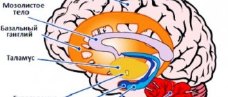
Rice. 66. A - Location of the basal ganglia in the volume of the brain. The basal ganglia are shaded red, the thalamus is shaded gray, and the rest of the brain is blank. 1 – Globus pallidus, 2 – Thalamus, 3 – Putamen, 4 – Caudate nucleus, 5 – Amygdala (Astapova, 2004).
B – Three-dimensional image of the location of the basal ganglia in the volume of the brain (Guyton, 2008)
Functional connections of the basal ganglia.
The basal ganglia
do not have input from the spinal cord, but do have direct input from the cerebral cortex
.
The basal ganglia are involved in motor functions, emotional and cognitive functions
.
Excitatory pathways
They go mainly to the striatum: from all areas of the cerebral cortex (directly and through the thalamus), from the nonspecific nuclei of the thalamus, from the substantia nigra (midbrain) (Fig. 67).
Rice. 67. Connection of the basal ganglia circuit with the corticospinocerebellar system for the regulation of motor activity (Guyton, 2008)
The striatum itself has a mainly inhibitory and, partially, excitatory effect on the globus pallidus.
From the globus pallidus the most important path goes to the ventral motor nuclei of the thalamus, from them the excitatory path goes to the motor cortex of the cerebrum. Some fibers from the striatum go to the cerebellum and to the centers of the brain stem (RF, red nucleus and then to the spinal cord.
Braking paths
from the striatum they go to
the substantia nigra
and, after switching, to the nuclei of the thalamus (Fig. 68).
Rice. 68. Nerve pathways secreting various types of neurotransmitters in the basal ganglia. Ax – acetylcholine; GABA – gamma-aminobutyric acid (Guyton, 2008)
Motor functions of the basal ganglia
In general, the basal ganglia, having bilateral connections with the cerebral cortex, thalamus, and brainstem nuclei, are involved in the creation of programs of targeted movements, taking into account the dominant motivation. In this case, neurons of the striatum have an inhibitory effect (transmitter - GABA) on neurons of the substantia nigra. In turn, neurons of the substantia nigra (transmitter - dopamine) have a modulating effect (inhibitory and excitatory) on the background activity of striatal neurons.
When dopaminergic influences on the basal ganglia are disrupted, movement disorders such as parkinsonism are observed, in which the concentration of dopamine in both nuclei of the striatum drops sharply. The most important functions of the basal ganglia are performed by the striatum and the globus pallidus.
Functions of the striatum
.
Participates in turning the head and body and walking in a circle
, which are part of the structure of indicative behavior.
Defeat
the caudate nucleus in diseases and when destroyed in experiments leads to violent, excessive movements (hyperkinesis: chorea and athetosis).
Functions of the globus pallidus
.
Has a modulating effect
to the motor cortex, cerebellum, RF, red nucleus. When stimulating the globus pallidus in animals, elementary motor reactions predominate in the form of contraction of the muscles of the limbs, neck and face, and activation of eating behavior.
Destruction of the globus pallidus
is accompanied by a decrease in motor activity -
adynamia
(pallor of motor reactions), and it (destruction) is accompanied by the development of drowsiness, “emotional dullness”, which
makes it difficult to carry out
existing
conditioned reflexes
and worsens
the development of new ones
(impairs short-term memory).
Pathologies in case of damage
In 1896, Ch. S. Sherrington described extreme muscle tension in an animal when the descending connections of the red nucleus are broken. If you cut the brain stem between the red and vestibular nuclei, then maximum tension occurs in the extensor muscles of the limbs, neck, and back.
Important Anankasty personality disorder
These muscles counteract gravity, which means this picture must be associated with the vestibular system. Indeed, the Deiters vestibular nucleus activates the extensors. The influence of the red nucleus on these neurons and the Deiters nucleus inhibits their activity. That is, muscle tone is created by the joint work of several nuclei.
Decerebrate rigidity in humans occurs after severe or traumatic brain injury and is a bad sign.
It looks like this: the arms are extended, as if stretched, and brought towards the body, the palms are turned outward (pronated), the fingers are bent, but the thumbs are abducted. The legs are extended and brought towards each other, the feet are turned inward. The toes are bent, as if in a suspended position. The jaws are clenched. It was described in 1912 by Dutch doctors R. Magnus and A. Klein (R. Magnus, A. de Klein).
Brain function can be disrupted due to injuries, infectious and vascular lesions of the brain, tumor processes, and aggression of the immune system.
Damage to the red nucleus and its connections in humans is manifested not only by decerebrate rigidity, but also by less severe pathology. The midbrain contains structures that give rise to nerves that control the muscles of the eyeball, pupil, and levator palpebral muscle. Therefore, damage to the red nucleus can be combined with “ocular” symptoms.
This happens with Claude syndrome and Benedict syndrome. They often develop after vascular accidents (stroke).
Claude syndrome was described by French neurologist and psychiatrist Henri Claude in 1912. In Claude's syndrome, the inferior part of the red nucleus, efferent fibers from the cerebellum to the thalamus, and the oculomotor nerve are affected.
Due to the damaged oculomotor nerve on the affected side, the upper eyelid droops, the pupil dilates and divergent strabismus appears. On the other side of the body, hand tremors occur when moving towards a goal (intention tremor), muscle weakness.
Eyelid drooping and strabismus in Claude syndrome
In this syndrome, the lesion involves the red nucleus, its connections with the cerebellum and the structures of the oculomotor nerve. On the damaged side the pupil dilates, on the opposite side intentional and erratic or writhing movements of the limbs (choreoathetosis) appear.
Latin name: nucleus ruber.
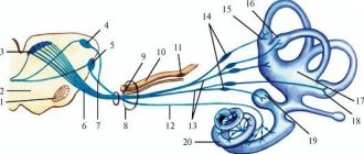
In the midbrain, the red nuclei are located in the very center. If we make a horizontal slice through the midbrain, then diagonally between and we will see two pale pink spots. These will be the red kernels. It is believed that they owe their color to iron, which they contain in two different forms - hemoglobin and ferritin.
In the following screenshot you can see a sagittal section of the brain stem. The bottom of the red nucleus lies on the ascending fibers of the superior cerebellar peduncles at the level of the top of the inferior cerebellum. From above - they reach the level of the hypothalamus.
You can learn more about where the red core is located on ours.
The red nucleus is motor, responsible for muscle tone and reflexes.
There are two parts:
- posterior large cell (magnocellular) - less developed in humans than in other vertebrates, because In humans, the cerebral cortex is much more developed, which takes away some of the functions from the magnocellular part.
- anterior small cell (parvocellular) - transmits information from the motor cortex to the cerebellum through the olives.
Some researchers isolate the posteromedial part separately.
Types of hypokinesis
If the lentiform nucleus is damaged, the following types of hypokinesis are also possible:
- Akinesia is a complete lack of motor activity, bradykinesia is a decrease in motor activity. This is most typical for Parkinson's disease, where the deviation is combined with increased muscle tone, mental disorders, stooping, and decreased facial activity. In addition, a similar pathology occurs in endocrinological diseases, namely hypothyroidism - decreased activity of the thyroid gland.
- Apraxia is the inability to make purposeful movements in the presence of normal elementary movements.
- Cataplexy is a sudden drop in muscle tone. Sometimes it leads to a fall and injury to the patient.
- Catatonia is the “freezing” of the patient for a long time in the position in which he was “left”, accompanied by high muscle tone. The patient can remain in this position for weeks or months.
- Muscle rigidity is increased muscle tone, which leads to a decrease in the number of movements.
The conditions listed above are not independent diseases. Typically, dyskinesia is just one of many symptoms that can lead to the correct diagnosis. The origin of many of these pathologies is not fully known, and disruption of the lenticular nucleus is only one of many causes. Therefore, the approach to the treatment of dyskinesias should be comprehensive; often, in addition to neurological care, psychiatric care is necessary.
Thus, the article provides an answer to the question of what it is - a lenticular nucleus. In short, this is a complex, complexly organized structure, one of the components of the extrapyramidal system, thanks to the presence of which we can perform precise actions.