It is important for every person to know how he works. And one of the most interesting organs to study is the brain, which has not yet been fully understood. Few people, after a school biology course, remember the functions of the midbrain and its purpose. The need to understand complex medical terms comes in adulthood, when a person begins to visit doctors or is about to enter a medical university.
If you want to know what the midbrain is and its location, you don't have to study complex medical encyclopedias or attend medical school. Conscientious patients, before going to a medical facility, want to learn more about the disease and what functions the diseased organ performs. Then hospital procedures will not seem so scary and incomprehensible.
Basic information
The central nervous system contains neurons with processes and glia. The brain has only five sections. First
– oblong – continuation of the dorsal.
It transmits information to and from other departments. Performs a regulatory function in relation to the coordination of movements. The second
is the bridge - here are the midbrain centers responsible for the assimilation of audio and video information.
This department stands for coordination of movements. The third
- the cerebellum - connects the posterior and anterior sections.
The fourth
- middle - is responsible for facial expressions, movements of the eyeballs, and the auditory pathways pass through it.
This is exactly what we will consider. The fifth
- the front - normalizes mental activity.
This is interesting. There is no connection between brain size and mental abilities in humans. The number of nerve connections is much more important.
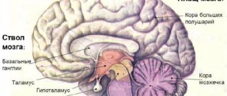
Brain stem
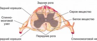
The brainstem consists of the medulla oblongata, pons, and midbrain and contains motor and sensory nuclei that perform motor and sensory functions for the face and head in the same way that the spinal cord performs these functions for the neck, trunk, and limbs. At the same time, the brain stem performs many special functions (including control functions: respirationcardiovascular systemGastrointestinal tractmany stereotypical body movementsbalanceeye movements) and serves as a hub for “command signals” from higher-lying centers. The vestibular and reticular nuclei of the brainstem play an important role in controlling body movements and balance.
Reticular nuclei
.
In Fig. Figure 14-3B shows the location of the reticular nuclei. They are divided into the reticular nuclei of the pons and the reticular nuclei of the medulla oblongata. These two systems of nuclei function antagonistically in relation to each other: the pons nuclei excite anti-gravity muscles
,
the nuclei the medulla oblongata inhibit them
.
Pontine reticular nuclei
transmit excitatory signals to the spinal cord through
the mostoreticulospinal tract
, localized in the anterior column of the spinal cord. The fibers of this tract activate spinal cord motor neurons, which send excitatory impulses to the muscles of the spinal column and extensor muscles of the limbs. The reticular nuclei of the bridge have high excitability. In addition, they receive excitatory impulses from both the vestibular nuclei and the deep cerebellar nuclei. Thus, the excitatory pontine reticular system causes a powerful activation of the anti-gravity muscles throughout the body.
Reticular nuclei of the medulla oblongata
transmit inhibitory signals to the same anti-gravity neurons of the spinal cord, but through a different pathway -
the reticulospinal tract of the medulla oblongata
, located in the lateral columns of the spinal cord. The reticular nuclei of the medulla oblongata receive collaterals from the corticospinal tract, rubrospinal tract and other motor pathways. The normal activity of the inhibitory reticular system of the medulla oblongata maintains a balance with the activity of the excitatory system of the reticular formation of the pons, as a result of which the muscles of the body do not have excessive tension. Commands from the upper parts of the brain can interrupt the inhibitory influence of the medulla oblongata system when the brain needs to excite the pontine system to control the vertical position of the body. Excitation of the reticular system of the medulla oblongata can inhibit the anti-gravity muscles in some parts of the body to perform any necessary movements.
The excitatory and inhibitory reticular nuclei are an essential part of the control system, which is controlled by signals from the motor cortex; In addition, these nuclei provide the primary level of tonic contraction to counteract gravitational forces and can inhibit specific muscle groups to provide other functions.
Vestibular nuclei
functionally connected with the reticular nuclei of the bridge, stimulating anti-gravity muscles.
The lateral vestibular nuclei
transmit strong excitatory signals to the lateral and medial vestibular tract. Without the participation of the vestibular nuclei, the reticular system of the bridge significantly weakens its exciting influence on the gravitational muscles of the neck, back, upper and lower extremities. The specific role of the vestibular nuclei is to selectively control the excitatory signals coming from the vestibular apparatus to various anti-gravity muscles to maintain balance.
Important Types of MRI examinations
It is important for every person to know how he works. And one of the most interesting organs to study is the brain, which has not yet been fully understood
Few people, after a school biology course, remember the functions of the midbrain and its purpose. The need to understand complex medical terms comes in adulthood, when a person begins to visit doctors or is about to enter a medical university.
If you want to know what the midbrain is and its location, you don't have to study complex medical encyclopedias or attend medical school. Conscientious patients, before going to a medical facility, want to learn more about the disease and what functions the diseased organ performs. Then hospital procedures will not seem so scary and incomprehensible.
How does the midbrain develop?
Children in their mother's womb must go through many stages of development. During the embryonic stage, the midbrain grows from a small vesicle and remains intact throughout life. Throughout development, more and more new cells appear in this part, they compress the cerebral aqueduct. If there are disturbances at this stage, problems with the cerebral aqueduct may develop - partial or complete blockage. One of the most dangerous consequences is such a dangerous disease as hydrocephalus.
Helpful information.
Every time a person remembers information, neural connections are formed. This means that the structures of various parts, including the midbrain, are constantly changing; it does not freeze in a certain state.
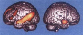
Pathologies in violation
It all started when science received a description of severe muscle tension in animals. Tension was created by breaking the connections of the red core. This break is called decerebrate rigidity. Based on this observation, a conclusion was made, the essence of which is that when the connection between the red and vestibular nuclei is lost, severe tension occurs in the skeletal muscles, muscles of the limbs, as well as the muscles of the neck and back.
The above muscles are distinguished by their ability to counteract gravity, so it was concluded that this development of events is associated with the vestibular system. As it turned out later, the vestibular nucleus of Deiters is capable of triggering the work of extensor motor neurons. The activity of these neurons is significantly slowed down under the influence of the red nuclei and Deiters nucleus.
It turns out that active muscle work is the result of the joint work of the entire complex. In humans, decerebrate rigidity occurs as a result of traumatic brain injury. You can also encounter this phenomenon after a stroke. It should be understood that such a condition is a bad sign. You can find out about its presence by the following signs:
- arms straight, spread in different directions;
- hands lie palms up;
- all fingers are clenched except the thumbs;
- legs extended and folded together;
- feet extended;
- toes curled;
- the jaws press tightly against each other.
In case of injuries, severe infectious diseases, all kinds of internal damage to organs, including the brain, as well as tumor processes and aggression of the immune system - all this leads to disruption of the brain. Thus, if connections with the red nuclei are disrupted, decerebrate rigidity may occur, as well as disruption of the eyeball and eyelid muscles, the latter being an easier reaction of the body to the rupture of connections.
What role does it play?
It is the middle section that regulates muscle tone. His role corresponds to his intermediate position. Due to the fact that the midbrain has a special structure, its functions include the transmission of information. It has a lot of different purposes:
- sensory
– to convey tactile sensations; - motor
– coordination depends on this part of the midbrain; - reflex
- for example, oculomotor, reaction to light and sound.
Due to the work of the middle section, a person can stand and walk. Without it, a person would not be able to fully move in space. Also, the work of the vestibular apparatus is controlled at the level of the midbrain.
Functions
In humans, the rubrospinal tract coming from the red nucleus partly controls gait and movements of the shoulder girdle. "Partly" means that it only controls large movements. The corticospinal tract is responsible for fine motor skills. If you “turn it off” and leave only the rubrospinal one, then the movements of such a person will become sharp and sweeping.
I also note that the rubrospinal tract is responsible for reflex movements.
Animal experiments show that electrical stimulation of the rubrospinal tract leads to excitation of flexor motor neurons and inhibition of extensor motor neurons. Thus, when the tract is cut at the level of the midbrain, the limbs are straightened and remain tense in this position. the head is thrown back.
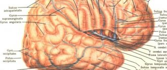
Organ structure
It is known that the average human brain has different parts, each of which performs its own role. Quadrigeminal - the structure consists of paired hills. The upper ones are visual and the lower ones are auditory.
The legs contain a black substance. Thanks to it, a person not only lies, but can make precise movements with his hands and eat food. At a certain point, the middle region processes information about when to bring a spoon to the mouth, how to chew food, and what function will allow it to be swallowed.
Useful to know: Brain: functions, structure
The ocular motor nerve originates between the crura and exits from there. It is responsible for the constriction of the pupil and some motor functions of the eyeball. To understand the structure of the midbrain, you need to know where it is located. It is composed of the intermediate and cerebral hemispheres of the cerebrum, its structure is simple and has only two sections. Quadrigemole on nearby two paired colliculi, which form the upper wall. They resemble a plate in appearance. Legs - conductive channels are located there, going to the hemispheres of the anterior section and connecting it with the lower parts of the nervous system.
Diagnosis and prognosis of pathology
In addition to neurologists, diagnostics is carried out by doctors from other offices (functional diagnostics). The main methods for identifying diseases of the basal ganglia are:
- analysis of the patient’s life, his anamnesis;
- objective external neurological examination and physical examination;
- magnetic resonance and computed tomography;
- study of the structure of blood vessels and the state of blood circulation in the brain;
- Ultrasound;
- visual methods for studying brain structures;
- electroencephalography;
Prognostic data depends on many factors, such as gender, age, general constitution of the patient, the moment of the disease and the moment of diagnosis, his genetic predispositions, the course and effectiveness of treatment, the pathology itself and its destructive properties. According to statistics, 50% of diseases of the basal ganglia have an unfavorable prognosis. The remaining half of cases have a chance for adaptation, rehabilitation and normal life in society.
Didn't find a suitable answer? Find a doctor and ask him a question!
How many parts does the middle section have?
There are three parts in total.
Dorsal - the roof of the middle section. It is divided into 4 mounds using grooves intersecting in pairs. The two upper hills are the subcortical centers for regulating vision, and the remaining lower ones are auditory. The ventral one is the so-called cerebral peduncle. The conductive channels to the anterior section are based here. The internal space of the brain looks like a hollow canal.
Helpful information.
If a person does not breathe oxygen for more than five minutes, the brain will be permanently damaged, resulting in death.
Cores
Gray matter accumulates inside the quadrigeminal tubercles, clusters of which are called nuclei. The main function of the nuclei is called the innervation of the eyes. They come in the following types.
Reticular formation - takes part in stabilizing the work of skeletal muscles. They activate the cells of the cerebral cortex of the head, and have an inhibitory effect on the spinal cord. Oculomotor nerve - contains fibers that innervate the sphincter and eye muscles. Trochlear nerve - supplies nerves to the oblique muscle of the organ of vision. Substantia nigra - color associated with the pigment melanin. The neurons of this substance themselves synthesize dopamine. Coordinate facial muscles and small movements. Red nuclei of the midbrain - activate neurons of the flexor and extensor muscles
What are the basal ganglia?
The basal ganglia of the brain are functionally and anatomically related accumulations of gray matter in the deep parts of the brain. These structures are deepened into the white matter, which functions as an information transmitter. Even in the embryo, the basal ganglia develop from the ganglion tubercle, then forming into mature brain structures that perform strictly specific functions in the nervous system.
The importance of puppet therapy as a psychological aid for a child
The basal ganglia are located at the base of the brain, lateral to the thalamus. Anatomically highly specific nuclei are part of the forebrain, which is located on the border of the frontal lobes and the brainstem. Often, by the term “subcortex”, experts mean precisely the set of basal ganglia of the brain.
Anatomists distinguish three concentrations of gray matter:
- Striped body. This structure means a set of two not entirely differentiated parts: The caudate nucleus of the brain. It has a thickened head, forming in front one of the walls of the lateral ventricle of the brain. The thin tail of the nucleus is adjacent to the bottom of the lateral ventricle. The caudate nucleus also borders the thalamus.
- Lenticular nucleus. This structure runs parallel to the previous accumulation of gray matter and, closer to the end, merges with it, forming the striatum. The lenticular nucleus consists of two white layers, each of which has its own name (globus pallidus, shell).
Corpus striatum received its name due to the alternating arrangement of white stripes on its gray matter. Recently, the lenticular nucleus has lost its functional meaning, and it is called exclusively in a topographical sense. The lenticular nucleus, as a functional compilation, is called the striopallidal system.
- The fence or claustrum is a small thin gray plate located near the shell of the striatum.
- Amygdala. This core is located under the shell. This structure also belongs to the limbic system of the brain. The amygdala usually means several separate functional formations, but they were combined due to their close location. This area of the brain has multiple connections with other brain structures, in particular with the hypothalamus, thalamus and cranial nerves.
The concentration of white matter is:
- Internal capsule - white matter between the thalamus and the lentiform nucleus
- Outer capsule - white substance between the lentil and the fence
- The outermost capsule is the white substance between the enclosure and the insula.
The internal capsule is divided into 3 parts and contains the following pathways:
Front leg:
- Frontothalamic tract - connection between the frontal cortex and the mediadersal nucleus of the thalamus
- Frontopontine tract - connection between the frontal cortex and the pons
Knee:
Corticonuclear tract - connection between the nuclei of the motor cortex and the nuclei of the motor cranial nerves
Rear leg:
- Corticospinal tract - conducts motor impulses from the cerebral cortex to the nuclei of the motor horns of the spinal cord
- Thalamo-parietal fibers - Axons of thalamic neurons are connected to the postcentral gyrus
- Temporo-parieto-occipital-pontine fasciculus - connects the pontine nuclei with the lobes of the brain
- Auditory radiance
- Visual radiance
Prevention of pathologies
The brain cannot function correctly without intellectual activity and physical activity. Typically, disruptions in the functioning of the central nervous system are observed in people over 70 years of age. But diseases of this group are diagnosed in those who, after retirement, no longer maintain their health and lead a healthy lifestyle. However, there are also congenital pathologies in the midbrain; you can get sick at any age.
Useful to know: Functions and structure of the cerebral pons, its description
Exercise regularly to the best of your physical ability, walk in the fresh air, do gymnastics in the morning. Quit tobacco and alcoholic beverages. Switch to a healthy diet, eat as many fresh vegetables and fruits as possible. Do not eat products with preservatives and emulsifiers. Train your mind - for this you can read books, solve crosswords, play chess, gain new knowledge in an area of interest.
To get rid of vitamin deficiency - take vitamins and antioxidants. Since the brain is 60% fat, you cannot give up oil, but it must be natural. For example, olive oil is perfect. Avoid stressful situations. Do not engage in monotonous work too often, take breaks, switching to other activities. Monitor your blood pressure levels - hypertension can cause a stroke.
Material from Wikipedia - the free encyclopedia
| Brain: Red Core | ||
| Cross section of the midbrain showing the location of the red nucleus. In the upper part of the image there are the quadrigeminal peduncle and the peduncle of the midbrain, the midbrain aqueduct, the substantia nigra and the nucleus of the oculomotor nerve are clearly visible. | ||
| Latin name | Nucleus ruber | |
| System | Extrapyramidal | |
Anatomy
This elongated sausage-shaped formation extends in the tegmentum of the cerebral peduncle from the hypothalamus of the diencephalon to the inferior colliculus, where it begins an important descending tract, tractus rubrospinal, connecting the red nucleus with the anterior horns of the spinal cord. This bundle, after leaving the red nucleus, intersects with a similar bundle of the opposite side in the ventral part of the median suture - the ventral decussation of the tegmentum. The red core contains a pigment that includes iron, which gives it a specific color.
Physiology
Nucleus ruber
is a very important coordination center of the extrapyramidal system, connected with its other parts.
Fibers pass to it from the cerebellum as part of the upper peduncles of the latter after their decussation under the roof of the midbrain, ventrally from the aqueductus cerebri
, as well as from
the pallidum
- the lowest and most ancient of the subcortical nodes of the brain that are part of the extrapyramidal system.
Thanks to these connections, the cerebellum and the extrapyramidal system, through the red nucleus and the tractus rubrospinal extending from it, influence the entire skeletal muscle in the sense of regulating unconscious automatic movements. The red nucleus has projections to the motor nuclei of the spinal cord, which controls the movement of the fore and hind limbs and is under the control of the cerebral cortex. Nucleus ruber
is an important intermediate authority for integrating the influences of the forebrain and cerebellum in the formation of motor commands to spinal cord neurons.
Participation in the corticorubral tract
The red nucleus receives a large number of nerve fibers directly from the primary motor cortex through the corticorubral tract, as well as many collaterals from the corticospinal tract as it passes through the midbrain. These fibers form synapses in the lower, magnocellular (magnocellular) part of the red nucleus, where large neurons are located, similar in size to Betz cells in the motor cortex. These neurons give rise to the rubrospinal tract, which crosses the opposite side in the lower part of the brainstem and descends into the lateral columns of the spinal cord, following in close proximity to and anterior to the corticospinal tract.
Red core
Red core
(lat.
Nucleus ruber
) - a structure in the midbrain involved in the coordination of movements. It consists of a caudal magnocellular and rostral parvocellular part. The red nucleus is located in the tegmentum of the midbrain next to the substantia nigra. The red nucleus and substantia nigra are the subcortical centers of the extrapyramidal motor system.
Pathophysiology
When the red nucleus and its pathways are damaged, the animal develops so-called decerebrate rigidity. When the red nucleus is damaged, various types of syndromes occur:
Claude's syndrome is an alternating syndrome with the localization of a pathological focus in the tegmentum of the midbrain, caused by damage to the lower part of the red nucleus, through which the root of the third nerve passes, as well as dento-rubral connections passing through the superior cerebellar peduncle. On the side of the pathological process there are signs of damage to the oculomotor nerve (ptosis of the upper eyelid, dilation of the pupil, divergent strabismus), and on the opposite side - intention tremor, hemiataxia, muscle hypotonia. Described in 1912 by the French neurologist N. Claude.
Benedict's syndrome - (M. Benedict, 1835-1920, Austrian neurologist) alternating syndrome occurs when the midbrain is damaged at the level of the red nucleus and the cerebellar-rednuclear tract: a combination of oculomotor nerve palsy on the affected side with choreoathetosis and intentional tremors on the opposite side.