Good afternoon, dear readers. Today we will continue the course of neuroanatomy, and on the agenda we have the anatomy of the medulla oblongata.
The medulla oblongata (myelencephalon) is a truly amazing part of the brain (encephalon). The length of the medulla oblongata does not exceed 2.5 - 3 centimeters, but the medulla oblongata is a critical part of the central nervous system. So much vital stuff is packed into these 3 centimeters that even a slight damage to the medulla oblongata can lead, at a minimum, to ending up in the intensive care unit. Today we will find out why this happens and why this happens, fortunately, very rarely.
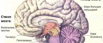
Medulla oblongata and brain stem
Before we study the internal structure of the medulla oblongata, we must study its external structure. The brain is traditionally divided into structures consisting of hemispheres - the cerebrum (cerebrum) and the cerebellum (cerebellum), as well as into structures that form the brain stem (truncus encephali).
If you take a specimen of a human brain and carefully remove the cerebral hemispheres and cerebellum, you will clearly see the brain stem (it really does look like a plant stem). The brain stem includes a group of unpaired components of the brain:
- Medulla;
- Varoliev bridge (pons);
- Midbrain (mesencephalon);
- Diencephalon.
I note that in different textbooks and atlases the brain stem includes different structures - the midbrain and, especially, the diencephalon are quite rare in the brain stem. But the medulla oblongata and the pons enter the brain stem from any source. The medulla oblongata is located below all other brain structures. In fact, the medulla oblongata is a direct continuation of the spinal cord.
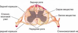
Therefore, knowledge of the anatomy of the spinal cord, which we discussed in great detail in the last four lessons, will be very useful to you while studying the medulla oblongata.
Topography of the medulla oblongata
The medulla oblongata is a small but very important component of the brain that is located between the pons and the spinal cord. The medulla oblongata has a dorsal (facies ventralis) and ventral (facies dorsalis) surface (posterior and anterior, respectively). Therefore, the boundaries of the medulla oblongata must be defined anteriorly and posteriorly.
In this illustration from Zolotko's atlas, the red arrow points to the ventral side of the medulla oblongata, and the blue arrow points to the dorsal side.
There is also a very good illustration from Sinelnikov’s atlas, in which the medulla oblongata is tinted blue:
The medulla oblongata is reliably protected by the bones of the skull and other parts of the central nervous system, for example, the cerebellum. We urgently need such multi-layer armor, because the medulla oblongata contains vital centers for breathing, swallowing and vascular tone. That is why traumatic injuries to the medulla oblongata are not a common problem, however, when this does happen, it puts the victim on the brink of life and death.
Front Borders
The upper anterior border of the medulla oblongata is the lower edge of the pons, and the lower border is the upper edge of the spinal cord. If we are talking about skeletotopy, that is, the projection of an organ onto the bones of the skeleton, then the lower edge of the spinal cord approximately corresponds to the edges of the foramen magnum occipitale. But this is not entirely accurate.
The most accurate landmark by which we can see the border of the spinal cord and brain is the decussation of the pyramids (decussatio pyramidum). A little further we will look at what the pyramids are and why they intersect, but for now let’s remember the anterior borders of the medulla oblongata:
- Upper - the lower edge of the bridge;
- The lower one is the intersection of the pyramids.
Here I have highlighted the upper border of the spinal cord in blue and the lower border in yellow.
Posterior borders
To see the posterior borders of the medulla oblongata, we need to remove the cerebellum, which completely covers them.
The upper posterior border of the medulla oblongata are bundles of white matter called the stria medullaris of the fourth ventricle (striae medullares ventriculi quarti). In fact, at the back, as well as at the front, the medulla oblongata borders the pons; the medullary stripes are simply an excellent landmark that visually separates the medulla oblongata and the pons. The inferior border of the posterior side of the medulla oblongata is a line parallel to the motor chiasm
So, remember the back boundaries:
- Upper - brain stripes;
- The lower one is a line parallel to the motor chiasm.
Main nuclei of the medulla oblongata
The nerve centers of the medulla oblongata organize pairs of cranial nerve nuclei:
- Pair IX - glossopharyngeal nerves, are composed of three parts: motor, affective and autonomic. The motor region is responsible for the movements of the muscles of the pharyngeal canal and the oral cavity. The affective region receives signals from the gustatory sensory system at the back of the tongue. Vegetative regulates the secretion of saliva.
- The X pair is the vagus nerve, which includes three nuclei: the autonomic one is responsible for the regulation of the larynx, esophagus, cardiovascular system, gastrointestinal tract and digestive glands. The nerve contains afferent and efferent fibers. The sensitive nucleus catches signals from receptors in the lungs and other internal systems. The motor nucleus controls contractions of the oral muscles during swallowing. There is also a mutual nucleus (n. ambiguus), the axons of which are activated when a person coughs, sneezes, vomits the contents of the stomach and changes the intonation of the voice.
- The XI pair is an accessory nerve, divided into 2 parts: the first is closely interconnected with the vagus nerve, and the second is directed to the muscles of the sternum, key and trapezius muscles. With pathology of the XI pair, disturbances in head movements occur - it throws back or moves to the side.
- XII pair is the hypoglossal nerve, responsible for the motility of the tongue. Regulates muscles such as the styloglossus, mentalis, as well as the rectus and transverse muscles of the tongue. The functions of the XII pair also include, in part, the reflexes of swallowing, chewing and sucking. The composition includes mainly motor neurons. The nuclei control lingual motor skills during the process of eating and grinding food, and the movement of the mouth and tongue during conversation.
Article on the topic: Diet after food poisoning - nutrition rules and menu for the week
The structure also contains the cuneate and tender nuclei, along the paths of which signals pass to the somatosensory area of the cortex. The cochlear nuclei regulate the auditory system. The nuclei of the underlying olivary control the transmission of impulses to the cerebellum.
In the underlying caudal region of the myelencephalon there is a hemodynamic center that interacts with the fibers of the 5th pair of nerves. It is assumed that it is from this area that excitatory activating signals of sympathetic fibers to the cardiovascular system are born. This fact is confirmed by studies on the intersection of the caudal areas of the medulla oblongata, after which the level of blood pressure did not change.
Also located within the structure is a section of the reticular formation. The axons of the locus coeruleus secrete the hormone norepinephrine, which affects the excitability of nerve cells. This center controls reactions such as tension and anxiety.
Respiration processes are controlled thanks to the respiratory center, which is located between the superior region of the pons and the underlying region of the medulla oblongata. Violations of this center lead to cessation of breathing and death.
External structure of the medulla oblongata
Now that we have learned the topography of the medulla oblongata, we can proceed to the external structure. Do not forget that the medulla oblongata has a ventral and dorsal surface, so we will analyze the external structure of the medulla oblongata, as well as the topography, from both sides.
Ventral (front) surface
If we look at the medulla oblongata from the front, we immediately see an anatomical formation that is very familiar to us. This is the anterior median fissure (fissura mediana anterior), exactly the same as on the anterior surface of the spinal cord.
To the right and left of the anterior median fissure we can see elongated projections. These are the pyramids of the medulla oblongata (pyramides medullae oblongatae). Remember the pathways of the spinal cord. In the anterior and lateral funiculi there are, respectively, anterior and lateral pyramidal tracts. Both are continuations of large bundles of white matter that run through the medulla oblongata. It is these bundles that are called pyramids.
Below these convexities turn into mounds, which are called the intersection of the pyramids (decussatio pyramidum). As you remember, it is the intersection of the pyramids that is the boundary between the spinal cord and the medulla oblongata. In this illustration from Sinelnikov’s atlas, I highlighted the pyramids themselves in red, and their intersection in green. The recess between the pyramids - the anterior median fissure - is clearly visible without any highlighting.
If we look a little laterally than the pyramids, we will find a couple more protrusions. These projections are called olives - it's easy to remember because they are really round and slightly elongated, like the fruit of an olive tree. I highlighted the olives in lilac:
Olives, unlike pyramids, are formed by gray matter, that is, by the bodies of neurons - we will look at this a little further. The last anatomical formation on the anterior surface of the medulla oblongata that we will study will be the anterior lateral sulcus (sulcus anterolateralis). This is a small depression that separates the olive and the pyramid. As you remember, the same groove adorned the ventral surface of the spinal cord.
Dorsal (posterior) surface
The posterior surface of the medulla oblongata has several features. We have already mentioned one of them - it is completely covered by the cerebellum, that is, without removing the cerebellum, we will not be able to see the posterior surface of the spinal cord.
The second feature is that the posterior surface of the medulla oblongata has the shape of a triangle. Adjacent to this triangle on top is another triangle, which is the posterior surface of the Varoliev bridge. Together, these two triangles form the rhomboid fossa (fossa rhomboidea) - a small diamond-shaped depression in which the nuclei of the cranial nerves are concentrated.
In this illustration from Gray's atlas we clearly see a diamond-shaped fossa. Pay attention to the cerebellar hemispheres - the left one is removed, and the right one is moved to the side so that we can see it.
So, what can we see on the back surface of the medulla oblongata? Similar to the spinal cord, in the very center here is the posterior median sulcus (sulcus medianus posterior). This groove, as in the spinal cord, separates thin bundles (fasciculus gracilis), which end in rounded convexities - thin tubercles (tuberculum gracilis). According to the authors, these formations will be called, respectively, Gaulle's bundles and Gaulle's tubercles.
Slightly more lateral are wedge-shaped bundles (fasciculus cuneatus), which also end in rounded convexities, wedge-shaped tubercles (tuberculum cuneatus). The author's names for these formations are Burdach's bundles and Burdach's tubercles, respectively.
In this picture from Sinelnikov’s atlas, the choroid plexus of the rhomboid fossa is colored as if it were a portal to another dimension:
But here we see perfectly:
- Wedge-shaped bundles (green);
- Thin bunches (blue color);
- Wedge-shaped tubercles (light green color);
- Thin tubercles (dark blue color).
Inferior cerebellar peduncles
The cerebellum connects to the brainstem through six anatomical structures called the cerebellar peduncles. These formations are processes of neurons united into parts of the pathways. There are lower, middle and upper pairs of cerebellar peduncles. You probably already guessed what the cerebellar peduncles are for - they connect the cerebellum with other parts of the brain:
- The superior cerebellar peduncles (pedunculi cerebellares superiores) connect the cerebellum and midbrain;
- The middle cerebellar peduncles (pedunculi cerebellares medii) connect the cerebellum and the Varoliev bridge;
- The lower cerebellar peduncles (pedunculi cerebellares inferiores) connect the cerebellum and medulla oblongata.
The inferior cerebellar peduncles are exactly what will interest us in this lesson, because they are most directly related to the medulla oblongata. It will be most convenient to show them on a horizontal section of the medulla oblongata, because on frontal images they are absent or indicated schematically.
Structure
The human medulla oblongata (lat. Myelencephalon) is just a part of the brain. This section is located between the dorsal and middle, in the posterior cranial fossa. It is a thickened continuation of the spinal cord. It looks like the head of an onion, which is compressed at the back and has a slight bulge at the front. This section connects the cerebellar part and the pons with the help of special processes.
Below, this area smoothly flows into the dorsal region. The lower boundary is determined by the point of exit of the upper root filament of the 1st cervical nerve. From above it borders with the pons. This part is separated from it by a perpendicular bulbar-pontine groove. The longitudinal size of this area is 2.5-3.2 cm, transverse - 1.5 cm, anteroposterior - 1 cm.
The structure of this section is heterogeneous; it consists of gray and white substance. There is a grayish substance inside. It is surrounded by tiny nuclei. The white matter is located on the outside. It surrounds a grayish substance. The white part consists of short and long fibers.
Long fibers are pathways that transit the spinal cord. They intersect in the area of the pyramids. In the nuclei of the posterior funiculi there are neuron bodies of upwardly extending fibers. The processes of these neurons go from the medulla oblongata to the thalamus. The fibers form a medial loop, which intersects in the medulla oblongata. In this section there are 2 intersections of long conducting paths.
The short ones include bundles of fibers that connect the nuclei of gray matter to each other. The nuclei of the medulla oblongata connect to neighboring parts of the brain.
External structure
The outer anterior part of the medulla oblongata is the ventral surface. It consists of paired cone-shaped lateral lobes that expand upward. They are formed by pyramidal tracts and have a median fissure. Olives are located near the pyramids. They are separated from the pyramids by a groove, which is a direct continuation of the anterolateral groove of the spinal cord. The transition of the groove from the spinal cord to the medulla oblongata is smoothed by external arcuate fibers.
The posterior outer part is the dorsal surface. It looks like two cylindrical thickenings that are separated by a median groove. This part consists of fibrous bundles that connect to the spinal cord.
On the dorsal side there are two bundles: thin and wedge-shaped. They end in tubercles of a thin and wedge-shaped nucleus. On the dorsal surface there is the lower part of the rhomboid fossa and the lower cerebellar peduncles. The posterior choroid plexus is also located here.
Between the ventral and dorsal surfaces are the lateral surfaces. They have grooves that originate in the spinal cord.
Internal structure
The internal structure coordinates the following functions: metabolic processes, blood circulation, breathing, movement, balance. A cross-section of the medulla oblongata, made at the level of the olives, shows grooves emerging from the spinal cord. Between them are pyramidal tracts.
Outside the pyramids there are small tubercles. These are olives. Inside them there are lower olive nuclei. They are convoluted plates of a gray substance. The olivary nuclei communicate with the cerebellar nuclei and are responsible for balance and activity of the vestibular apparatus. There are fibers between them. Between the pyramid and the olive is the anterior groove.
In the posterolateral sections there are ascending pathways that connect the lower part of the brain with the upper parts. In the dorsal part of the medulla oblongata there are nuclei of the vagus, glossopharyngeal, and accessory cranial nerves.
The ventral part of the medulla oblongata is a reticular formation. It is formed due to the interweaving of nerve fibers and the nerve cells present between them. The motor part of the reticular formation contains centers that control breathing and blood circulation.
Internal structure of the medulla oblongata
Any part of the central nervous system is a combination of white matter and gray matter, as well as auxiliary structures. Supporting structures are represented by wound-like cell populations - for example, supporting cells, macrophages and others, diffusely scattered throughout the white and gray matter. Epideminal cells line cavities in the brain called ventricles. We will definitely analyze these cells in a specially dedicated topic.
The main components of any part of the central nervous system are gray and white matter. Gray matter is the cell bodies of neurons, which are typically grouped into clusters called nuclei. White matter is the processes of neurons. Many branches are formed into components of conductive paths, much like an electrical cable carrying current.
When we studied the internal structure of the spinal cord, we studied the nuclei (gray matter) and pathways (white matter). When studying the medulla oblongata, the principle will remain the same, because it also consists of gray and white matter.
Gray matter of the medulla oblongata
Although the external structure of the medulla oblongata is very similar to the external structure of the spinal cord, the structure of the gray matter of these two parts of the central nervous system is very different. This is clearly visible microscopically, because in the medulla oblongata the butterfly of the gray matter of the spinal cord is lost, and the nuclei are located in a different order. In which? Let's figure it out.
The gray matter of the medulla oblongata is grouped into four categories:
- Cranial nerve nuclei;
- Nuclei of the thin and wedge-shaped tubercles;
- Olive kernels;
- Nuclei of the reticular formation.
Sometimes the neurons of the reticular formation, scattered throughout the medulla oblongata, are placed in a separate category, the processes of which anastomose with the fibers of the nuclei that control the autonomic nervous system (for example, these are the autonomic nuclei of the vagus nerve).
Cranial nerve nuclei
Almost all cranial nerve nuclei are located in the rhomboid fossa. Of these, the nuclei of cranial nerves 9, 10, 11 and 12 are located on the lower triangle of the rhomboid fossa. Let's remember the names of these cranial nerves:
- 9th - glossopharyngeus (nervus glossopharyngeus);
- 10th - vagus (nervus vagus);
- 11th - additional (nervus accessorius);
- 12th - sublingual (nervus hypoglossus).
The glossopharyngeal and vagus nerves are mixed - that is, they have motor, sensory and autonomic nuclei. The accessory and hypoglossal nerves are motor only.
In this illustration, I have marked in pink the location of one of the nuclei of the accessory nerve (the second is located in the gray matter of the cervical spinal cord), in red the nucleus of the hypoglossal nerve, and in gray the approximate location of the nuclei of the remaining cranial nerves. The choice of colors is not random - I try to adhere to the anatomical rule, according to which the motor nuclei and tracts should be colored in shades of red, and the sensory ones in shades of blue. I colored the nuclei of the 9th and 10th cranial nerves gray because they contain motor, sensory, and autonomic neurons.
I also used a long red line to indicate the fibers of the hypoglossal nerve, because a pair of these fibers fit ideally into the anterior lateral grooves and serve as a good guide.
At the beginning of the article, signing the Latin translation of the term “medulla oblongata”, I used the most popular version - “medulla oblongata”. But there is another translation option for this phrase, which sounds like “bulbus”, or “bulbus cerebri”, that is, “bulb” or “brain bulb”. This name is obviously related to the shape of the medulla oblongata, which really looks like an inverted onion.
The term “bulbus” is quite important because it gives the name to bulbar syndrome, a dangerous condition that indicates damage to the medulla oblongata. Bulbar syndrome consists of symptoms of damage to the nuclei of the cranial nerves located in the medulla oblongata, and it includes:
- Dysphagia (impaired swallowing);
- Dysarthria (impaired pronunciation of words with intact written speech);
- Aphonia (inability to speak at the usual volume until it becomes a barely audible whisper).
Thin and wedge-shaped nuclei
The accurate nucleus and the cuneate nucleus form the tubercles of the same name, which we have already covered a little higher. That is, if we cut the medulla oblongata at the level of these tubercles, we will see the gracile nucleus and the cuneate nucleus. To see both nuclei at once, we will have to make an oblique cut, as the thin and wedge-shaped tubercles are located at different heights.
In this picture, I highlighted thin nuclei in blue, and wedge-shaped ones in blue. To understand the function of these nuclei, we need to recall the components of the conscious propriceptive pathways. Let me remind you that this sensitivity is responsible for how well we understand the position of our body parts in space. A typical example is that you are probably sitting in a chair right now, reading my blog on your mobile device. Now, without taking your eyes off the screen, think about the position of your legs. You felt them crossed, folded or straightened, didn't you? The pathways of conscious proprioceptive sensitivity are responsible for this.
The fibers of conscious proprioceptive sensitivity, which carry information along the spinal cord in the afferent direction, are divided into two groups:
- The first group of fibers carries information from the lower extremities and lower torso. These fibers are grouped into a thin bundle (Gaulle's bundle). The fibers of the Gaulle bundle pass into the medulla oblongata and form synapses with the neurons of the thin nucleus (Gaull nucleus);
- The second group of fibers carries information from the upper limbs, neck and upper torso. These fibers are grouped into the wedge-shaped fasciculus (Burdach's fascicle), which pass into the medulla oblongata and form synapses with the neurons of the sphenoid nucleus (Burdach's nucleus).
To help you avoid confusion, I decided to match the illustration for this step with the illustration from the lesson on the spinal cord pathways. The arrow indicates the afferent direction of the impulse:
Olive kernels
Olives are an excellent landmark that is noticeable both on a whole myelencephalon and on a cut one, as in our drawing. I painted the olive kernels bright pink, since this is an element of the motor, or, more precisely, extrapyramidal (unconscious) pathways.
The processes of the cell bodies of the neurons that form the olivary nuclei are sent directly to the spinal cord in the form of the olivospinal tract (tractus olivospinalis). On the other hand, the olives are connected to the cerebellum, which controls unconscious movements.
We haven't covered the cerebellum yet, so I'll only depict the pathway that connects the olives and the spinal cord. This is the olivo-spinal tract, and it is, of course, efferent:
Reticular formation
The reticular formation (formatio reticularis) is a rather unusual anatomical formation. The word “reticulum” is translated as “network,” and this well reflects the essence of the reticular formation, because the reticular formation consists of numerous neurons, most of which are not formed into large nuclei.
The reticular formation covers the spinal cord, medulla oblongata, pons and midbrain. I will not draw the reticular formation on the diagram of our section, because these numerous lines should cover the entire drawing, and you and I still have to draw parts of the pathways.
The reticular formation performs several functions, the main one of which is participation in the conduction of somatic afferent impulses (such as severe pain) and autonomic efferent impulses, such as the control of swallowing, salivation and breathing. The reticular formation is also involved in the regulation of circadian rhythms using a nucleus called the locus coeruleus or nucleus coeruleus. We will touch on this core when we study the Varoliev bridge, since the main part of it is located precisely at the level of the bridge.
White matter of the medulla oblongata
As you know, white matter is represented by processes of nerve cells. These processes group together and form parts of the pathways. Now we will find out which parts of the pathways are located in the medulla oblongata.
Pyramids
It was not for nothing that I began the analysis of the external structure of the medulla oblongata with the pyramids, because they are an excellent landmark in both the external and internal structure.
But you and I must agree on one thing: the pyramids are NOT the beginning of the pyramid paths!
This is a very serious mistake, for which my anatomy teacher immediately gave me a bad grade. And I understand it perfectly, because if a student says that the pyramidal paths begin from the pyramids, he demonstrates a lack of understanding of what is the difference between the pyramidal system and the extrapyramidal one.
So, let's remember - the pyramidal tracts start from the cerebral cortex, because they carry out conscious movements, that is, movements that require preliminary comprehension in the cortex. As you understand, there can be no comprehension, no analysis or synthesis of information in the medulla oblongata, therefore the pyramidal tracts begin only from the cortex.
Why are they called that, since the pyramids of the medulla oblongata are not their beginning? The fact is that the first neurons of the pyramidal tracts are Betz pyramidal cells, which lie in the fifth layer of the cortex of the precentral gyrus of the cerebral hemispheres. We will definitely look at this in the next lessons.
Now let's note the pyramids themselves - small sections of pyramidal fibers that run through the medulla oblongata.
As we already know, the pyramids at the level of the foramen magnum of the occipital bone make a cross. Most of the fibers cross and diverge laterally, forming the lateral corticospinal tract (tractus corticospinalis lateralis). A small part of uncrossed fibers follow medially and form the anterior corticospinal tract (tractus corticospinalis anterior).
Red spinal tract
The motor tracts do not end with the pyramidal tracts, do they? Of course, otherwise we would have to think for quite a long time and use the cortex in order, for example, to move our leg forward and take a step. Unconscious, habitual movements in our body are controlled by the extrapyramidal system, one of the centers of which is the red nucleus (nucleus ruber). Since this nucleus is located in the midbrain, we will not study it now.
However, we definitely need to note the fibers that go from the red nucleus towards the spinal cord through the medulla oblongata. Let's note the approximate location of these bundles, which are called the red spinal tract (nucleus rubrospinalis):
These same tracts continue in the spinal cord, without changing either name or direction:
Medial loop (lemniscus medialis)
As you remember, the pathways of conscious proprioceptive sensitivity entered the medulla oblongata as part of the gracilis and cuneate fasciculi, stopping at the gracilis and cuneate nuclei, respectively. After this, bundles moved away from the thin and wedge-shaped nuclei towards the cerebral hemispheres. But there is a peculiarity in how exactly they are located.
The fact is that the fibers extending from the thin and wedge-shaped bundles towards the brain are combined into one solid tract on each side. The overwhelming majority of the resulting fibers form a cross, moving to the opposite side (anatomists call this direction contralateral) and upward. Before the crossover, these fibers are called internal arcuate fibers (fibrae arcuatae internae), and after the crossover - the medial loop (lemniscus medialis). The medial lemniscus rises towards the thalamus, that is, in the rostral direction.
It is very important to remember what these fibers are and where they come from. Therefore, I decided to use the already familiar illustration of the pathways of conscious proprioceptive sensitivity, supplementing it with arcuate fibers and the medial lemniscus:
An important point is that a small part of the arcuate fibers does not reach the chiasm, but shifts laterally and goes to the cerebellum as part of the posterior cerebellar peduncles. These processes of neurons are called external arcuate fibers (fibrae arcuatae externae). Let's draw these fibers, and at the same time draw the fibers to emphasize their upward direction:
Spinocerebellar tracts
If we return again to the spinal cord, we remember that there were not only pathways of conscious sensitivity located there. We also saw tracts that carried unconscious sensitivity - for example, these are the Flexig and Govers paths. When we talk about spinocerebellar tracts, we always mean unconscious sensitivity from proprioceptors. The simplest example is that you slip on the street, and information about the deviation of your body from the vertical axis, that is, that you will fall, immediately runs to the cerebellum along the spinocerebellar tracts.
In the spinal cord, we studied two spinocerebellar tracts—the anterior (Goversa) and the posterior (Flexiga). Both of these bundles enter the spinal cord near the inferior cerebellar peduncle.
In this case, the anterior spinocerebellar tract is directed upward, passes to the border between the Varoliev bridge and the midbrain, and then, as part of the superior cerebellar peduncles, enters the cerebellum.
The posterior spinocerebellar tract is much simpler - having entered the medulla oblongata, it turns in its direction and, as part of the inferior cerebellar peduncles, joins the cerebellum.
Spinothalamic tract
Information from proprioceptors, which control the position of our body in space and are responsible for the sense of balance, passes through the medulla oblongata. But we need more than just information from proprioceptors, don't we? Of course, we also need information about what our skin comes into contact with - tactile, temperature and pain sensitivity. Impulses from skin receptors enter the spinal cord and pass through fibers that are grouped into spinothalamic tracts - anterior and lateral.
When we went through the spinal cord, we studied both tracts. We now look at the medulla oblongata and we can trace the fibers of the anterior and lateral spinothalamic tracts.
So, both of these tracts enter the spinal cord separately, but almost immediately they merge into a single bundle of fibers, which is called the spinothalamic tract, without any adjectives (tractus spinothalamicus). Let's mark this bundle in our schematic drawing:
And let us schematically designate the connection between the spinothalamic tracts of the spinal cord and the single spinothalamic tract of the medulla oblongata:
Roof-spinal tract
You may have noticed that I followed a logical sequence in this lesson - or rather, I did. First we analyzed the motor pathways, and then the sensory pathways. However, due to the fact that I forgot about them due to technical difficulties, I will again return to the motor pathways and analyze a few more, because they are very important.
One of these important pathways is the roof-spinal tract (tractus tectospinalis). The roof-spinal tract begins from the tegmentum of the midbrain, into which information about sound and visual stimuli flows. There, this information is processed very quickly (unlike the cortex) and a motor response is triggered in the form of a wince or some other motor action.
The roof-spinal tract, starting from the midbrain, goes down through the pons, medulla oblongata and spinal cord, leaving the latter through the anterior roots. Therefore, now, when we pass the medulla oblongata, we can safely mark the roof-spinal tract:
Let's connect the sections of the roof-spinal tract that are located in the spinal cord and in the medulla oblongata:
Medial longitudinal fasciculus
The medial longitudinal fasciculus (fasciculus longitudinalis medialis) is a group of motor extrapyramidal fibers that coordinate the joint rotation of the head and eyes. Anatomy teachers at universities often associate the work of this pathway with students who cheat - a head turned in one direction and a gaze directed in the other direction speaks of a pathology that interrupts the impulse running along the longitudinal fasciculus. Well, or that the student is simply trying to cheat, directing his gaze towards his knees, with his head turned towards the teacher.
Also, according to Gaivoronsky’s textbook, the fibers of the longitudinal fasciculus transmit impulses that coordinate joint turns of the eyes in one direction. The main nuclei of the longitudinal fasciculus are located in the reticular formation at the level of the midbrain. The fibers of this pathway descend down through the brain and medulla oblongata, forming anastomoses with the 3rd, 4th and 6th cranial nerves and reaching the spinal cord. More precisely, to the nuclei, the fibers of which innervate the neck muscles. Therefore, we can with a clear conscience note the medial longitudinal fasciculus at the level of the medulla oblongata:
Due to the too small cross-sectional size of the longitudinal beams, I had to paint over their entire contours.
What are the functions of the medulla oblongata?
The medulla oblongata regulates important manifestations of the body and brain; even a minor insignificant violation of any area will lead to serious pathologies.
Sensory
The sensory department regulates the reception of afferent impulses that are perceived by sensory receptors from the external or internal world. Receptors may consist of:
- sensoroepithelial cells (taste and vestibular process);
- nerve fibers of afferent neurons (pain, pressure, temperature changes).
The signals from the respiratory centers are analyzed - the structure and composition of the blood, the structure of the lung tissue, based on the results of which not only respiration, but also metabolic processes are assessed. Sensory functionality also means control over the sensitivity of the face, taste, hearing, and receiving information from the food processing system.
The result of the analysis of all these indicators is the resulting further reaction in the form of reflex regulation, which is activated by the centers of the medulla oblongata.
For example, the accumulation of gas in the blood and a decrease in oxygen becomes the reason for the ensuing behavioral manifestations: negative feelings, lack of air, and others that motivate the body to find a source of air.
Conductor
The presence of conductivity facilitates the transmission of nerve stimuli from the medulla oblongata to nerve tissues of other areas of the central nervous system and to motor nerve cells. Information arrives in the myelencephalon along fibers of 8-12 pairs of nerves from various receptors.
Next, the information is transmitted to the nuclei of the cranial nerves, where processing and occurrence of counter reflex signals occurs. Motor signals from neural nuclei can be transmitted to the following nuclei of other sections to produce the following complex manifestations of the central nervous system.
Through the myelencephalon, pathways stretch from the dorsal region to such regions as the cerebellum, optic thalamus and nuclei of the brain stem.
The following types of pathways are activated here:
- thin and wedge-shaped in the posterior region;
- spinocerebellar;
- spinothalamic;
- cortico-dorsal in the ventral region;
- descending olivospinal, tectospinal, Monakov's bundle in the lateral section.
White matter is the location of the listed pathways; most of them fall in the opposite direction in the area of the pyramids, that is, they intersect.
Integrative
Integration involves the interaction of the centers of the medulla oblongata with parts of other types of the nervous system.
This relationship is manifested in complex reflexes - for example, the movement of the eyeballs during head oscillations, which is possible due to the joint work of the vestibular and oculomotor centers with the intervention of the posterior longitudinal fasciculus.
Reflex
Reflex functionality is manifested in the regulation of muscle tone, body position, and defensive reactions. The main types of reflexes of the medulla oblongata:
- Straightening - restore the posture of the body and skull. They act thanks to the vestibular centers and muscle distortion receptors, as well as mechanoreceptors of the epidermis.
- Labyrinth - help in fixing a certain position of the skull. These reflexes are tonic and phasic. The former fix the pose in a certain form for a certain period of time, and the latter do not allow the given pose to be disturbed in the absence of balance, regulating instant transformations of tension in the muscles.
- Cervical - coordinate the activity of the muscles of the arms and legs with the assistance of proprioceptors of the efferent center of the cervical spine.
- Tonic reflexes of posture are noticeable during the rotation of the head to the right and left. They arise due to the presence of the vestibular center and muscle stretch receptors. Visual centers are also involved.
Defensive reactions are another central function of the medulla oblongata, which is noticeable from the first days of life. Defensive reflexes include:
- Sneezing occurs during a sharp exhalation of air in response to physical or chemical irritation of the nasal cavity. There are two stages of this reflex. The first stage is nasal, activating at the moment of direct impact on the mucous membranes. The second stage, the respiratory stage, is activated in a situation where the impulses entering the sneezing department are sufficient to cause motor nervous reactions to occur.
- Eruption of stomach contents - vomiting . It occurs in a situation when the neurons of the vomiting center receive sensitive impulses from taste receptors. The response of this reflex is also possible thanks to the motor nuclei, which are responsible for contracting the muscles of the pharynx.
- Swallowing is accomplished by passing food mass mixed with saliva. This requires contraction of the lingual and laryngeal muscles. This reflex occurs due to complex joint contractions and tensions of many muscles, as well as clusters of neurons that represent the swallowing center in the medulla oblongata.
How to work with this lesson?
I have finally completed this medulla oblongata anatomy guide and can now focus on making a guide to the guide. In fact, there are several points that will greatly facilitate your study of neuroanatomy in general, and the structure of the medulla oblongata in particular:
- First learn the anatomy of the spinal cord, read my spinal cord guides carefully;
- Correlate the external structure of the medulla oblongata with the external structure of the spinal cord;
- Be sure to draw the pathways that connect the spinal cord and medulla oblongata;
- Remember not only the names and location, but, above all, the functions of each nucleus and each pathway;
- Use Latin, it makes memorization much easier;
- Use video tutorials from masters of normal anatomy such as Professor Izranov and Professor Kafarov.
Explanation of point 3
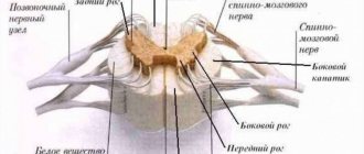
As you can see, I drew the pathways in this lesson very schematically, deliberately omitting very important features - first of all, this concerns the nuclei of the spinal cord. For example, if we were to completely draw a segment of the spinothalamic tract, we would have to draw this in detail:
- The impulse is met by the first neuron, which is located in the spinal ganglion (1);
- Then, along the process of this neuron it enters its own nucleus, which lies in the dorsal horn (2);
- Next, along the neuron process from its own nucleus, the impulse enters the medulla oblongata and follows to the thalamus, where the third nucleus is located.
As you can see, this would create unnecessary clutter, since it would be necessary to add anatomical structures that are not directly related to the medulla oblongata. I will most likely do a separate lesson on pathways, but to do that we need to study all parts of the CNS.
The lines that connect the medulla oblongata and the spinal cord in this lesson very roughly and schematically show the pathways. I drew them so that, while studying the medulla oblongata, you would constantly correlate each new element with the spinal cord. This will help you a lot in the future.