Hemangioma
– a common benign tumor of vascular tissue. Outwardly it resembles a bright crimson or red formation that rises above the skin. Its surface is lumpy and uneven, sometimes it heats up on its own for a short period of time.
Hemangioma in adults cannot provoke any serious disturbances in the functioning of the body, but everything depends on the location of the growth and its size. Hemangioma located on internal organs, such as the kidneys or liver, can cause mechanical compression, which leads to impaired functional activity.
Where does hemangioma appear?
In 80% of cases, hemangioma in adults, a photo of which you can easily find on the Internet, occurs in the form of a single structure; in the remaining cases, several such elements are observed in one place at once. Very rarely, the lesion occurs in the area around the ear.
There are cases when hemangioma occurs on the lip of adults, which causes a lot of inconvenience and discomfort. Also, such lesions can be located in the genital area. This course of the disease is considered the most dangerous, as it is often accompanied by bleeding, infection, and ulceration. Most often, this lesion occurs in the neck or head:
- On the eyelids and around the eyes.
- On the cheeks: both on the outer and inner sides.
- In the region of the nose.
- Near the hairline.
Often vascular hemangioma in adults spreads throughout the body. It can also affect internal organs, which can be detected by ultrasound and computed tomography. If you begin to notice that a hemangioma has begun to grow on your skin, consult a doctor immediately. The sooner you start treatment, the easier and faster it is to get rid of the problem.
Symptoms and signs
Meningioma of the brain (ICD 10 - D32.0) is characterized by relatively slow growth, so it can develop asymptomatically for a long time.
One of the first symptoms is a headache - dull, bursting or aching. It is distinguished by its diffuse nature and localization in the area of the back of the head, forehead or temples.
The occurrence of other symptoms is associated with the localization of the tumor (compression of certain brain structures). Such symptoms are called focal.
Brain meningioma may be suspected if the following symptoms are present:
- paresis of the limbs (severe weakness, decreased sensitivity, the appearance of pathological reflexes);
- loss of visual fields and other visual disorders (decreased visual acuity, double vision). A characteristic symptom is ptosis - drooping of the upper eyelid;
- deterioration of hearing function;
- reduction or complete loss of smell, olfactory hallucinations;
- epileptiform seizures;
- psychoemotional disorders, behavioral disorders - these symptoms most often manifest themselves as meningioma of the frontal lobe of the brain;
- thinking disorders;
- impaired coordination and gait;
- increased intraocular pressure;
- nausea and vomiting that does not bring relief.
If the outflow of cerebrospinal fluid is disrupted, hydrocephalus and cerebral edema occur, as a result of which patients develop persistent headaches, dizziness, and mental disorders.
Causes
To date, doctors have not been able to determine the exact cause of why hemangioma occurs in adults on the arm or any other part of the body.
The nature of the congenital form of the lesion is as follows: during the period of growth, the body produces an excessive amount of connective tissue, which is why it has to become denser. The luminaries of modern medicine do not have the necessary equipment that would allow them to identify hemangioma on the body in adults in the early stages, a photo of which you can easily find on the Internet.
A hemangioma on the leg of an adult, which manifests itself as a bright red tumor, will not grow over time or torment you with unpleasant sensations. It is not known for certain what causes an adult body to manifest such an ailment on the skin. Common causes of hemangioma in the adult population include:
- Hereditary predisposition.
- Environmental influence.
- Diseases of internal organs, in particular the endocrine system.
- Disorders in the cardiovascular system.
- Exposure to sunlight or ultraviolet radiation.
Symptoms of hemangioma in adults
Hemangioma of the skin is quite difficult to recognize in the initial stages. At first, you will be able to notice how your skin begins to gradually turn red, small spots of irregular shape appear on it, which can be easily picked out. Over time, they unite into one large formation with a large tumor point in the center, from which all the small blood capillaries diverge.
It should be noted that hemangioma on the head of an adult develops quite quickly; it grows to its peak state in 2-3 weeks.
There are several types of hemangiomas, which are differentiated by location and manifestation of symptoms. Most often, simple, cavernous, combined and mixed types are distinguished. Only a qualified specialist can determine what has affected your skin. Sometimes a hemangioma can occupy significant areas on the human body, causing serious discomfort.
Treatment
Hemangioma on the skin does not pose any danger to the body other than aesthetic discomfort. However, if such a formation begins to grow, it can lead to some negative consequences, such as infection, blood loss, impaired blood clotting, and an effect on some internal organs. In rare cases, tumors disappear on their own; in others, measures need to be taken.
At the Healthy Skin Center, experienced specialists using modern diagnostic methods will easily select for you an effective and safe treatment that will save you from hemangioma on the skin forever.
At the Healthy Skin Center, you will be offered a radio wave surgery method to get rid of hemangioma. This effect allows you to achieve both clinical and cosmetic effects. After exposure to a demato-oncological magnifying glass, no scars or cicatrices remain on the skin. Under the influence of high-frequency radiation, the affected cells completely evaporate, which protects the treated area of skin from the subsequent development of hemangioma.
Contact the Healthy Skin Center, they will always help you.
Doctor's comment
Does the upcoming removal of a hemangioma cause you many questions? First of all, it is necessary to determine whether there is a risk of injury to the tumor or damage to nearby organs. In some cases, it is possible to use conservative treatment methods: taking hormonal drugs and cytostatics, compression therapy. If the tumor is located near organs and blood vessels, if there is a risk of complications, then removal of the hemangioma in a more radical way is indicated. There are several methods to solve the problem: radiation therapy, diathermocoagulation, sclerotherapy, laser therapy, excision of hemangioma. Which one is right for you can only be determined during a medical consultation and after an examination. Our clinic has the opportunity to undergo all the necessary tests - quickly and without tiring queues. Based on the results obtained, the most suitable treatment method will be selected for you, based on the individual characteristics of your body. Make an appointment and together we will discuss the details of the upcoming treatment. Get rid of this defect quickly and with excellent cosmetic results!
Hemangioma
Hemangioma is a benign vascular tumor. Depending on the damage to the vessels, capillary, arterial, arteriovenous and cavernous forms are distinguished.
- Capillary hemangioma
- Arterial hemangioma
- Arteriovenous hemangioma
- Cavernous hemangioma
Capillary
– presented in the form of spots, slightly protruding above the skin level, of varying diameters, from bright pink to bluish-purple. A variant is telangiectasias (“spider veins”), in which rays – stripes, like cobwebs – diverge from the spot.
Arterial
– is a spot from pale to deep pink, clearly demarcated, with superficial localization a pulsation is detected.
Arteriovenous
The form is distinguished by a large affected area (up to 10 cm in diameter), superficial location, bluish-purple color, tortuous bundles of blood vessels, thinning and easy bleeding.
Cavernous
– develops in newborns and increases as the child grows. It is presented in the form of a single formation, which has a lobular structure and protrudes above the skin level like a “mulberry”, bluish-purple in color. There is no favorite localization; it can spontaneously regress.
Answers to frequently asked questions
What does a hemangioma look like?
Externally, the tumor is a red or bluish spot that merges with the skin or rises above its surface. The size of the spot in diameter ranges from 1-2 cm to 10-20 cm. The tumor has different shapes. In children, temperature asymmetry appears; the defect is always warm to the touch.
Is it necessary to remove the hemangioma?
A skin hemangioma must be removed if it causes discomfort, itches or bleeds. It is necessary to remove the tumor if it is located in difficult places that are constantly rubbed by clothing.
Can a soft tissue tumor go away on its own?
It is not always necessary to remove hemangioma surgically. The tumor can regress on its own. There are several stages in the extinction process. It begins with the formation of a pale spot in the center of the tumor, then the shade changes and becomes normal towards the periphery. The process of disappearance is long, up to several years.
Why is hemangioma dangerous?
If treated incorrectly and untimely, complications may occur that pose a threat to human life and health:
- germination with subsequent destruction of nearby organs;
- destruction of bone and muscle tissue;
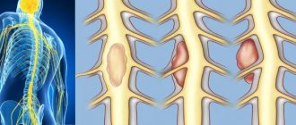
- spinal cord compression;
- ulceration of the tumor, infection;
- malignancy;
- development of pathologies of the vascular system;
- cosmetic defect.
Diagnosis and treatment of hemangioma
Diagnostics: Skin ultrasound, deep dermatoscopy, cytology.
Treatment: removal using radiosurgery.
In our center, tumor removal is performed by a dermato-oncologist using radio wave surgery under local anesthesia.
In order to remove the formation more efficiently, the doctor uses a dermato-oncological magnifying glass, which allows for more detailed and complete removal of skin tumors without leaving pathological cells in the wound. Therefore, after removal, we can say with complete confidence that the formation has been completely removed and you will no longer have it. Read more about tumor removal in our center...
The cost of removing a single hemangioma starts from 900 rubles.
Table of contents
- Etiology and pathogenesis
- Clinical manifestations
- Treatment methods
- Medicinal methods
- Surgical methods
- Hardware methods
Hemangioma (capillary hemangioma, capillary hemangioma) is a benign tumor that develops from hyperplastic vascular endothelium. It is often absent at birth, but appears in childhood and is characterized by progressive growth.
In our company you can purchase the following equipment for removing hemangiomas:
- M22 (Lumenis)
- AcuPulse (Lumenis)
- Fraxel (Solta Medical)
- UltraPulse (Lumenis)
A feature of hemangiomas is the possibility of spontaneous involution, therefore the treatment tactics for these neoplasms are selected individually for each patient, based on the current state of the tumor and its dynamics. Spontaneous regression distinguishes hemangiomas from other vascular malformations, such as port-wine stains.
According to statistics, capillary hemangiomas occur in 1–2% of newborns, with about 50% of neoplasms forming on the head (often in the eye area) and neck. 30% of patients say that their parents or doctors recorded signs of hemangioma immediately after birth, but the majority note the onset of manifestation of this neoplasm at the age of 6 months. The ratio of men to women in the incidence of hemangioma is 1:3.