The medulla oblongata is localized in the posterior part of the brain and connects it with the spinal part of the central nervous system. Formed from the structures of the rhombencephalon and is part of the brainstem. Controls the primary processes of the body - breathing and blood circulation.
Since the medulla oblongata connects the cerebrum and the spinal part of the central nervous system, it combines the functions and structure of both structures.
Brain in section
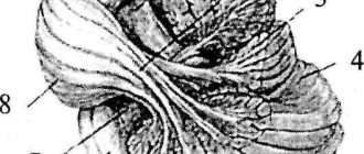
Structure of the medulla oblongata
The oblongata section is located at the junction of the spinal and cephalic sections of the central nervous system. Anatomically, it is located in the area of exit of the roots of the 1st cervical spinal nerve, and is limited from above by the bridge. In appearance it resembles an onion. Its height in an adult is approximately 2-3 cm.
Despite its relatively small size, it performs many functions without which the individual could not exist.
Its structures consist of white and gray matter, which determines its dual functions - it combines the characteristics of the spinal and telencephalon with minor differences: its internal part consists of compactly located clusters of gray matter, and the external part is made of white matter. From the back of the oblong section, 2 cords stretch to the dorsal section, which unite them.
As you know, the cerebral hemispheres control opposite sides of the body: the left side responds to the right side and vice versa. This feature of the central nervous system is achieved due to the characteristic structure of the medulla oblongata: it implements a conductor function, which consists of transmitting excitation impulses from its own nuclei to the nervous tissue of other parts, but not in a direct way, but crosswise.
The size and structure of the medulla oblongata change with age. For example, unlike adults, in infants this part of the central nervous system is larger relative to other parts. It acquires its final appearance by the age of 7 years.
Location boundaries
External structure
The structure of the medulla oblongata is divided into 2 parts: internal and external. Let us take a closer look at the appearance of this department and the features of its structure from an anatomical point of view.
The medulla oblongata is an inverted cone: its wide end is at the top, the narrow end is at the bottom. In the central part of the front side there is an anterior median fissure. It is a continuation of the central sulcus of the dorsal part of the central nervous system.
On the sides of it there are pyramids that pass into the facial cables of the spinal cord, consisting of a cluster of nerve cells. On the posterior side of the medulla oblongata, the posterior middle sulcus is visible, which passes into the dorsal region.
Detailed structure
Next to it are the caudal cords, which are connected to the grooves of the spinal part of the central nervous system. These cords are the lower boundary line of the oblong section, and the basal boundary, passing in the area of the Varoliev bridge, has become its upper boundary.
Internal structure
The internal structure of the medulla oblongata is more effectively viewed in a cross section made at the level of the olives. If you look at it from the front side, the anatomy looks like this: tracts of intentional motor impulses extend from paired elevations (pyramids) on the outer side into the depths of the structure.
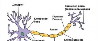
Next to them are olives, covering the basal ganglia, the roots of the hypoglossal nerve going to the lingual muscles, axons and nerve cells grouped into nuclei. Long fibers pass from each nucleus to the cerebellum, connecting them to each other. They form the olivocerebellar tract.
Near the olive tree there are also:
- Fibers of the glossopharyngeal nerve;
- Vagus nerve fibers;
- Accessory nerve fibers.
The efferent conduction tracts lie on the lateral (side) side of the medulla oblongata:
- Rubrospinal;
- Tectospinal;
- Reticulospinal;
- Vestibulospinal.
At the level of the pyramids, most of the axons (up to 80%) that form the efferent pathways intersect with each other. The rest also form a decussation and move down to the other side, to the spinal part of the central nervous system.
In the posterior part of the medulla oblongata, 4 sensory tracts stretch from the spinal cord to the upper parts of the central nervous system:
- Thin;
- Wedge-shaped;
- Spinothalamic;
- Spinocerebellar.
They are a natural continuation of similar structures of the spinal cord.
Structure
The medulla oblongata consists of white and gray matter, like the entire brain. The structure of the medulla oblongata can be divided into internal and external. The lower border ( dorsal ) is considered to be the exit point of the roots of the first cervical spinal nerve, and the upper border is the pons of the brain.
External structure
Externally, an important part of the brain looks like an onion. It measures 2-3cm. Because This part is a continuation of the spinal cord, then this part of the brain includes the anatomical features of both the spinal cord and the brain.
Externally, one can distinguish the anterior midline, which separates the pyramids (a continuation of the anterior cords of the spinal cord). Pyramids are a feature of human brain development, because they appeared during the development of the neocortex. In younger primates, pyramids are also observed, but they are less developed. On the sides of the pyramids there is an oval extension “olive”, which contains the cores of the same name. Each nucleus contains the olivocerebellar tract.
Internal structure
The nuclei of gray matter are responsible for vital functions:
- Olive nucleus - connected to the dentate nucleus of the cerebellum
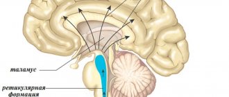
- Reticular formation - regulates contact with all sense organs and the spinal cord
- Nuclei of 9-12 pairs of cranial nerves, accessory nerve, glossopharyngeal nerve, vagus nerve
- Circulatory and respiratory centers that are associated with the nuclei of the vagus nerve
Long pathways are responsible for communication with the spinal cord and neighboring sections: pyramidal and the pathways of the wedge-shaped and thin fasciculi.
Functions of the medulla oblongata centers:
- Locus coeruleus - axons of this center can release norepinephrine into the intercellular space, which in turn changes the excitability of neurons
- Dorsal nucleus of the trapezius body - works with auditory apparatus
- Nuclei of the reticular formation - affects the nuclei of the cerebral and spinal cord through excitation or inhibition. Forms vegetative centers
- Olive kernel - is an intermediate center of balance
- Nuclei 5-12 pairs of cranial nerves - motor, sensory and autonomic functions
- The nuclei of the cuneate and gracilis fasciculus are associative nuclei of proprioceptive and tactile sensitivity
Main nuclei of the medulla oblongata
The gray matter of the medulla oblongata processes data arriving in the central nervous system from all receptors of the human body. It is a cluster of neuron structures (bodies and partly processes), compactly located in the thickness of the white matter.
Highlight:
- Nuclei of 9-12 pairs of cranial nerves;
- Circulatory and respiratory centers;
- Olive kernel;
- Reticular formation.
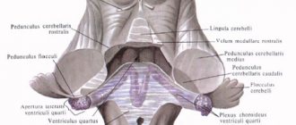
In the medulla oblongata there are 5 pairs of cranial nerve nuclei. They are concentrated in the caudal part of the cerebrum below the bottom of the 4th ventricle:
- Vestibulocochlear nerve (VIII pair). Consists of the cochlear and vestibular parts. Responsible for conducting auditory impulses, as well as signals originating from the vestibular part of the inner ear.
- Glossopharyngeal nerves (IX pair). Includes sensory, motor and secretory fibers. Motor axons implement motor function - they control the activity of the muscles of the pharyngeal canal and oral cavity. With the help of sensory fibers, the nucleus receives information received from the food-gustatory sensory system of the back of the tongue. Parasympathetic fibers are responsible for the secretory function in the oral cavity: they control salivary processes.
- Vagus nerve (X pair). Consists of 3 types of nuclei: vegetative, sensory and motor. The autonomic nucleus functions as a connecting link between the cerebrum and the organs of the head, neck, chest and abdominal cavity. Incoming and descending fibers extend from it in different directions. The sensitive core receives information from the lungs, heart and other important systems of the body. The motor nucleus controls contraction of the oral muscles during swallowing. The reciprocal nucleus is also located here, the axons of which perform a protective function - they activate the processes of coughing, sneezing, and vomiting.
- Accessory nerve (XI pair). It is represented by 2 nuclei: one lies in the medulla oblongata at the bottom of the rhomboid fossa, the other in the anterior horn at the level of 1 segment of the spine. Fibers from it enter the foramen magnum of the skull, unite with the cranial roots, forming the trunk of the accessory nerve. They are then brought out from the cranial cavity through the jugular foramen. The accessory nerve performs a controlling function - it is responsible for the motility of the sternocleidomastoid and trapezius muscles.
- Hypoglossal nerve (XII pair). Controls the motor function of the tongue, as it is associated with its styloglossus, mentalis, rectus and transverse muscles. Unconditioned reflexes depend on its work: swallowing, chewing, sucking. The nuclei of the hypoglossal nerve control the motor skills of the tongue during chewing, speaking, and other mouth movements. Information from the hypoglossal nerve is sent to the sensorimotor region of the cerebral cortex through the sphenoid and tender nucleus.
The hemodynamic center is located in the lower caudal part of the oblongata. Its fibers are closely connected with the nuclei of the V pair of nerves. Scientists suggest that it is in the hemodynamic center that excitatory activating signals of sympathetic fibers to the cardiovascular system arise.
Core kernels
Sometimes the hemodynamic center is suppressed by shock, trauma, stroke, poisoning, and metabolic disorders.
Olive kernel. It is a serrated plate, the functions of which are determined by its structure - it consists of gray matter. Curved in the shape of a horseshoe. Interacts with the dentate nuclei of the cerebellum, that is, they are the main structures of balance.
The olivary kernel is the best developed of all mammals in humans, since humans, more than anyone else in the animal world, are in an upright position and need a good vestibular apparatus. In addition, the olive nuclei help to navigate in space: they compare the volume of sound waves penetrating the right and left ear. Analyzing them, a person turns to face the source of the sound.
In the lower part of the medulla oblongata there is the so-called “blue spot” - the zone of the reticular formation. The fibers of neurons of this structure secrete a specific hormone that affects the degree of excitability of nerve cells.
The “blue spot” is responsible for feelings of anxiety and internal tension, and also ensures the department’s connection with the sensory organs, the spinal cord and other departments, serving as a regulator of nervous activity.
What are the functions of the medulla oblongata
The functions performed by the human medulla oblongata are important for the body. Without this part of the brain, a person cannot live, since it controls the vital organs of the body - the lungs and heart muscle. Therefore, when this cerebral part of the central nervous system is injured, instant death occurs. Nuclei - accumulations of gray matter - are responsible for all this activity.
Table of functions of the nuclei of the medulla oblongata:
| Structure name | Tasks it performs |
| Nuclei of the V-XII pair of cranial nerves | Sensory, motor, autonomic functions of the hindbrain |
| Gray matter of the gracilis and cuneate fasciculus | Responsible for tactile and proprioceptive sensitivity |
| Olive kernels | Are part of the center of balance |
| Dorsal nucleus of the trapezius | Participate in the analysis of auditory information |
| Gray matter of the reticular formation | Depending on the situation, they activate or inhibit neurons of the spinal cord and the cerebral cortex of the central nervous system, forming autonomic centers |
| blue spot | The structures are capable of releasing a specific hormone that affects the excitability of individual zones of the cerebrum cortex |
Main functions
Thus, the functions of the human medulla oblongata are divided into several categories:
- Implementation and control of unconditioned reflexes (reflex function);
- Motor skills of protective and orienting actions (sensory function);
- Automaticity of the process of breathing and blood circulation (integrative function);
- Maintaining a balance of torso position and muscle tone (conductive function).
Sensory
The sensory department controls and processes the reception of incoming impulses that come to it from receptors throughout the human body. These receptive structures consist of 2 units:
- Sensoepithelial cells;
- Axons of afferent neurons.
There, for example, impulses from the respiratory center are processed - blood counts and the structure of the lung tissue are assessed. Based on the results of these data, blood saturation with oxygen and metabolic processes in the body subsequently occur. The sensory section of the medulla oblongata receives and transmits impulses from the digestive organs, hearing, vision, and tactile points for further processing.
The medulla oblongata perceives tactile information using the following structures:
- When facial skin receptors are activated, the neurons of the trigeminal nerve are excited;
- Taste receptors - glossopharyngeal nerve;
- Sound receptors – cochlear nerve;
- If stability is violated - olives.
Based on the data obtained, a further reaction of the body follows, which is expressed in the implementation of unconditional reflex reactions. They are also carried out by the medulla oblongata of the central nervous system. With insufficient oxygen saturation in the blood, a person begins to breathe deeply and look for a source of fresh air, thereby activating the motor function of the department.
Conductor
The oblongata section is the connecting link between the dorsal and terminal sections of the central nervous system. Afferent and efferent tracts pass through it: corticospinal, spinothalamic, rubrospinal.
With their help, information is transmitted to the higher structures of the cerebrum and returned back to the organs in the form of impulses. The olivospinal, vestibulospinal and reticulospinal tracts begin here. Through them, muscle tension and coordination of movements of the arms and legs are stabilized.
In the medulla oblongata, the corticoreticular tracts from the cortex, as well as the upward spreading fibers of proprioceptive susceptibility from the spinal cord, end. This feature allows you to feel parts of your own body relative to each other in space.
Integrative
The integrative function of the medulla oblongata is to carry out body reactions unrelated to reflex activity. Their appearance is associated with the work of terminal neurons, which are programmed to carry out regulatory processes in a certain order. They are implemented through other NS centers or directly.
The movements of the eyeballs when the head is tilted are realized on the basis of the joint activity of the nuclei of the vestibular and oculomotor systems of the brain. Some of the nerve cells of the reticular substance of the medulla oblongata control the tone and coordination of other nerve centers of the cerebrum.
Psychophysical development of children in senior preschool and primary school age.“A well-built brain is worth more than a well-filled brain.”
(M. Montaigne)
In older preschool and primary school age, there is rapid development and restructuring in the work of all physiological systems of the child’s body: nervous, cardiovascular, endocrine, musculoskeletal. At this age, the body develops very intensively. The child quickly gains height and weight, and body proportions change.
During the first seven or eight years, all internal organs enlarge and their functions improve. Significant changes in higher nervous activity occur. In terms of its characteristics, a child’s brain at this age is more similar to that of an adult’s brain. During this period, the body indicates its readiness to move to a higher stage of age-related development, which involves more intense mental and physical stress.
It is during these years that the child becomes the most mobile and resilient, and his movements become more coordinated. Children have high motor activity, they are better at movements that require speed and flexibility, but their strength and endurance are still low. Motor activity has a great influence on the formation of the psychophysiological status of the child: it stimulates and develops perception of the world, memory, intellectual processes, rhythmic movements, trains the pyramidal system (supports complex and fine coordination of movements) and the extrapyramidal system (participates in the control of movements, maintains muscle tone and postures) person). It is also very important that at this age the formation of posture and the arch of the foot occurs. During this period, it would be very useful and correct to practice: children walking barefoot, performing special exercises, massage, water procedures. In older preschool age, against the background of general physical development, mobility, balance, stability of nervous processes improve, a health reserve accumulates: the frequency of diseases decreases, they proceed relatively easily, most often without complications.
In a child under 7-8 years old, the right hemisphere dominates, which is responsible for imaginative perception, the emotional sphere, processing in the brain of a holistic picture based on images, movement, rhythm, emotions, intuition, external speech, integrated thinking, etc. Therefore, children need to be taught through play, the development of body coordination, musical and motor rhythm.
By the age of 8, the frontal lobes are formed in children, which leads to the improvement of fine motor skills (holding a pen correctly, the quality of writing improves), and the development of internal speech. Development and coordination of eye movements: tracking and focusing
And by the age of 9, the left hemisphere is formed, which is responsible for speech, logical thinking, detailed and linear processing of information, improving the skills of speech, reading and writing, counting, drawing, dancing, and music perception.
Changes also occur in emotional development: a sense of humor develops, children themselves come up with simple jokes and repeat them with pleasure again and again.
The child subordinates his behavior to rules. In joint activities, information exchange, planning, division and coordination of functions are carried out.
Strives for contacts, shows goodwill in communicating with adults and peers, adequacy in behavior, and emotional responsiveness. Shows interest in learning new things (he himself finds information, games, and methods of action that are interesting to him).
To raise a healthy and happy child, use the recommendations that will help you.
1 Raising right-hemisphere and left-hemisphere children, boys and girls, must be carried out using various methods
2 Early symbolic voluntary learning (numbers, letters) is unacceptable. It promotes the formation of minimal brain dysfunction.
3 Children under 7 years of age should be raised and taught as right-brained, as this is appropriate for their age stage of development
4 An immobile child does not learn. Any new information should be reinforced by movement (speaking out loud, writing down or sketching on paper, fingering a rosary, etc.), and be emotionally charged.
5 Girls can be capricious due to fatigue (exhaustion of the right “emotional” hemisphere). In this case, boys are depleted of information (decreased left rational-logical hemisphere).
6 A child’s laziness is often a signal of trouble. In pedagogical activities, the methods of working with this child were incorrectly chosen.
7 Children coordinate movements better and are very mobile, but at the same time children still quickly get tired, “exhausted” and when overloaded, protective inhibition occurs. Therefore, you should not give your child very intense exercise. Alternate moments of activity and calm, thereby protecting your child from nervous tension
8 At this age, children become more independent and are not afraid to break away from their parents. Encourage independence in children.
9 During this period, it is very important that basic trust in the world, autonomy and initiative in children’s independent activities be formed.
10 Your children still need physical closeness and support from their parents.
Teacher-psychologist Kazakevich E.N.