Useful articles
Spinal stroke is a severe pathology caused by an acute disruption of blood flow to the spinal cord. It often becomes a cause of disability, especially if the patient is not provided with medical care in a hospital setting. Recovery after a spinal stroke can be quite lengthy.
Causes
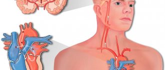
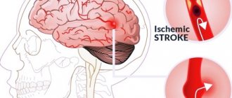
Ischemia is a cessation of blood flow due to complete or partial blockage of blood vessels. Cerebral and spinal ischemia have a similar mechanism of development.
Disturbance of the spinal blood supply occurs for the following reasons:
- Vascular lesions of the spinal cord (congenital vascular malformations, coarctation of the aorta, atherosclerosis, thromboembolism).
- Compression of spinal vessels from the outside (compression of herniated intervertebral discs, enlarged lymph nodes, tumor, fragments of an injured vertebra, pregnant uterus).
- Iatrogenic causes, that is, caused by medical actions (paravertebral blockades, epidural anesthesia, rough manual therapy techniques).
Diastematomyelia (a type of tethered spinal cord syndrome)
Hidden malformation of the spine and spinal cord characterized by the formation of a bone, cartilaginous or fibrous septum or cord emanating from the arches, vertebral body and separating the spinal cord completely or partially into two halves (hemichords). The reason for this anomaly is a disruption of the gastrulation process (the laying of the ecto, meso, endoderm) from 13 to 16 days after ovulation, which leads to the formation of diastematomyelia or diplomyelia. This pathology is more common in women than in men (5:3). According to the Pang classification, two forms are currently distinguished: Type I - two hemichords are located in their own dural sacs, separated by an extradural bone spine or cartilaginous bridge; Type II - both halves of the spinal cord are located in a single dural sac, separated by an intradural fibrous cord. Both types of spinal cord bifurcation involve dorsal and rarely ventral, paramedian, nonfunctional aberrant nerves arising from the medial surface of the hemichords and penetrating the septum. A feature of septal anomaly is the inclusion of abnormal vessels and vestigial nerves. In the vast majority of cases, diastematomyelia is located in the lower thoracic - lumbar spine. Diastematomyelia in 40–50% of cases is combined with myelomeningocele, dermal sinus, and thickened filum terminale. In 85% there was a combination with spinal defects (segmentation of the vertebrae, hypoplasia of the vertebral bodies).
Clinical picture of diastematomyelia
The key factor in the development of symptoms of the disease is the fixation of spinal cord structures by bone and cartilaginous tumors of the spinal canal. The period of asymptomatic disease and the manifestation of neurological symptoms depends on the anatomical features of the defect and the growth rate of the child. In most cases, primary neuroorthopedic symptoms appear between 3.5 and 7 years of age. The symptom complex of spinal cord duplication forms a triad of syndromes - skin, orthopedic, neurological. The syndrome of skin developmental stigmas in this form of dysraphism is a pathognomonic manifestation and is represented by local hypertrichosis (increased hair growth) in the area of formation of the osteocartilaginous septum with a frequency of 65–90%, often combined with dermal sinuses and capillary hemangioma. Local hypertrichosis in the projection of the thoracic and lumbosacral spine during the asymptomatic period should prompt a diagnostic search. Suffering of the segmental and conductive apparatus of the spinal cord during fixation by the bone septum and, as a consequence, tension during the growth of the child, leads to the development of neurological symptoms. The main symptoms are moderate paraparesis, mainly in the feet and asymmetry, which is associated with the severity of damage to one or another half of the spinal cord. Hypotrophy of all muscles of the affected limb, with rare sensory disturbances. In advanced cases and the rapid growth of the child, dysfunction of the pelvic organs occurs, from episodes of urinary incontinence to complete retention. Orthopedic disorders of the axial skeleton (scoliosis of the spine, abnormalities of the vertebral bodies and arches, shortening and deformation of the feet, shortening of the lower extremities) on the one hand, act as a concomitant pathology of the skeletal system, and on the other hand, is a consequence of neurogenic dysfunction of muscle innervation. Timely detection of pathology makes it possible to prevent further deterioration of skeletal deformity and the possibility of conservative correction.
Diagnosis of diastematomyelia
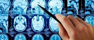
The imaging method for diastematomyelia is MSCT and MRI of the spine. These methods complement and clarify the relationship between bone and neural structures in the spinal canal, identifying concomitant and indirect signs of spinal dysraphism. According to MRI data, diastematomyelia in 70% of cases is combined with a thickened filum terminale, the development of syringomyelia was detected in 30-40%, and is often combined with a dermal sinus. MSCT is used to visualize the features of the bone spine in the spinal canal and accompanying bone pathology with the possibility of 3D reconstruction. Complex visualization and image analysis makes it possible to develop optimal and safe surgical treatment tactics.
Surgical treatment of diastematomyelia
A diagnosed bone spine dividing the spinal cord and spinal canal into two halves is a direct indication for surgical treatment in both asymptomatic patients and in patients with mild neurological and orthopedic problems. The use of an operating microscope, microinstruments, and an ultrasonic bone scalpel with neurophysiological control makes it possible to perform resection of the bone septum with complete safety. The operation is performed using economical skin incisions, with minimal trauma to soft tissues. If it is necessary to restore normal anatomical parameters of the spinal canal, modern plastic materials are used. Timely correction prevents the development of neurological deficits and creates conditions for the restoration of lost functions.
Clinical manifestations
Spinal ischemia or ischemic spinal stroke occurs equally frequently in men and women aged 40 years and older.
The clinical picture of ischemic spinal cord stroke is varied. Depending on the level at which the stroke occurred, certain symptoms will appear. For example, a circulatory disorder in the upper cervical level causes a sharp disorder of movements in the arms and legs, and respiratory failure with symptoms of spinal shock may develop. Below the level of ischemia, hyposthesia also occurs, loss of all types of sensitivity: temperature, tactile, pain.
When the lesion is localized in the cervical region, flaccid paralysis of the arms and spastic paresis of the legs develop.
The lumbar region of the spinal cord is most often affected. Ischemia of this zone leads to paralysis of the legs with dysfunction of the pelvic organs.
With any form of spinal stroke, bedsores and trophic disorders develop.
By analyzing the symptoms, it is possible to determine which vascular basin has been damaged, or which artery has lost patency.
The rate of development of the listed symptoms is different; the pathological process can develop suddenly or gradually over the course of a day. Sometimes an acute disorder of spinal circulation is preceded by warning symptoms: pain in the spine, intermittent claudication, transient paralysis.
During the course of the disease, neurologists distinguish the following stages:
- period of harbingers;
- stage of acute stroke;
- symptom regression stage;
- stage of residual (residual) phenomena.
Ischemic spinal stroke
Men and women get sick with equal frequency between the ages of 30 and 70 years and older.
The course of the disease can be divided into several stages:
- stage of harbingers (distant and close);
- stage of stroke development;
- stage of reverse development;
- stage of residual effects (if complete recovery has not occurred).
Precursors of ischemic spinal stroke are paroxysms of transient spinal disorders (myelogenous, caudogenic or combined intermittent claudication, transient pain and paresthesia in the spine or in the projection of the branching of certain spinal roots, disorders of the functions of the pelvic organs).
The rate of occurrence of a stroke varies - from sudden (with embolism or traumatic compression of the vessels supplying the spinal cord) to several hours or even days.
It has already been mentioned that spinal infarction is often preceded by pain in the spine or along individual roots.
It is typical for this pain to cease or significantly subside after the development of myelischemia. This occurs due to an interruption in the passage of pain impulses through sensitive conductors at the level of the focus of spinal cord ischemia.
Clinical picture
ischemic spinal stroke is very polymorphic and depends on the extent of ischemia both along the length and across the diameter of the spinal cord. Depending on the extent of ischemia across the diameter of the spinal cord, the following variants of the clinical picture occur.
Ischemia syndrome of the ventral half of the spinal cord
(
anterior spinal artery occlusion syndrome, Preobrazhensky syndrome
).
It is characterized by the acute development of paralysis of the limbs, dissociated paraesthesia, and dysfunction of the pelvic organs. If ischemia is localized in the cervical segments of the spinal cord, paralysis (paresis) develops in the arms, flaccid, in the legs - spastic. Ischemia of the thoracic segments is manifested by lower spastic paraparesis, myelischemia of the lumbosacral localization is manifested by lower flaccid paraparesis. The upper limit of dissociated paranesthesia helps to navigate the extent of the ischemic focus along the length of the spinal cord. Articular-muscular and tactile sensation is not impaired. Ischemia of the lumbosacral thickening is manifested by lower flaccid paraplegia with areflexia, dissociated paraanesthesia, urinary and fecal retention. This symptom complex is called Stanilovsky-Tanon syndrome.
Anterior ischemic poliomyelopathy syndrome
is one of the variants of partial damage to the structures of the ventral half of the spinal cord. It is characterized by the rapid development of flaccid paresis of certain muscle groups of the upper or lower extremities with areflexia and muscle atrophy and changes in EMG indicating ischemia within the anterior horns of the spinal cord. This syndrome has to be differentiated from polio, in which signs of infection and the stage of gastrointestinal disorders are revealed.
Ischemic Brown-Séquard syndrome
Occurs occasionally. It differs from a typical compression lesion of half of the spinal cord in that during ischemia the posterior cords remain preserved, so joint-muscular sensation on the side of the central limb paralysis is not impaired. The anatomical validity of this variant of myelischemia has already been mentioned; it is due to the fact that individual sulcal-commissural arteries supply only one, right or left, half of the diameter of the spinal cord.
Centromedullary ischemia syndrome
It is characterized by acute or subacute development of segmental dissociated anesthesia with the loss of corresponding segmental deep reflexes and mild peripheral paresis of the same myotomes. The clinical picture resembles syringomyelia ( ischemic syringomyelic syndrome
).
Ischemia syndrome of the marginal zone of the anterior and lateral cords
It manifests itself as spastic paresis of the limbs, cerebellar ataxia and mild conductive parahypesthesia. The acute onset of the disease and the subsequent possibility of an intermittent course resemble the spinal form of multiple sclerosis. Diagnosis is aided by monitoring the further development of the disease.
Ischemic amyotrophic lateral sclerosis syndrome
Most often develops in the upper arterial basin of the spinal cord. The clinical picture is characterized by weakness of the distal parts of the upper extremities, atrophy of the small muscles of the hands, increased deep reflexes, and pathological hand and foot signs. Fascicular twitching of the muscles of the shoulder girdle is possible. With this syndrome, there is no spread of paretic phenomena to the bulbar muscle group (tongue, larynx and pharynx).
Ischemia syndrome of the dorsal part of the spinal cord (Williamson syndrome)
It is rare and is associated with occlusion of the posterior spinal artery. In such patients, sensitive ataxia acutely appears in one, two or more limbs, moderate spastic paresis of the same limbs, segmental hypoesthesia indicating the level of ischemia, vibration sensitivity in the legs is lost.
Spinal cord transverse ischemia syndrome
It develops when the large radiculo-medullary artery, which is involved in the formation of both the anterior and posterior spinal arteries, is turned off. Almost always, such a topography of the lesion is observed when the venous outflow from the spinal cord is disrupted (thrombosis or compression of the spinal and radicular veins). Details of the clinical picture vary depending on the level of involvement (cervical, thoracic or lumbar segments).
Knowledge of typical variations in the distribution of the radicular spinal arteries in some cases makes it possible to clinically determine the affected area of such an artery. Let us give a brief clinical picture of myelischemia when individual spinal arteries are switched off.
Occlusion syndrome of the large anterior cervical radiculo-medullary artery (cervical thickening artery)
It manifests itself as flaccid or mixed paresis of the upper extremities and spastic lower extremities, segmental and conduction sensitivity disorders, dysfunction of the pelvic organs of the central type.
When turning off the superior accessory radiculo-medullary artery
lower paraparesis and dissociated paraesthesia with an upper border on the ThI-LIV segments develop acutely. Urinary retention occurs. Initially, the knee and Achilles reflexes usually fade. However, Babinski's sign is always caused. Over the next 5-6 days, lower paraparesis acquires the features of central paraparesis (muscle tone increases, deep reflexes are revived). Sensory disorders are usually concentrated in the area of the upper thoracic dermatomes. In the residual stage, along with signs of damage to the ThI-ThIV segments, extinction of deep reflexes in the hands and wasting of small muscles of the hands are sometimes observed. Mild signs of peripheral motor neuron damage are confirmed by electromyography. These symptoms can be considered as distant symptoms.
Adamkiewicz artery shutdown syndrome (lumbar enlargement artery)
The clinical picture can be quite varied. It depends on the stage of the disease. In the acute phase of a stroke, flaccid lower paraparesis (paraplegia), dissociated or rarely total paraanesthesia with an upper limit varying from the ThI-SI segment is always detected. The function of the pelvic organs (incontinence or retention of urine and feces) always suffers. Bedsores often quickly develop.
Treatment
Treatment of ischemic spinal stroke is carried out taking into account the causes of the pathology. The following measures are common to all types of circulatory disorders:
- Hospitalization in a specialized neurology department, bed rest;
- Prevention of bedsores and trophic disorders;
- Regular emptying of the bladder;
- Maintaining basic life functions;
- Therapeutic exercise, massage of affected limbs.
Drug therapy for spinal cord ischemia is similar to the treatment for cerebral stroke:
- Neuroprotectors - Gliatilin, Cerakson, Actovegin, Mexidol.
- Drugs that improve blood supply to the spinal cord - Cavinton, Sermion, Instenon.
- Medicines to thin the blood and prevent blood clots - Trental, Curantil.
- Anticoagulants – Fraxiparin, Heparin.
- Vitamins, antioxidants.
If there is no effect from conservative therapy or if the cause of the stroke is compression by a tumor or hernia, neurosurgical intervention cannot be avoided.
Acute spinal circulatory disorders are easier to prevent than to cure. Prevention of the disease consists of timely treatment of chronic diseases (atherosclerosis, arterial hypertension), osteochondrosis, and spinal injuries.
Functional neurosurgery
Neuromodulation is a therapeutic change in the functions of the nervous system using non-destructive electrical and neurotransmitter effects. Currently, neuromodulation belongs to high-tech medicine and is actively used to help patients.
The most common syndrome treated with neuromodulation techniques is spasticity syndrome. The onset of spasticity at an early age leads to impaired motor development and the formation of myogenic and subsequently fixed contractures. Clinical signs include: – muscle tension and limitation of movement – difficulty in carrying out activities of daily living (including walking, eating, self-care) – development of unexpected and painful muscle spasms Severe spasticity in particular can: – interfere with adequate and comfortable life activities – leads to loss of independence due to the inability to independently carry out daily activities - reduces the quality of life of the patient and his loved ones and other caregivers - interferes with effective motor control The most common diseases in which spasticity syndrome is observed are cerebral palsy syndrome (CP), consequences of a traumatic brain injury, consequences of an intracranial hemorrhage. In the treatment of spasticity today, an integrated approach is used, including the use of conservative therapy, rehabilitation, orthopedic surgery and neurosurgical treatment. The goal of treatment is to reduce spasticity, correct disorders of the musculoskeletal system and form the correct motor stereotype. According to international studies, conservative therapy is not effective enough to adequately correct spastic syndrome.
Neurosurgical medications used in the treatment of spasticity: - Intrathecal baclofen therapy (ITB) - Chronic electrical stimulation of the spinal cord
Intrathecal baclofen therapy A method of continuously injecting baclofen directly into the spinal cord spaces using a special pump.
The programmable infusion system is designed to continuously deliver precise doses of medications to people requiring long-term treatment for muscle spasticity. Such infusion therapy will not cure the underlying disease, but will help cope with its symptoms. This method allows you to reduce the total dose of the drug and thereby reduce side effects from oral Baclofen therapy. Currently, intrathecal baclofen therapy is the only treatment option for spastic hemiparesis and tetraparesis.
The implantation procedure consists of two stages: 1. Conducting a screening test The screening test consists of a single endolumbar injection of Lioresal and assessment of changes in muscle tone after administration. If the result is positive, the next step is implantation of the infusion system 2. Implantation of the infusion system The system consists of: - drug pump (volume 20 or 40 ml depending on the physique and age of the patient)
An implantable pump is a battery-powered device that supplies a prescribed drug and is completely implanted under the skin. There are 2 pump sizes: 20 ml and 40 ml, which corresponds to the amount of medication that the pump can hold. In the center of the pump there is a raised area called a diaphragm through which the doctor injects medication to fill the pump. The diameter of the pump is 7.62 cm, and its thickness is 2.5 cm. - a catheter directly inserted into the lumbar space
A catheter (tube) is connected at one end directly to the pump, and the other end is inserted into the spinal canal. A constant dose of medication is slowly and continuously delivered through a catheter directly to the spinal cord. - programmable device.
The control device is a portable, battery-powered computer that allows the physician to adjust the dose of the drug and control the operation of the pump via wireless radio communication.
The pump is manufactured using advanced technologies, thanks to which the operating life of this device with precise administration of the drug reaches approximately 8 years. When the batteries run out, the pump must be surgically replaced. Based on the amount of drug remaining in the pump reservoir, as well as the programmed delivery rate, the pump informs your healthcare provider of the recommended refill date. In addition, the pump signals a decrease in reserve. The pump is implanted during surgery under general anesthesia. This procedure takes from 1 to 2 hours. The pump is placed under the skin in the lower abdomen
When the pump is first implanted, and each time it is refilled, the pump will notify your physician of the recommended next refill date. Depending on the capacity of the pump, the concentration of the Lioresal solution and the programmed rate of administration of the drug, the interval between repeated fillings can be from 1 to 8 months - on average 2-3 months. Refill the pump several days before the expected date of the end of the drug supply - this will ensure continuous relief of symptoms. Refilling your pump is as easy as giving an injection. The whole procedure takes 10-15 minutes. Your doctor or nurse inserts a needle through the skin and membrane into your MEDSTREAM pump to remove any remaining drug from the pump. You will feel a slight prick as the needle passes through the skin. The doctor will then connect a syringe with fresh medication to the already installed needle and fill the pump reservoir. When refilled, the tank expands and the next working cycle starts.
It is very important to keep a schedule of appointments with your doctor so that the drug in your pump does not run out. If you are unable to keep your appointment, contact your doctor as soon as possible to reschedule your appointment.
Electrical stimulation of the spinal cord Chronic electrical stimulation of the spinal cord is based on the effect of high-frequency electric current on the processes of conducting nerve signals through the posterior structures of the spinal cord. As a result, a blockade of the pathological reflex occurs, which is involved in maintaining increased muscle tone, and as a result, spasticity decreases. Stimulation is carried out using an implantable system consisting of electrodes and a neurostimulator. The indication for the use of this technique is spastic lower paraparesis.
The neurostimulation system consists of two main implanted parts: - neurostimulator - electrode
A neurostimulator is an isolated device consisting of a battery and electronics. It is implanted subcutaneously and produces the electrical impulses needed for stimulation. The impulses are carried through extension cables and electrodes into the spinal cord. Electrodes – an electrode is a thin wire with contacts at the tip. For stimulation, electrodes are implanted in the epidural space (the space between the vertebra and the dura mater). When the neurostimulation system is turned on, it generates electrical impulses that are carried through extension cords and electrodes to the corresponding segments of the spinal cord. Non-implantable components of the system: - doctor programmer - patient console - charger for recharging the neurostimulator batteries. Clinician Programmer Used to program the stimulator. The parameters of the generated pulses can be non-invasively changed. The programmer transmits the settings to the neurostimulator remotely.
Patient control panel. A portable device that allows the patient to turn the neurostimulator on/off when necessary or to check the battery charge status.
The operation to install a neurostimulator consists of three parts: electrode implantation, test stimulation, and neurostimulator implantation. 1. Implantation of electrodes
2. Test stimulation. Rg, CT or MRI control.
3. Neurostimulator implantation. Selection of stimulation program To effectively reduce muscle tone, in most cases, a periodic stimulation mode is used: 3–6 times a day for 10–60 minutes. Electrical stimulation parameters are selected individually. Expected results for surgical treatment of spasticity:
Reduced muscle tone in the legs and specific muscle groups Improved self-care Improved gait Improved functional capacity and independence Reduced pain associated with spasticity Improved mobility Improved sleep Increased participation in rehabilitation activities Prevented or reduced the risk of spasms and contractures Facilitated hygiene care Ease of care patients
Discussion
When determining the indications for patients with FMS to undergo operations that eliminate spinal cord fixation, they proceed from the concept of reversibility of neurological deficit and improvement of the prognosis of the disease when restoring the mobility of the spinal cord by surgically eliminating the factors causing deformation and stretching of its caudal parts and cauda equina roots [5].
In our series of observations, stabilization of the condition after surgery was achieved in all cases, and regression of clinical manifestations was noted in a significant proportion of patients. Since FMS is considered a dynamic condition in which neurological deficits tend to progress, stabilization of the condition can be considered a positive treatment outcome [1-3]. It seems important to identify the factors that determine the reversibility of the clinical manifestations of FMS. They can be caused by both damage to the gray matter of the caudal segments and pathology of the spinal cord conductors. Traditionally, instrumental diagnosis of the pathology of spinal conduction systems is reduced to the analysis of sensory and motor evoked potentials, however, final clarification of the nature and prognosis of the disease requires determination of the structural and functional organization of the pathological process as a whole. Modern neuroimaging methods make it possible to identify structural changes in the spinal cord, differentiating the pathology of the gray matter and the conduction apparatus. It is believed that diffusion-weighted MRI imaging allows one to obtain data on the movement of water molecules in tissues [10]. Considering that the unidirectional movement of water molecules in the central nervous system is primarily due to the presence of axonal membranes, volumetric images obtained with spinal diffusion-weighted MRI tractography are, with a certain degree of convention, considered to be the conductive tracts of the white matter of the spinal cord [11-15] . At the same time, a decrease in FA, which characterizes the degree of unidirectional diffusion of water molecules, is currently considered as one of the signs of damage to the spinal cord pathways in various pathological conditions [16-18]. It is believed that normally this indicator for the caudal parts of the spinal cord is 0.2-0.5 [13, 16]. There is little experience with the use of MRI tractography for FMS in adults [19]. We also presented the results of MRI tractography in children with consequences of correction of MMC in the lumbosacral region [20]. It is believed that the clinical manifestations of FSM are formed as a result of its tension, which has a negative effect on blood circulation and cellular metabolism [5]. It is known that neuronal axons, due to their viscoelastic properties, have significant resistance to mechanical deformation [21].
Experimental studies by a number of authors have demonstrated a high degree of compensation in long conductors (posterior columns) during stretching and ischemia [22, 23]. It was found that a violation of the conductive function of the ventral cord of the spinal cord of a pig in the form of disappearance of evoked potentials develops when its fragment is stretched with an increase in length by 2 times [24]. In an experiment on cats, when stimulating the posterior sacral roots and removing excitation from the anterior ones against the background of stretching of the caudal parts of the spinal cord, a decrease in the amplitude and an increase in the latency of evoked potentials were observed at the level of interneurons of the segmental apparatus [5].
Based on the data obtained, an idea was formed of FMS as a pathological condition caused by tension, characterized by a violation of cellular metabolism and the conductive function of gray matter neurons. Considering that approximately 97% of the gray matter of the caudal parts of the spinal cord is represented by interneurons of the propriospinal system, giving rise to more than 60% of the white matter fibers at this level [25-27], there is reason to believe that the development of FSM is associated, among other things, with damage to the short propriospinal fibers which, due to their relatively shorter length, are largely susceptible to stretching. The result of their excessive tension can be either a disruption of conduction or irreversible changes leading to degeneration of nerve fibers. This assumption is indirectly confirmed by clinical signs of disruption of the integrative function of the propriospinal system of the caudal parts of the spinal cord [25] in patients of the first group, as well as loss of segmental functions distal to the UPT in patients of the second group.
The abrupt complete interruption of tracts on MRI tractograms in these patients is apparently due to a sudden critical decrease in FA. The latter may be caused by damage to the conductors of the propriospinal system of the caudal parts of the spinal cord. A characteristic neuroimaging feature in this case is the absence of MRI tractographic signs of retrograde degeneration of the long conductors of the spinal cord proximal to the UTA [28, 29]. The revealed dependence of the level of PA on the age of patients in the 1st group of patients is probably associated with the ongoing process of formation of the myelin sheaths of the spinal cord conductors.
Thus, there is obviously a connection between changes in the spinal cord conductors, detected during MRI tractography, and the clinical manifestations of FMS. Severe neurological deficit against the background of tract interruption, according to MRI tractography, suggests irreversible structural damage to the spinal cord, including the structures of the propriospinal system. Along with this, the clinical signs of FMS may be associated with other manifestations of myelodysplasia (syringomyelia, diastematomyelia, etc.), change against the background of concomitant pathologies (hydrocephalus, Chiari malformation, etc.), the consequences of any “external” influence (trauma, ischemia, cicatricial adhesive process) [4], and MRI tractography data, in turn, can vary depending on the age of the subjects, and can also be distorted against the background of pulse movements of the spinal cord and the movement of cerebrospinal fluid in the terminal cistern [18].
Vertebral artery syndrome (Barre-Lieu) - symptoms and treatment
For chronic neck pain, surgical and pharmacological treatment methods are used, as well as various methods of traction therapy —spinal traction with the help of special devices (blocks, belts, rings) [12].
The effectiveness of manual therapy methods - soft techniques, post-isometric relaxation, muscle stretching - is still being studied. To this end, two studies were conducted in 2015. A meta-analysis by Chinese scientists showed that manual techniques are less effective in eliminating pain than cervical spine traction [13]. However, a study by Canadian scientists found that manual therapy becomes more effective when combined with another method of active treatment for acute and chronic neck pain. Also, manual techniques cope better with chronic pain than massage, and more effectively combat acute and subacute neck pain than drug treatment. However, due to side effects from taking medications, manual therapy is preferable. In terms of effectiveness, manual techniques are similar to mobilization of the cervical spine, but mobilization as a separate intervention does not reduce pain [14].
As for the non-drug treatment method of acupuncture , a 2021 meta-analysis showed its effectiveness over sham acupuncture (acupuncture of non-acupuncture points) and inactive treatment [15].
An undoubted effective method of treating the syndrome is physical exercise . Their effectiveness in relieving acute and chronic pain is supported by a 2005 review. In this case, it is worth focusing on stretching the cervical region, shoulder girdle and chest. And the combination of exercises with mobilization and manual techniques on the cervical spine helps reduce pain in the short and long term [16].
Surgical methods include :
- puncture interbody fusion - combining and fixing several vertebrae to avoid their displacement;
- fenestration - partial removal of the arch of the intervertebral disc;
- autodermoplasty of intervertebral discs - replacement of the disc with one's own tissues.
During the operation, porous explants made of an alloy of titanium and nickel are implanted. Due to their porosity, bone tissue quickly grows in them. This makes the fixation strong and significantly reduces the patient’s period of incapacity for work and the neck being in a stationary position.
In addition to stabilizing operations of the cervical spine, other types of interventions are also performed:
- decompressive-stabilizing - removal of compression with subsequent fixation of the spine;
- decompressive-plastic (laminoplasty) - elimination of compression by enlarging the spinal canal while maintaining the integrity of the posterior elements of the vertebrae;
- decompressive operations - removal of the intervertebral disc or its arch, compressing the artery, etc. [9].
There is considerable experience in reconstruction of vertebral arteries . The following methods are used:
- transposition - displacement of the vertebral artery into the subclavian (common carotid);
- angioplasty - dilation of a vessel using an expanding balloon;
- Stenting is the expansion of a vessel by installing a stent.
If the first and second segments of the vertebral artery are simultaneously affected, bypass surgery is performed—creating a bypass at the level of the third segment [17].