2.Why are bone marrow biopsies and spinal taps performed?
A bone marrow biopsy and puncture is performed to:
- Find the cause of changes in red blood cells, white blood cells, or platelets in patients with thrombocytopenia, anemia, or abnormal white blood cell counts.
- Identify blood disorders such as leukemia, anemia, or factors affecting the bone marrow (multiple myeloma or polycythemia).
- Make sure that Hodgkin lymphoma or another type of lymphoma has not spread to the bone marrow.
- Look for infections or tumors that may have spread to the bone marrow.
- Find the best treatment methods for bone marrow diseases.
- Collect bone marrow samples for medical procedures such as stem cell transplants or chromosomal analysis.
Risks and side effects
The following are noted as consequences and possible complications after lumbar puncture:
- bleeding and hemorrhage;
- infections and inflammations;
- circulatory disorders and fainting;
- passing numbness and paresis.
In patients suffering from epilepsy or migraines, the procedure may provoke an attack. In addition, the risks of such a study include the so-called liquor hypotension syndrome, in which the patient experiences headaches, neck stiffness, tinnitus, nausea and photosensitivity.
Common side effects are:
- headache;
- back pain;
- nausea, vomiting;
- pain in the injection area.
3.How is a biopsy performed?
These procedures are performed by a hematologist, oncologist, internist, pathologist, or specially trained physician. In adults, a sample of bone marrow fluid is usually taken from the back of the pelvic bone. In rare cases, a fluid sample is taken from the chest or the front of the pelvic bone. In young children, samples are obtained from the front lower part of the lower leg, just below the knee. A bone marrow biopsy is taken only from the pelvic bone. The puncture is performed using a needle. A biopsy uses a special instrument that is screwed into the bone.
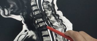
Lumbar puncture. Indications for use
Using cerebrospinal fluid sampling, intracranial pressure is measured, subarachnoid hemorrhages, their degree and severity are determined, and puncture is also used for:
- diagnosis and treatment of pathologies after brain injury and treatment of spinal injuries;
- diagnosis and treatment of neurological diseases;
- determination of sugar content;
- reducing intracranial pressure in the treatment of benign cranial hypertension or normal pressure hydrocephalus;
- administration of anesthesia, painkillers or chemicals, antibiotics;
- administration of contrast agents during radiography, myelography, encephalography or iodine-containing agents, oxygen, nitrogen, ozone, etc.
The spinal cord is protected by several layers of membranes (soft, arachnoid and hard), the space between which is filled with fluid. The epidural space, the space between the spinal cord and the spinal canal, is also filled with spinal substance. The fluid is taken, as a rule, from under the arachnoid layer. Normally, it should be transparent in color and slightly viscous in consistency.
Indications for cerebrospinal fluid collection
Lumbar puncture is performed:
- in the treatment of spinal diseases of chronic etiology and determination of subarachnoid hemorrhages in traumatic injuries of the spinal column, spinal cord, brain;
- in the treatment of infectious diseases of the central nervous system (encephalitis, meningitis, myelitis, tuberculosis, syphilis, etc.);
- for neurological diseases (multiple sclerosis, polyneuropathy, neuroleukemia, etc.);
- in the treatment of liquorrhea (leakage of cerebrospinal fluid from the nose and ears after injury to the bones of the base of the skull or spine, after neurosurgical operations on the spine);
- in the treatment of hydrocephalus (hydrocephalus) – excessive accumulation of cerebrospinal fluid in the brain or in tumors of varying quality in the brain and spinal cord;
- in the diagnosis and treatment of arachnoiditis - serous inflammation of the arachnoid layer of the cerebral or spinal cord cortex.
Contraindications for lumbar puncture
Contraindications to cerebrospinal fluid sampling may include:
- intracranial hematoma or abscess of the brain or spinal cord (accumulation of pus in the brain matter) after traumatic injuries to the spine or skull;
- pronounced signs of pinching of the spinal cord, for example, with spinal stenosis, the treatment of which has just begun;
- compression of the spinal cord by an intervertebral hernia, the treatment of which requires surgery;
- combination of spinal injury with traumatic shock, severe bleeding and extensive soft tissue damage;
- bedsores, infectious, purulent and inflammatory processes in the lumbosacral region.
Lumbar puncture technique
Lumbar sampling of cerebrospinal fluid is performed with the patient lying on his side with his legs pulled up to his stomach, or sitting with his back slightly bent forward. The puncture site is treated with an antiseptic. Then a thin needle is gradually inserted into the intervertebral space, through which an anesthetic (Novocaine, Lidocaine) is injected. Next, a thicker puncture needle or Beer needle with a mandrel is inserted into the hole already made.
The manipulation is carried out strictly along the midline between the spinous processes of the vertebrae, making a slight upward slope and gradually moving inward through the muscular and ligamentous apparatus. A sign of a needle entering the subarachnoid space is a feeling of failure. If, on the contrary, an obstacle (bone) is felt along the path, then the needle is pulled back a little and its direction is slightly changed. The flow of fluid will indicate that the puncture was performed correctly.
The total amount of fluid required for the study of an adult is approximately 120 ml.
After the procedure, the puncture site is sealed with a sterile napkin and the patient needs to lie on his stomach for 2-3 hours to restore the meninges and soft tissues. A lumbar puncture may cause a mild, dull headache or pain in the spine, which is caused by a decrease in intracranial pressure; it usually goes away 5-7 days after the procedure. Author: K.M.N., Academician of the Russian Academy of Medical Sciences M.A. Bobyr
4. Bone marrow biopsy results
Bone marrow biopsy results are usually ready within a week.
The following indicators are considered the norm:
- Bone marrow has normal amounts of fat, connective tissue, and iron. Normal ratio of adult and growing bone marrow cells.
- There are no signs of infection.
- There are no cancer cells such as leukemia, lymphoma or multiple myeloma.
- There is no spread of cancer cells from other affected areas.
Deviation from the norm:
- Bone marrow cells with pathology.
- The ratio of the number of different cells is disturbed.
- Bone tissue with pathology.
- Too much iron or too little iron (iron deficiency anemia).
- There are signs of infection.
- Cancer cells (leukemia, lymphoma, or multiple myeloma) are present.
- The bone marrow is replaced by scar tissue.
Depending on the results of a bone marrow biopsy or spinal tap, the doctor may prescribe additional examinations, select or adjust a treatment regimen, or, conversely, make sure that everything is in order with your health.
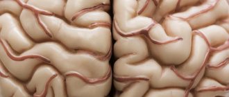
Signs of developing a brain tumor
The primary clinical picture is unremarkable, so it is almost impossible to detect cancer. The first signs appear when the tumor has already reached a significant size and compresses healthy brain tissue. Symptoms of malignancy depend on its location:
- Occipital lobe: Loss or deterioration of vision in one eye, and vision may become blurred.
- Frontal region: behavioral disorders, character changes.
- Cerebellum: loss of coordination, nausea and vomiting, weakened muscle fibers at the back of the head, involuntary movements of the eyeballs.
- Parietal lobe: loss of coordination, difficulty speaking and understanding speech.
- Brain stem: bifurcation of the visible image, poor orientation in space, difficulty swallowing, slurred speech, unsteady gait.
- Temporal region: memory impairment, unexplained feelings of fear or anxiety, frequent loss of consciousness, difficulty pronouncing words, hallucinations.
In addition, oncology may be accompanied by:
- partial or complete loss of sensation in the lower or upper extremities;
- general weakness;
- ringing in the ears;
- violation of writing;
- severe headache that is not relieved by medications, often worsening with sudden movements or changes in body position, as well as in the morning;
- problems maintaining balance;
- increased fatigue.
Find out the price
Find out the price
Error! Please fill in all required fields
Thank you! We will contact you shortly
✕
Can't fly to Turkey right now? Sign up for an online consultation with an Anadolu specialist.
Pleural puncture in children
In childhood, the procedure for therapeutic purposes is indicated:
- For aspiration of liquid or gas components from the pleural cavity in order to facilitate breathing.
- With exudative pleurisy and pleural ampyema.
- For tumor diseases in the chest.
- In case of hemothorax and pneumothorax.
For diagnostic purposes, a puncture is performed to obtain analysis from the pleural cavity.
The procedure is carried out directly in the manipulation rooms. The child should lie on his side (back) or sit on a chair. The puncture site is the 5th-6th intercostal space (nipple level) or the deepest point of the effusion. First, local anesthesia is performed with a solution of novocaine (0.25%). A “lemon peel” is made with a thin needle, after which it is changed to a needle with a large lumen, which first pierces the skin, and then the subcutaneous fat and muscles. Having moved the needle to the level of the upper edge of the underlying rib, the surgeon punctures the chest wall and infiltrates the tissue with novocaine. Puncture of the pleura gives the feeling of the needle falling into emptiness.
The pleural cavity is anesthetized with two to three milliliters of novocaine, after which liquid is sucked out of it with a syringe for testing. If there is blood, pus or air in it, the doctor connects the needle to the adapter tube and aspirates the contents of the cavity. The contents are removed from the syringe into a previously prepared container, and the syringe is disconnected from the tube with a special clamp. After evacuating the contents, the cavity is washed with antiseptics. The procedure ends with the introduction of an antibiotic, but only after it has been possible to achieve maximum vacuum in the pleural cavity (“collapse” of the rubber tube).
If the first puncture has a positive effect, the manipulations are repeated until complete recovery. If the result of the procedure is unsuccessful (thick pus or an unsuccessful puncture site), one-time punctures are performed in other places until a positive result is obtained.
In the absence of positive results, passive drainage according to Bulau, or active, is indicated by creating a vacuum when connecting the drainage tube to a water jet or electric suction. Also in modern medicine, microdrainage is increasingly being practiced - the use of a venous polyethylene catheter with a diameter of 0.8-1.0 mm, inserted after removing the needle. Its advantages: elimination of organ injury and the possibility of repeated lavages of the pleural cavity with the administration of antibiotics.
To protect the child from a state of shock due to the loss of a large volume of fluid, as well as to prevent the development of infection and the formation of a fistula at the site of the canal, special care is required for him. Upon completion of the manipulation, the patient is placed on the punctured side and, in order to facilitate breathing, the upper part of the body is placed in an elevated position. Basic vital processes are monitored, in particular, respiratory function is monitored first every quarter of an hour, then every half hour, and then every 2-4 hours. Also make sure that there is no bleeding.
In what cases is a thyroid puncture necessary?
Usually, nodules in the thyroid gland are identified by an endocrinologist during palpation (palpation), or they are detected during an ultrasound. Sometimes there are large nodes that are noticeable under the skin, deform the neck, and impair breathing and swallowing.
For some patients, it is enough to undergo an ultrasound examination, a computed tomography scan of the neck and a blood test for hormones. These diagnostic methods can show that the nodule is not cancerous. But in most cases, the diagnosis has to be clarified using puncture and fine-needle biopsy.
You can suspect the malignant nature of a node during an ultrasound based on some signs:
- low echogenicity (dark color in the images);
- heterogeneous structure;
- absence of a dark Halo rim around the node - it is usually detected with benign formations;
- uneven, unclear boundaries;
- irregular shape;
- the presence of compactions - calcifications;
- abundant or, on the contrary, poor blood supply;
- enlarged lymph nodes - this may indicate that they are also affected by a malignant tumor.
The risk that the nodule will be malignant is increased in people with a family history (thyroid cancer in close relatives), in those who previously suffered from cancer of the endocrine organs, or were exposed to ionizing radiation.
In accordance with modern recommendations, a diagnostic puncture is indicated when a node with a diameter of 1 cm or more is detected in the thyroid gland, which is determined by touch.
The doctor decides whether it is necessary to perform a puncture of the thyroid gland and a biopsy of the node, in each case individually, after studying the results of ultrasound and other studies. Some criteria play an important role:
| Factors that indicate a high risk of thyroid cancer | Factors that indicate a low risk of malignancy (puncture can be postponed) |
|
|
You can get advice from a competent specialist at the international clinic Medica24. Our doctors work in accordance with modern international standards.
Request a call back. We work around the clock
Message sent!
expect a call, we will contact you shortly
Diagnosis of brain tumors
With the help of an initial examination, an experienced oncologist or neurologist determines the most obvious symptoms of brain damage. To do this, the patient's coordination, behavior, sensitivity to pain, and various reflexes are tested. To determine the presence of a malignant formation, laboratory and instrumental diagnostic techniques are also used:
- radiography;
- checking blood for tumor markers;
- angiography;
- vision check by an ophthalmologist;
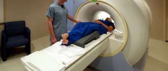
- magnetic resonance, positron emission or computed tomography;
- spinal tap;
- Ultrasound;
- electroencephalography.
A histological analysis of tumor tissue provides a 100% accurate examination result. If the brain is affected by a malignant process, performing a biopsy becomes a complex neurosurgical intervention. It can be performed using a craniotomy or stereotactically, where a needle is inserted into a small hole and monitored by a computer.