Synonyms: hydrocephalic syndrome, hypertensive-hydrocephalic syndrome, HGS. ICD-10 code: G91
Medical editor: Druzhinkina V.Yu., neurologist. November, 2020.
Hydrocephalic syndrome is a pathological condition characterized by excess production of cerebrospinal fluid (CSF), which accumulates under the meninges and in the ventricles of the brain.
The clinical picture of hypertensive-hydrocephalic syndrome can be explained by two concepts:
- hypertension (increased intracranial pressure);
- hydrocephalus (increased amount of cerebrospinal fluid in the brain).
Important! Hydrocephalic syndrome as a term is accepted only in the former USSR (in the CIS) and in modern Russia. Western doctors attribute hydrocephalic syndrome to a manifestation of brain pathologies.
The syndrome is often diagnosed by pediatric neurologists, and, as a rule, without reason. HHS is a fairly rare pathology, and in 97% of cases the diagnosis of hydrocephalic syndrome has no right to exist.
Causes
There are congenital causes of hydrocephalic syndrome (HHS in newborns) and acquired ones.
Congenital causes of hydrocephalic syndrome
- course of pregnancy and childbirth with complications;
- hypoxic (intrauterine hypoxia and intrauterine growth retardation, their manifestation in fetal bradycardia) and ischemic (injuries during childbirth) brain damage;
- premature birth (up to 36 weeks);
- head injuries during childbirth (subarachnoid hemorrhages);
- intrauterine infections (toxoplasmosis, influenza, cytomegalovirus infection and others);
- congenital abnormalities of brain development;
- delayed birth (at 42 weeks and later);
- long water-free period (more than 12 hours);
- chronic maternal diseases (diabetes mellitus, chronic pyelonephritis, arterial hypertension and others).
Acquired causes of hydrocephalic syndrome:
- tumors, abscesses, hematomas, parasitic cysts of the brain;
- foreign bodies in the cranial cavity;
- fractures of the skull bones with the introduction of fragments into the brain;
- causeless intracranial hypertension;
- infectious diseases (malaria, tick-borne encephalitis);
- post-stroke disorders;
- metabolic disorders.
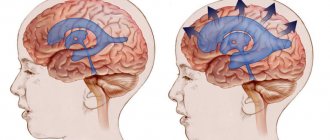
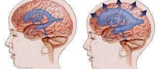
Photo: cerebrospinal fluid pressure on the brain with hydrocephalus.
Treatment of hydrocephalus
The option of surgical intervention for hydrocephalus is quite effective. Two types of operations are used: neuroendoscopy or liquor shunting. They are performed by a neurosurgeon, a specialist who can correct problems in the brain or spinal cord, as well as the peripheral nerve ending system.
Bypass surgery
In this type of surgery, a thin tube is inserted into the brain ventricle. Excess cerebrospinal fluid flows through it to another anatomical zone of the body. If the second end of the drainage system is placed in the peritoneum, the shunt is called ventriculoperitoneal. Then the excess cerebrospinal fluid is absorbed by the abdominal cavity and then enters the blood.
When the draining end is directed into the cardiac chamber, the shunt is called ventriculoatrial. This option is practiced during surgery for children, because their increase in height does not affect the functioning of the organ as much, so the drainage does not have to be changed as often as the ventriculoperitoneal system.
Occasionally, lumboperitoneal shunts are also used. With the help of such drainage, cerebrospinal fluid is drained from the lumbar region into the peritoneal cavity.
Any drainage system contains a valve that is set to a certain capacity. It is selected for a specific patient, taking into account the patient’s cerebrospinal fluid pressure. The doctor performing the surgery determines these values in advance by installing a suitable valve, which then looks like a “bump” protruding under the skin on the head.
Currently, neurosurgeons have two types of valves for drainage systems:
- With predefined characteristics that define a specific throughput.
- Adjustable magnetic fixtures. When choosing them, the doctor remotely, using special equipment, without performing additional incisions, changes the pressure in the device to achieve an optimal result. Thanks to this, it is possible to neutralize adverse side effects in the form of insufficient or excessive cerebrospinal fluid pressure.
Bypass surgery is performed under general anesthesia. The procedure lasts 1–2 hours and after it the patient is kept in the hospital for several days. If the outflow through the shunt is disrupted or the patient becomes infected, a repeat procedure is sometimes performed.
Neuroendoscopy
This operation is an excellent alternative to bypass surgery. Without taking steps to install drainage, the neurosurgeon forms a hole directly in the wall of the cerebral ventricle, after which he creates a bypass path from the vessel through which cerebrospinal fluid flows freely to the surface. There it is absorbed by the brain tissue without obstacles.
This procedure is not universal - it cannot be done on all patients, although this method is often used if the natural cerebrospinal fluid pathways are blocked. In this case, the cerebrospinal fluid flows through the created hole, successfully bypassing all blocked vessels.
The endoscopic procedure is done under general anesthesia. First, the neurosurgeon makes a burr hole with a diameter of 1 cm on the skull. Then, using an endoscope, he examines the inside of the cerebral ventricles. Through the internal channel of the instrument, surgical instruments are inserted, which are used to perform an operation inside the brain. When the endoscope reaches the third ventricle, the doctor creates a hole in its lower wall through which the cerebrospinal fluid penetrates into the subarachnoid cavity. After removing the device, first the aponeurosis is sutured, and then the skin on the skull. The duration of the operation is approximately an hour.
The risk of infection with such an intervention is significantly lower than with cerebrospinal fluid bypass. Neuroendoscopy does not have significant advantages over drainage surgery over long periods of patient observation. After such an intervention, hydrocephalus can also develop again after a few years.
Actions for normal pressure hydrocephalus
When normal pressure hydrocephalus (a common occurrence in older people) is diagnosed, the patient's condition can be improved by performing a cerebrospinal fluid shunt. Although surgery is not effective for every patient. Due to the considerable risks that accompany all operations, certain tests are required to assess how much the potential benefit outweighs the risk of negative consequences.
Almost 80% of patients diagnosed with normal pressure hydrocephalus experienced significant improvement after preliminary testing, so ventriculoperitoneal shunting was recommended for them. Significant clinical postoperative improvement occurs after a few weeks, sometimes even months. Only a timely correct diagnosis guarantees successful recovery even with many years of development of hydrocephalus.
Symptoms of hydrocephalic syndrome
Signs of hydrocephalic syndrome in newborns
Parents note that the child does not latch on well, constantly cries for no apparent reason, and sometimes moans.
The child has:
- decreased muscle tone (“seal feet” and “heel feet”);
- weak innate reflexes (swallowing, grasping);
- tremor (trembling of the limbs, chin) and convulsions may occur;
- spitting up like a fountain;
- strabismus;
- when examined by a doctor, positive Graefe's sign is observed (the eyeball is directed downward - a white stripe is visible between the pupil and the upper eyelid) and the rising sun sign (the iris is almost half hidden behind the lower eyelid)
- opening of the sutures of the skull (in particular the sagittal);
- bulging, tension of the fontanelles;
- in dynamics there is an increased increase in head circumference (by 1 cm every month), which is significantly ahead of age standards;
- head thrown back, which is especially noticeable during sleep;
- cyanosis, pale skin;
- increased respiratory movements;
- When examining the fundus, swelling of the optic discs is observed.
Clinical manifestations of HGS in children
Symptoms of hypertensive-hydrocephalic syndrome in older children usually develop after an infection or brain injury.
A characteristic symptom is headache, which often occurs in the early morning hours, nausea and vomiting, which follow it and do not bring relief. The pain is dull, aching or bursting; localized in the area of the temples, forehead and brow ridges.
Children complain that it is difficult for them to raise their eyes and lower their heads. Dizziness often occurs (young children define it as “swinging on a swing” or “instability of objects”).
During an attack of pain, pale skin, weakness and lethargy are noted. Bright light and loud sound cause irritation and increase unpleasant sensations.
Walking “on tiptoes” due to increased tone of the leg muscles, squint, drowsiness and slow thinking, poor memory and attentiveness are also typical. Such children do not eat well because they feel nauseous.
Hydrocephalic syndrome in adults
HGS in adults develops as a result of traumatic brain injuries, tumors, neuroinfections and after a stroke.
Signs of hydrocephalic syndrome are similar to those in older children:
- visual impairment (double vision, strabismus);
- severe headaches;
- nausea and vomiting, especially severe in the morning;
- disturbance of consciousness up to coma and convulsions.
Complications
Excess cerebrospinal fluid dilates the ventricles, compresses the medulla - swelling occurs. A closed cranial cavity provokes the development of irreversible changes in blood vessels - in the absence of the necessary treatment, this condition is life-threatening.
Children who develop hydrocephalus are mentally behind their peers. With complications, mental disorders, mental retardation, personality disorders, and inhibited reactions are likely. Irritability, attacks of euphoria or anger appear. Due to brain damage, speech problems, vision or hearing problems occur.
Adult patients complain of a bursting headache and nausea. Only vomiting sometimes alleviates the condition. The organs of vision experience internal pressure, and there is a feeling as if sand has gotten into the eyes. As pressure increases, drowsiness occurs.
Chronic hydrocephalus provokes an increase in intracranial pressure. The pathology develops slowly, appearing several months after the onset of the disease. Its main symptoms:
- cognitive disorders;
- walking disorders.
Diagnostics
Diagnosis of hydrocephalic syndrome is difficult. Not all instrumental methods help establish a diagnosis in 100% of cases. In infants, regular measurement of head circumference, checking reflexes, dynamics of body weight gain, and neurological examinations are important.
Also in the definition of GGS is used:
- electroencephalography;
- lumbar puncture of the spinal cord to take cerebrospinal fluid to measure its pressure (the most reliable method);
- neurosonography (ultrasound examination of the anatomical structures of the brain, in particular the size of the ventricles);
- assessment of fundus vessels (swelling, congestion or vasospasm, hemorrhage);
- computed tomography () and nuclear magnetic resonance (NMR).
Causes
The substance of the brain and spinal cord is constantly washed by cerebrospinal fluid. It protects the brain from damage and provides nutrients. The circulation of cerebrospinal fluid occurs in the subarachnoid space between the pia mater and the choroid along the surface of the cerebral hemispheres and the cerebellum. Under the base of the brain there are cisterns in which cerebrospinal fluid accumulates. They connect to the subarachnoid space.
Liquor is concentrated in the three ventricles of the brain. They communicate with the fourth ventricle, which is located between the brain stem and the cerebellum. From the fourth ventricle, cerebrospinal fluid enters the spinal canal through two lateral openings and spreads down to the lumbar region. When the cerebrospinal fluid outflow pathways are blocked, hydrocephalus develops.
Normally, the volume of cerebrospinal fluid is about 150 milliliters. The same amount of cerebrospinal fluid is absorbed back. The processes of formation and absorption are in a state of dynamic equilibrium. In case of excess production or insufficient reabsorption of cerebrospinal fluid, its volume increases, which leads to increased intracranial pressure and the development of hydrocephalus.
Replacement hydrocephalus of the brain in older people develops due to atrophy of the brain matter and the accumulation of excess fluid in the freed space. Atrophy of the brain substance is the result of physiological aging of the body. Brain volume in older people may decrease after infectious diseases (syphilis), traumatic brain injuries, or as a result of alcohol intoxication.
Treatment of hydrocephalic syndrome
Treatment of hydrocephalic syndrome is carried out by neurologists and neurosurgeons with the involvement of ophthalmologists. Patients with HGS need to be observed and treated in a specialized neurological center.
Treatment in newborns
Children require outpatient or surgical treatment, depending on the cause. Main therapeutic measures:
- prescribing a diuretic drug - diacarb (reduces the production of cerebrospinal fluid and removes excess fluid from the body, primarily from the cranial cavity);
- it is possible to take nootropics - some experts claim that these drugs improve blood supply to the brain (Encephabol, Actovegin), but there are no convincing large-scale studies on this topic;
- for hyperexcitability, sedatives are indicated;
- massage.
Treatment for infants is quite long, taking several months.
Treatment of HGS in older children and adults
In adults and older children, therapy also depends on the cause of hydrocephalic syndrome. If it is the result of a neuroinfection, then appropriate antiviral or antibacterial therapy is carried out.
In case of traumatic brain injuries and tumors, surgical intervention is indicated. Diuretics and nootropics are also used.
Diagnosis of hydrocephalus
Signs of the disease are often detected while the fetus is still in the womb during ultrasound. Then the disease is diagnosed by a doctor after the baby is born. The main indicator for determining pathology is the large diameter of the skull. In older children, as in adults, the doctor evaluates the patient’s thinking, his gait, the presence of signs of urinary incontinence, and the condition of the ventricles using MRI images.
To verify the presence of congenital hydrocephalus, CT or MRI are used, which allow:
- assess the state of the brain;
- identify excess cerebrospinal fluid volume;
- detect increased cerebrospinal fluid pressure;
- see structural changes in brain tissue.
Normal pressure hydrocephalus is more difficult to identify because its symptoms are largely similar to neurodegenerative processes. This variant of pathology is diagnosed according to the following signs:
- walking disorder;
- mental disorders;
- urinary incontinence;
- excess volume of cerebrospinal fluid.
With any variant of the disease, it is important to make an accurate diagnosis in a timely manner so as not to be late with surgical intervention that will eliminate the severity of symptoms or reduce their intensity.
To find out how effective the operation will be, some additional measures are necessary:
- Lumbar puncture. A small amount of cerebrospinal fluid is removed through a puncture made in the lower back. The cerebrospinal fluid pressure is then measured. The procedure allows you to slightly reduce it in the cerebral ventricles in order to neutralize severe symptoms.
- Lumbar drainage. It is necessary when the previous procedure was ineffective. A catheter is installed between the lumbar vertebrae, through which the cerebrospinal fluid comes out for several days. All this time the patient is observed in the hospital. Antibiotics are administered to prevent infection.
- Infusion test. This is a stress diagnostic option that helps assess how much the brain is able to absorb cerebrospinal fluid.
- ICP measurement. To perform the procedure, a hole is drilled into the skull bone. A sensor in the form of a fiber-optic cable is installed through it. The event is carried out while the patient is in the hospital for at least 24 hours. The sensor detects surges in intracranial pressure, sending values to a recording device.
When an event involves the need to measure ICP, various sensor options are used:
- Intraventricular catheter. This is the most accurate way to measure. The device is brought to the lateral cerebral ventricle through a hole drilled in the skull bone. The instrument helps to simultaneously ensure the drainage of excess fluid.
- Subdural sensor. The device is attached under the dura mater of the brain. It is used if there is an urgent need to check ICP. The method allows you to quickly identify changes in ICP.
- Epidural sensor. The instrument is attached between the dura mater of the brain and the cranial bone, having previously drilled a hole in it. The procedure is less traumatic than others, but has a significant drawback - excess cerebrospinal fluid cannot be drained during the procedure.
When installing sensors, local anesthesia is required. It is also sometimes necessary to additionally inject the patient with sedative medications in order to reduce anxiety and relax the patient as much as possible.
Typically, sensors used to measure ICP are administered to people in intensive care, often in the most critical condition. The necessary indications for such an event are severe trauma to the skull, pathologies of the brain that cause swelling and depression of consciousness up to a coma. In patients who have undergone brain surgery, such a monitoring sensor helps detect increasing swelling.
Drainage of cerebrospinal fluid also helps to reduce high ICP. It is performed by installing a ventricular catheter, intravenous injection of certain medications, and changing the method of ventilation (in a situation where the patient breathes through a tube inserted into the trachea). Normal ICP is 1–20.
Installing sensors is a risky undertaking, as it can cause the following complications if done incorrectly:
- bleeding;
- irreversible damage in a situation with prohibitive ICP values;
- “herniation” of the brain;
- damage to brain tissue during catheter insertion;
- infectious complications;
- inability to accurately determine the location of the ventricle in order to place a catheter to it;
- other risks present during general anesthesia of the patient.
Complications and prognosis
Complications of hypertensive-hydrocephalic syndrome are possible at any age:
- delayed mental and physical development;
- blindness;
- deafness;
- coma;
- paralysis;
- epilepsy;
- urinary and fecal incontinence;
- death.
The prognosis is most favorable for hydrocephalic syndrome in infants. This is because they experience transient increases in blood pressure and cerebrospinal fluid, which stabilize with age.
In older children and adults, the prognosis is relatively favorable and depends on the cause of HGS, the timeliness and adequacy of treatment.
Sources:
- Bitterlich L.R. On the issue of differential diagnosis, classification and differentiated treatment of hydrocephalus and hirdocephalic syndromes in children. - International Neurological Journal, No. 1 (79), 2021.
- Order on approval of the standard of specialized medical care for children with hydrocephalus. — Ministry of Health of the Russian Federation, 2013.
- Federal clinical guidelines for the provision of medical care to children with consequences of perinatal damage to the central nervous system with hydrocephalic and hypertension syndromes. — approved Union of Pediatricians of Russia February 14, 2015
Symptoms of hydrocephalus
Typically, the manifestations of the problem depend on the age of the patient and the type of hydrocephalus. Newborns have noticeable characteristic appearance features:
- increased head diameter;
- the skin is thin, the veins are visible;
- the fontanel protrudes;
- eyes are directed downwards.
Clinically, this pathology manifests itself:
- frequent vomiting;
- lack of appetite;
- excessive irritability;
- drowsiness;
- decrease or increase in muscle tone of the limbs.
The acquired pathology is manifested by frequent headaches, especially severe after a night's sleep. This is due to the fact that when positioned horizontally, cerebrospinal fluid flows more slowly, it accumulates, bursting the brain tissue. The condition improves after standing up, although this improvement gradually disappears as the disease progresses.
Other manifestations include:
- neck pain;
- drowsiness;
- mental disorders;
- memory impairment;
- the appearance of a “veil” before the eyes;
- lack of coordination;
- nausea;
- walking disorder;
- epilepsy;
- urinary incontinence, sometimes involuntary bowel movements.
Normal pressure hydrocephalus is a disease of older people. It is characterized by the following symptoms:
- walking disorders;
- deterioration of intellectual abilities;
- urinary incontinence.
The first thing to break is gait. It becomes difficult to start moving. As the disease progresses, instability becomes more pronounced and a person may suddenly fall when changing direction. Walking disorder is accompanied by urinary incontinence: first there is a frequent urge that requires immediate emptying, then control of this function is completely lost.
The disease also affects thought processes:
- reaction worsens;
- the patient is not able to quickly respond to changing situations;
- a failure occurs when trying to process the received information.
Kinds
Hydrocephalus can be congenital or acquired. According to the mechanism of development of the pathological process, three main types of the disease are distinguished: occlusive, communicating and hypersecretory hydrocephalus. Sometimes neurologists identify external or mixed hydrocele of the brain. It is classified as brain atrophy, since the pathogenesis of the disease is determined by a decrease in brain tissue.
Depending on the speed of development and progression of the symptoms of the disease, acute hydrocephalus (develops within three days from the onset of the disease), subacute hydrocephalus (develops within one month from the onset of the disease) and a chronic form of the disease, the formation of which may take several weeks up to six months. Based on the level of cerebrospinal fluid pressure, normotensive, hypertensive and hypotensive hydrocephalus is distinguished.
Hydrocephalus - what is it?
The literal translation of the term “hydrocephalus” suggests that this disease is expressed by the presence of excess water in the head. This interpretation is not entirely correct. We are not talking about water, but about cerebrospinal fluid.
Unlike the heart, the brain is not a muscle. It is a delicate substance consisting of nervous tissue. A kind of protective “airbag” for it is cerebrospinal fluid, which provides:
- delivery of nutrients to the brain and reverse removal of metabolic products;
- stability of intracranial pressure.
Like blood circulation in the cardiovascular system, this fluid, vital for the proper functioning of the brain, circulates in the cavity of the cranium. The place of its formation is the ventricles, connected to each other by special channels. After washing the surfaces of the spinal cord and brain, the cerebrospinal fluid, the composition of which is constantly renewed, is absorbed into the bloodstream. Hydrocephalus is an excess volume of cerebrospinal fluid in the head. It is formed due to an imbalance between the amount of cerebrospinal fluid produced and its absorption.
Hydrocephalus in adults
The main signs of cerebral hydrocele in adults are expressed in symptoms of increased intracranial pressure. The acute form is accompanied by:
- paresis;
- headache that is not relieved by painkillers (analgesics), most severe in the morning;
- nausea and vomiting;
- drowsiness;
- oculomotor disorders.
In the case of the chronic form, the main manifestations of hydrocephalus are:
- dementia;
- enuresis;
- uncertainty, gait disturbance.


