Most likely, you know that MRI is magnetic resonance imaging. Perhaps this is all you know about this technique. Then there are several facts about how brain MRI is done that will be useful for you to know. Tomography is a modern diagnostic method that allows you to obtain the clearest possible image of internal organs. In the case of an MRI of the brain, the image shows all the structures of the brain - tissues, blood vessels, cerebrospinal fluid. Why does a doctor prescribe magnetic resonance imaging of the brain? In order to confidently diagnose or exclude suspected pathologies.
Benefits of examining the brain using MRI
First of all, it should be noted that MRI is safe, because the patient is not exposed to ionizing radiation. Therefore, MRI can be safely performed even at short time intervals (for example, to monitor the dynamics of treatment). Other advantages of this diagnostic method include the following facts:
- The pictures are as informative and clear as possible: this result cannot be achieved by other methods;
- Magnetic resonance imaging is a non-invasive study that does not involve the use of any medications, etc.;
- Ability to monitor the functional activity of all parts of the brain;
- Quick receipt of study results, the possibility of consulting a doctor immediately after the MRI;
- If necessary, the diagnostic process can be recorded on video.
What will the MRI results show?
A study using an angiographic protocol is a harmless, non-invasive procedure for which the patient does not even need to prepare. Using diagnostics, doctors can:
- establish the state of the vascular system;
- identify abnormalities, injuries, arterial occlusions;
- study problems that arise due to a malfunction of the circulatory system, determining their true cause;
- detect oncopathologies and benign neoplasms.
Indications for MRI of the brain
Brain diagnostics are carried out according to the doctor’s indications. As a rule, MRI is prescribed in the following cases:
- Suspicion of the presence of benign or malignant neoplasms, as well as metastases in the brain;
- Manifestation of epilepsy of various types;
- Previous head injuries, suspected concussion or functional damage;
- MRI of the brain is performed after operations, for example, after removal of tumors, to track dynamics;
- Suspicion of meningitis and other inflammatory diseases located near the brain;
- Symptoms of cerebrovascular accident.
Also, a diagnostic procedure can be carried out if the patient complains of the following symptoms:
- Unreasonable fainting;
- Absent-mindedness, memory lapses;
- Frequent causeless headaches;
- Disorientation in space;
- Impaired sensitivity of the upper or lower extremities, face;
- A sharp decrease in hearing or vision;
- Dizziness accompanied by nausea and vomiting.
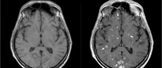
Do they do MRI of brain vessels with contrast?
Typically, tomography of arteries and veins is carried out without the use of an intravenous intensifier, since the blood itself can become a dye. But in some cases, MRI of the head is prescribed with contrast. Typically, screening is needed to obtain accurate and detailed images of the vascular bed when there is a likelihood of the formation of tumors of unknown origin, as well as to detect factors indicating the development of multiple sclerosis.
To conduct a scan with an additional enhancer, the substance is administered intravenously before the start of the examination. Usually a drug containing gadolinium salts is used. Contrast has the ability to accumulate in affected tissues, which makes them stand out in photographs against the background of healthy structures. Gadolinium-based substances rarely provoke allergies in patients. The diagnostic procedure has its limitations, which include:
- pregnant women;
- persons with individual intolerance to the components of the contrast agent;
- patients suffering from severe kidney disease.
After diagnosis, nursing mothers will need to transfer the baby to artificial nutrition for at least two days or express milk in advance.
How to properly prepare for manipulation
Magnetic resonance imaging of the brain does not require special preparation. There is no need to follow special diets, stop taking medications, etc. The only rule is to abstain from alcohol before the study. It is also not recommended to smoke at least 2-3 hours before the test. Experts recommend avoiding increased physical activity. Before the study, you need to relax as much as possible, avoid stressful situations, etc.
If you are worried or even afraid of your reaction to the confined space in the tomograph, your doctor may recommend taking mild sedatives. It is better to avoid taking strong tranquilizers, because they can cause distortion of the clinical picture. Before the examination, you need to remove all metal objects and jewelry. Before an MRI, it is better not to drink a lot of water so that during the examination you will not want to go to the toilet - you cannot move during the examination.
It is recommended to take your passport, money (if MRI is performed for a fee), and a personal outpatient card with you for the study. If a study has already been carried out previously, take the results of previous diagnostics with you, this will help the doctor determine the dynamics of changes in the brain.
Preparatory stage
The doctor, based on the test results, determines whether to perform a tomography with or without the introduction of a contrast agent. If the first option is chosen, then the patient does not take food or liquid 5 hours before the diagnosis. Before the procedure, you must remove all jewelry and accessories, watches and objects with metal elements.
During a consultation with a doctor, the patient reports the presence of chronic diseases, allergies to medications, claustrophobia, and pregnancy.
When preparing a child for a head tomography, it is not recommended to give him anything to drink or eat 3 hours before the diagnosis. If an MRI is performed with the administration of contrast, the examination is performed on an empty stomach. Before the procedure, the child should be shown to an allergist to check for an allergic reaction to the injected substance.
Diagnostic algorithm
Let's look at how magnetic resonance imaging of the brain is performed:
- First, the patient must remove all jewelry, as well as elastic bands and hairpins, if they have a metal insert. If your clothes have metal buttons, hooks, etc., you need to remove your clothes; if necessary, you will be given a robe.
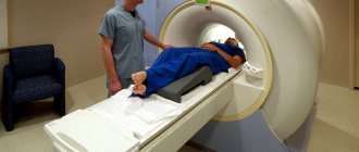
- The patient lies on his back on a special retractable couch. A radio-receiving RF coil is placed above the head, receiving the signal, which, when scanned, is converted into an image.
- The specialist secures the legs and arms with special straps. This is necessary to ensure that the person does not move during the examination, as this may affect the quality of the images.
- The couch moves into the tomograph, where the scan is performed.
- During the manipulation, noise is heard - do not be alarmed, this is the norm. To reduce the intensity of sounds, patients may be offered earplugs.
- There is nothing to worry about - even if you feel ill or very scared from a confined space, you can always press the panic button or contact a doctor through the microphone.
- In general, diagnosis usually takes no more than half an hour. At the end of the manipulation, the couch moves out again, and the doctor releases the patient from the straps.
- The patient receives diagnostic results after 30-40 minutes. If necessary, you can immediately consult a doctor.
All that is required of the patient is to lie as still as possible, preferably not to yawn or talk (although if you really need to contact the doctor, you can do this). Experts recommend wearing loose, comfortable clothes, lying down comfortably and relaxing. Only if this rule is followed will the diagnostic results be as informative as possible.
After the diagnosis, the patient does not feel any changes, the state of health does not change, since the body is exposed to harmless magnetic radiation that does not cause changes in the functioning of organs and the condition of tissues. For this reason, various health consequences and complications are excluded.
What are the contraindications to MRI?
Tomographic screening has a minimal list of limitations, which is the main advantage of this technique. Almost all contraindications are associated with exposure to magnetic radiation on the body. Diagnostics is not carried out in the following cases:
- the patient has metal structures in the body;
- the patient has electronic devices in the body;
- the patient is in the first trimester of pregnancy;
- the person under study is under five years old;
- The patient has excessive body weight, exceeding 130 kg, and the body diameter is more than 150 cm.
What pathological processes can be identified?
During the diagnostic procedure, many pathologies can be identified even at an early stage. With the help of research it is possible to discover:
- Inflammatory processes;
- Aneurysms;
- Multiple sclerosis;
- Strokes and micro-strokes;
- Cysts, lipomas and other benign neoplasms;
- Malignant tumors;
- Hydrocephalus;
- Encephalopathy;
- Epilepsy;
- Vascular atherosclerosis;
- Meningitis;
- Hemorrhages in the brain structures.
If you conduct a study with a contrast agent, you can also clearly see various vascular pathologies, for example, narrowing of the lumen, cerebral varicose veins, venous malformations, etc.
How is a head MRI performed?
An MRI of the head is performed as soon as possible. It usually only takes a few minutes. If necessary, a contrast agent is first injected into the vessels. The patient's body is fixed on the couch to prevent accidental movements. Communication with staff is carried out through a special loudspeaker. The doctor may ask the patient to hold his breath.
MRI of the head is easily tolerated by all categories of patients. If necessary, the patient is first given a light sedative to help him relax. Thanks to this, head examinations can be performed even on patients with claustrophobia.
Diagnostic results
A highly qualified doctor interprets the images. The photographs and conclusion can be provided in printed form or recorded on electronic media. If magnetic resonance imaging does not reveal any abnormalities, the report contains approximately the following information:
“The sub- and supratentorial structures were visualized in axial, sagittal and frontal projections. The median structures are without features, the localization corresponds to the norm. The cortex and white matter are well developed, with normal MR signal intensity. Convexital grooves of the cerebellum and cerebrum are within normal limits. The ventricles of the brain have the same shape and size. The basal cisterns and subarachnoid spaces are normal. There are no symptoms of impaired outflow of cerebrospinal fluid and increased intracranial pressure. Neoplasms and other pathological processes were not identified.”
Do not try to interpret the diagnostic results yourself - entrust this to specialists. The doctor will competently decipher the conclusion, tell you whether there are pathological processes, whether any treatment is required, what the prognosis is, etc.
If necessary, effective therapy will be prescribed. Be healthy!
Contraindications to magnetic resonance imaging
MRI of the head is not performed if:
- exacerbation of serious mental pathologies;
- heart, kidney and liver failure;
- general serious condition;
- the presence of hearing and other implants, pacemakers, insulin pumps;
- with body weight over 250 kg.
Also, examination is not prescribed for severe damage to the respiratory system. MRI of the head is not recommended for pregnant women or children. However, if there is an urgent need for diagnosis, it can be carried out.
Important! Each case of MRI of the head is considered individually. The final decision to perform an MRI of the head is made by the attending physician. If necessary, the patient is first referred for consultation to specialists. If necessary, additional diagnostics are prescribed.
Description of the procedure
- The procedure takes up to 60 minutes, depending on the scope of the examination
- it is possible to perform MRI using a contrast agent, which increases the information content of the study
- at the Yauza Clinical Hospital, MRI is performed on the Ingenia 1.5T digital system (Philips), which provides stable and high image quality
- After the procedure is completed, the doctor decrypts the data, the images are recorded on a disk or flash card
During an MRI of the head, the doctor receives information about the state of brain tissue, can look at its individual structures, assess cerebral blood flow, and see vascular disorders and pathologies. An MRI of the head can detect diseases such as stroke or cerebral ischemia, see vascular aneurysms, determine Parkinson's disease, encephalopathy, pituitary adenoma, bruise or concussion.