Symptom Description
With a unilateral decrease in visual acuity, which is observed against the background of intraocular hypertension, inflammatory eye disease, hemorrhage in this area, the optic nerve may also be affected. A bilateral process is often associated not with local changes in the eyes, but with a systemic disease (cardiovascular pathology, renal failure, severe blood loss, diabetes mellitus).
Blurred vision may be constant or appear towards the end of the day. Decreased vision can occur both in the central parts and in the periphery.
All this must be taken into account when describing complaints - this will help to quickly understand the diagnosis.
Other causes of dizziness
If everything is swimming before your eyes, then this condition is not necessarily caused by stress. Sometimes the symptoms that arise are completely unrelated to VSD (although they can be provoked by it), and the VSD person also needs to know about them.
- Lack of glucose. Hypoglycemia causes blurred vision and a feeling that the world is floating before the eyes.
- Poisoning. When the body is intoxicated, the functioning of almost all organs and systems malfunctions, so dizziness and lightheadedness are justified.
- Some medications may have the side effect of dizziness.
- Extreme fatigue or poor routine. Poor nutrition, lack of sleep, and fatigue at work lead to the body breaking down and starting to give alarm signals with various symptoms, including a feeling of fluidity in the situation.
If everything starts to swim before your eyes and you feel dizzy, then it is better to first consult a neurologist to rule out various physical diseases. If no significant health problems are found, then the symptoms are due to nervousness and then the problem needs to be solved through work with a psychologist.
Why does fog appear before my eyes?
Common reasons
The appearance of fog in the eyes can be associated with severe toxicosis in pregnant women, poisoning with alcohol products (ethyl and methyl alcohol), overdose of quinine, nicotine, lead and other active substances.
Quite rarely, blurred vision is a consequence of vasospasm of the retinal vessels. In this case, a functional disturbance of blood flow through these arteries occurs, which does not lead to organic changes in the vessels. Typically, patients with hypertension and frequent migraine pain are susceptible to vasospasm. In addition, arterial spasm occurs in pregnant women with eclampsia or preeclampsia, as well as against the background of chronic stress and severe poisoning. Fog in the eyes in this case can be combined with the appearance of floating black dots or flickering spots, and visual acuity is also temporarily (up to several hours) reduced.
If the doctor suspects angiospasm in the patient, then an ophthalmoscopy should be performed, which will show in which area of the fundus the local narrowing of the arteries has occurred. Usually the fundus itself is not changed during ophthalmoscopy, except in situations where the cause of vasospasm is atherosclerotic damage to the vascular stack (in this case, a picture of sclerosis of the retinal arteries is revealed).
Blurred vision can be a sign of a stroke, high or low blood pressure, high or low blood sugar [2].
Eye diseases
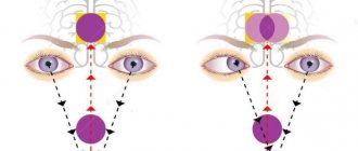
A disturbance in one of the light-conducting structures of the eye—the cornea, lens, or vitreous body—leads to decreased vision [3]. The veil can also be caused by a lesion in the light-receiving part (retina) or due to diseases of the optic nerve (neuritis).
Fig. 2 Light-conducting media of the eye, light-receiving apparatus and optic nerve
Glaucoma is considered one of the most common causes of foggy eyes in older people. As a result of an increase in the level of intraocular pressure in primary, post-traumatic, and secondary glaucoma, a veil appears before the eyes and overall visual acuity decreases. Other symptoms characteristic of glaucoma are pain in the eyes themselves, in the head (temporal, superciliary areas), the appearance of rainbow circles or glare around point light sources, as well as fog in the eyes [4].
During an ophthalmological examination, which is carried out in patients with such complaints, corneal edema and a decrease in the depth of the anterior chamber of the eye can be detected. With advanced changes, pallor of the optic nerve head and its excavation also occur.
Another common cause of foggy eyes in older people is DVT - destruction of the vitreous body - age-related clouding of the biological lens of the eye (lens). The process develops gradually over several years. Cataracts can appear in young people due to trauma, radiation (“radiation cataract”), or diabetes [5].
Violation of the structure and transparency of the vitreous body (the gel-like substance that fills the eye from the inside) leads to the appearance of fog, floating “cobwebs” and “worms before the eyes.” This condition is called DVT - destruction of the vitreous body, which is often accompanied by posterior vitreous detachment (PVD).
Fig. 4 Changes in the vitreous body, leading to decreased vision
Fog in the central field of vision can be caused by age-related macular degeneration (AMD) - both dry and wet forms, macular edema due to a number of reasons (diabetes, central serous chorioretinopathy, etc.).
Retinal detachment, when the inner retina detaches from the wall of the eyeball and quickly dies, poses a great danger. Without emergency surgery, vision is lost forever [6].
Inflammatory eye diseases - conjunctivitis, retinitis, uveitis can also lead to blurred vision. At the same time, signs of inflammation are not always expressed clearly enough. Quite often, there is only a gradual decrease in visual acuity and the appearance of floating spots before the eyes, which gradually increase in size. Acute signs of inflammation, including pain, photophobia, redness, are more typical for the acute course of iridocyclitis or iritis. The main sign of anterior uveitis, rather, can be called narrowing of the pupillary opening or hyperemia in the area located near the limbus.
Foggy eyes after surgery
Blurred vision after surgical interventions - laser vision correction, cataract removal, etc. for several days (up to a week) is a completely normal phenomenon and is associated both with the presence of inflammatory edema of the cornea in response to surgery, and can also be caused by the antibacterial and anti-inflammatory drops used.
Fig. 5 Eye on the first day after cataract removal - corneal edema and hyperemia (redness) of the conjunctiva
What factors cause blurry vision?
The inability to focus is almost always related to underlying problems present in the body. They are destructive, inflammatory or tumor in nature. Without an examination, it will not be possible to determine why there is floating in the eyes. After the interview and examination, the patient needs to undergo hardware and radiation diagnostics. This will help you find out why your vision periodically becomes blurry, your head starts to hurt, and related symptoms occur.
Anemia
Also in the official medical literature it may be called “anaemia”. The disease is characterized by a low level of hemoglobin, which is facilitated by blood loss, low-quality, irregular nutrition. Anemia develops equally often in both sexes.
In women, anemia often occurs as a result of monthly blood loss, if it is massive and uncompensated.
Pathology can only be identified based on laboratory testing. The patient will have to donate blood for clinical analysis.
Symptoms of the condition:
- General weakness occurs.
- Blood pressure levels decrease.
- Extremities are cold to the touch.
- Sensation in the hands is impaired.
- It can sometimes get dark before your eyes, especially when moving from a horizontal to a vertical position.
Against the background of low pressure, the patient complains that objects seem blurry and it is difficult to concentrate. Anemia can rarely go away on its own - the patient needs improved nutrition, proper rest, and walks in the fresh air.
Cerebrovascular accident
It occurs suddenly, for example, due to a blow to the head, or increases gradually. In the second case, the pathology is a consequence of disturbances in the systemic circulation. This occurs due to medication, stress, fasting, and alcohol abuse.
Signs of impaired cerebral circulation:
- The image is not clearly visible and may disappear for a short time.
- After a trip in public transport, you may begin to feel sick.
- It hurts and I feel dizzy.
- It's hard to focus on a specific object.
- Sometimes vomiting occurs.
These signs are grounds for immediate consultation with a doctor. In 95% of cases, the patient is indicated for immediate hospitalization in a hospital department. With the listed symptoms, you need to lie down on the bed with the head end elevated. This will minimize the load on the blood vessels of the brain.
Hormonal disorders
Nausea, blurred vision, dizziness, decreased performance are symptoms indicating pregnancy. The reason for the change in condition and the appearance of these signs is the sharp hormonal fluctuations that occur in the female body immediately after fertilization.
Violation of the daily routine
Exhaustive sports training, fasting, overwork, lack of sleep - all these factors negatively affect the blood supply to the brain. If everything suddenly swims before your eyes, you feel dizzy, nauseous, or general anxiety arises, you need to reduce physical activity, reconsider your diet and daily routine.
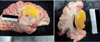
A brain tumor
One of the common reasons why everything floats in the eyes is the presence of a neoplasm inside parts of the brain. The reasons for the development of the tumor process are polymorphic.
Symptoms:
- Pain syndrome. The head hurts and begins to feel dizzy from minor irritation - for example, due to a trip in transport. Discomfort is not always localized on the side of the tumor.
- Insomnia due to severe headache.
- Nausea and vomiting occurs.
- The voice changes slightly.
- Vision and hearing are impaired - when the tumor compresses the nerves that transmit impulses to the eyes, or if the tumor is located near the ear.
- General weakness, which sometimes leads to an inability to assume an upright position.
Often, the patient explains the symptoms of a brain tumor by fatigue or other factors, and seeks help at least at stage 2 of tumor development.
For brain tumors, treatment is carried out by a neurosurgeon with the participation of an oncologist.
Hypertensive crisis
A condition in which blood pressure reaches high limits, which contradicts the person’s normal well-being.
Symptoms of hypertension:
- Noise in ears.
- A pulsating headache that can radiate to the ears or spread to the eyes.
- Weakness due to which the patient is forced to assume a horizontal position of the body.
During a hypertensive crisis, it is impossible to concentrate and focus your gaze on a specific object. The condition is characterized by lacrimation and a feeling of sand getting into the eyes.
A direct sign of increased blood pressure is nosebleeds.
Before the patient feels normal, he needs to be helped to assume a horizontal body position and provide him with access to fresh air.
Osteochondrosis of the cervical spine
The main reasons why everything floats before your eyes is pinched nerves or blood vessels running inside the neck. A similar thing happens with osteochondrosis of this part of the spine. Signs of the condition are pain when turning the head, nausea, motion sickness in transport. As the nerve fibers are compressed, the affected area begins to go numb. What should not be done until the presence of osteochondrosis is confirmed:
- Apply a compress to the troubling area of the body.
- Take antibiotics.
- Apply a heating pad to the area of inflammation.
If after minor physical activity you feel dizzy and immediately have blurry vision, you need to visit a neurologist. Already during the examination and interview, a presumptive diagnosis is established: the clinical manifestations of the disease are specific.
Hypotension
A condition in which blood pressure levels are reduced. Signs of hypotension:
- My head hurts and I feel dizzy.
- It is problematic to get out of bed, because there is unsteadiness on the legs, and it is dark before the eyes.
- There is general drowsiness.
Worsening of health may occur in the evening or in the morning - it does not depend on the time of day. When the cause of hypotension is VSD, the condition worsens on the eve of a change in weather conditions.
A standard pain reliever provides a short-term effect. Initially, you should undergo diagnostics, identify the cause of low blood pressure, and act on it with medication or another method.
Diagnostics
To diagnose the primary disease that has led to the appearance of fog before the eyes, you need to perform, at a minimum, biomicroscopy using a slit lamp and examine the fundus with a wide pupil. In this case, it is often possible to identify signs of damage to the cornea, lens, vitreous body or retina [7].
Testing vision with and without correction, as well as determining refraction in conditions of a dilated pupil (cycloplegia) will help identify disorders such as spasm of accommodation, myopia, farsightedness and astigmatism.
Determination of intraocular pressure (IOP) excludes or confirms glaucoma.
Fig. 6 Basic examination (biomicroscopy and ophthalmoscopy) allows in most cases to identify the cause of the appearance of fog
In some cases, additional examinations are necessary to clarify the diagnosis—primarily optical coherence tomography of the retina (including with angiography function) [8]. This method allows you to see minimal changes in the central zone of the retina and the optic nerve head (ONH), accurately and quickly make a diagnosis and prescribe treatment. This test is available in our clinic on the same day of your visit.
| Study | Signs | Disease |
| Vision testing with and without correction | Descending into the distance | Myopia, farsightedness, astigmatism, accommodation spasm |
| Measuring intraocular pressure | Promotion | Acute attack of glaucoma |
| Biomicroscopy of the cornea | Swelling, cloudiness | Keratitis |
| Biomicroscopy of the lens | Cloudiness | Cataract |
| Vitreous ophthalmoscopy | Cloudiness, peeling | DST, ZOST |
| Retinal ophthalmoscopy | Swelling, ruptures, detachment | Macular degeneration, retinal detachment and tears |
| Ophthalmoscopy of the optic disc | Swelling, pallor | Optic neuritis, glaucoma, atrophy |
Table 1. Research method, signs and final diagnosis
Principles of treatment of a pathological condition
A specialist determines which methods help eliminate this unpleasant symptom of blurred vision. If refractive error occurs as a result of a certain ophthalmological disease, wearing specialized optical lenses or glasses will help.
In this case, a surgical intervention such as LASIK can also significantly help a patient whose eyes are blurry, relieving him of refractive problems.
If the pathological phenomenon is provoked by dry eye syndrome, medications that imitate natural tears should be used. A variety of moisturizing gels and ointments are also suitable in this case. If your eyes are blurry as a result of cataracts, the problem can be corrected with surgery. For glaucoma, drops are used that lower intraocular pressure. In complex cases of glaucoma, surgical intervention is also performed.
It should be noted that ophthalmic factors of clouding in the eyes must be treated without fail. If some pathological processes are triggered, this can even lead to absolute blindness. Other diseases that cause blurry vision are treated differently. Each specific case has its own method of therapy.
What should you do if you only sometimes float before your eyes?
Treatment
Blurred vision can only be cured by identifying the cause of this condition. Often, a patient with such a symptom requires emergency hospitalization and immediate surgical intervention (for example, during an acute attack of glaucoma). Due to the high probability of serious complications, at the first appearance of fog in the eyes, you should consult an ophthalmologist.
When choosing a center where you want to receive medical care, you need to pay attention to the ophthalmology clinic that has fairly modern equipment, experienced specialists, and does not require long waiting in line. It is important not to put off taking care of your vision too long!
What to do if you feel dizzy and have dark vision?
If these symptoms occur regularly, you should get tested. There are a lot of diseases that are accompanied by darkening of the eyes and dizziness. To identify the true cause, the following may be prescribed:
- Ultrasound of the cervical spine and cerebral vessels;
- MRI;
- CT scan;
- measurement of intraocular and blood pressure;
- fundus examination;
- blood tests;
- neurological tests.
A person who is systematically bothered by dizziness and blurred vision must be able to help himself so as not to get injured. When these signs appear, you need to stop, lean against a wall or sit down. If symptoms are caused by a lack of glucose, you should eat something sweet. If you have high blood pressure, you need to take a pill to lower it. The method of treatment directly depends on the underlying disease.
List of sources used to prepare the material
- Changes in the organ of vision in common diseases. Eye diseases. Fundamentals of Ophthalmology: Textbook / Ed. V. G. Kopaeva. — 2012. — 560
- Zolotareva M.M., Rabinovich M.G. Ophthalmological symptoms in various diseases of the body. - Minsk. - 1965. - 171 p.
- Yartseva N.S., Deev L.A. Selected lectures on ophthalmology. Volume I Lecture No. 7 Pathology of the cornea. Moscow, 2007.
- Egorov E.A. and others. International guide to glaucoma. Volume 2. Glaucoma clinic. Moscow, 2021.
- Chkonia E.A. On the issue of diabetic cataract // Ophthalmol. magazine — 1968.- No. 1.-P.31
- Shkvorchenko D.O., Kakunina S.A., Pismenskaya V.A., Belousova E.V., Rusanovskaya A.V. Analysis of the timing of restoration of visual functions in patients with rhegmatogenous fresh retinal detachment after local episcleral filling. Modern technologies in ophthalmology No. 1 2014
- Ophthalmoscopy in the diagnosis of fundus diseases. Shchuko A.G., Yuryeva T.N., Akulenko M.V. “Laser surgery for vascular pathology of the fundus” // Moscow, 2014.
- Application of OCT in clinical practice. New in ophthalmology No. 1 2021. Heidelberg Engineering Academy.