The most common symptoms of damage to the vascular system of the brain and neck are headaches, unsteadiness of gait or weakness in the arm or leg. If symptoms are present, the patient should be examined using MRI of the brain vessels. The purpose of diagnosis is to exclude congenital pathology, namely:
- arterial aneurysm;
- arteriovenous malformation (AVM).
Moreover, symptoms can go undetected for too long, up to 45 years.
The essence of the technique
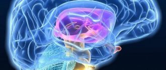
The study is based on hydrogen atoms, which are affected by a magnetic field, forcing them to be an “exchanger” of energy. This is due to the fact that hydrogen atoms are part of any human organ, including liquids.
Hydrogen atoms rotate around a magnetic axis. When a person is placed in a tomograph, energy is absorbed by hydrogen atoms, and upon completion of magnetic resonance imaging, energy is released. The degree of release by different tissues is somewhat different, since their chemical composition and density play a role. The computer captures this emitted energy, amplifies and re-digitizes it in various degrees of gradation in brightness. Next, the information is displayed on the monitor and recorded on all supported media.
The study is harmless to the patient's health. This is the fastest diagnostic method that painlessly displays all structural and morphological changes in the vascular system of the brain, head and neck.
Preparatory measures
Let's consider how to properly prepare for the upcoming diagnosis:
- The main rule is to avoid alcohol and energy drinks 3 days before the upcoming test. Ideally, nicotine should be eliminated for a day, but if this is difficult for you to do, stop smoking for at least 4-5 hours before the upcoming diagnosis.
- Be sure to tell your doctor about all medications you take.
- It is advisable to come to the procedure on an empty stomach. If the diagnosis is scheduled for the evening, you need to go without food for at least 5-6 hours. Moreover, eating should be light.
- You should not drink water or other liquids for 3 hours, so that while you are in the tomograph you will not have the desire to empty your bladder.
- The mobile phone must be turned off or left outside the diagnostic room.
- If you have a mild form of claustrophobia, ask your doctor about sedatives that will help improve your mental well-being.
- Remove all jewelry at home, including hair clips and hair ties with metal elements. It is better to do this at home so as not to waste time in the diagnostic room.
Your doctor will tell you more detailed information about preparation measures.
Diagnostics MRI of cerebral vessels
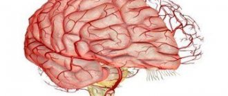
MRI of head and neck vessels is an informative neuroimaging method. Give a chance:
- assess the condition of the cerebral hemispheres;
- identify the presence of space-occupying formations;
- determine the presence and development of focal pathological changes;
- assess the condition of the cerebellum and brain stem.
During MRI of the vessels of the head and neck, the entire structure is analyzed with the exception of the skull bones. At the stage of such a procedure, the following occurs:
- assessment of the structure of the cerebral vascular system;
- the presence of an aneurysm in the substance is excluded or confirmed;
- confirmation or refutation of the pathological state of the structure of the vascular system.
Preparing for an MRI of the brain
Usually, there is no need to do any complicated steps before the examination. Sometimes the specialist may ask the patient to refrain from eating and drinking before the procedure. The person being examined must remove all metal jewelry. In many centers, the patient is given a loose shirt, in some they are allowed to remain in their own clothes, provided that they are loose and free of metal parts. Before a brain MRI, women should inform their doctor about the possibility of pregnancy. The patient is also obliged to inform the doctor about recent operations and diseases, allergies and the presence of metal implants. If the person being examined suffers from claustrophobia or is very worried before the procedure, he may ask the doctor for a sedative.
Possible diagnosable diseases, pathologies
The diagnostic method will help confirm or refute:
- development of deviations due to previous traumatic brain injuries;
- any possible brain infections;
- tumor formations;
- stretching of the walls of the artery with subsequent deformation (most often, immediately after the bed);
- non-standard connection, as well as the location of the capillaries of the vascular system;
- autoimmune diseases;
- carotid artery syndrome;
- artery abnormalities;
- blockage of deep veins.
Such diagnostics can be carried out without the introduction of a contrast agent.
MRI of the vessels of the head and neck is relevant in cases where the patient has been experiencing for a long time:
- headache;
- dizziness;
- attacks of loss of consciousness;
- epilepsy attacks;
- other convulsive conditions.
The patient needs to contact a neurologist with this problem. The latter, using generally accepted medical techniques, will be able to determine how important MRI of the vessels and arteries of the neck is for the patient.
Interpretation of the results of an MRI study of the brain
MRI is a progressive diagnostic method. With its help, you can even identify anomalies that are not visible with other examination methods. Interpreting an MRI of the brain is a complex process. It can only be performed by a specialist with sufficient experience. Correct interpretation of the results will help make the correct diagnosis and develop a treatment program.
What diseases can be seen on MRI images?
Such an examination helps to identify all abnormalities of the brain and blood vessels. The procedure is prescribed for the following diseases:
- Malformations of the brain.
- Visual or hearing impairment.
- Malignant and benign neoplasms.
- Infectious diseases, such as meningitis or encephalitis.
- Hydrocephalus.
- Hematomas after injury.
- Stroke.
- Epilepsy.
- Multiple sclerosis and some other diseases of the nervous system.
- Aneurysm, venous thrombosis and other vascular disorders.
- Dementia.
For these diseases, MRI will become the only reliable diagnostic method.
How are the results deciphered?
After the picture is taken, the doctor immediately begins to examine it. At the very end, he draws up a paper conclusion with all the results of his research and gives it to the patient. If desired, the examination result can be recorded on any electronic medium. This will help the patient show the pictures to various doctors and get a more accurate diagnosis.
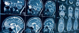
Interpretation of brain MRI results consists of the following steps:
- The magnetic resonance tomograph transmits the examination results to a special computer. They are displayed as pictures of the brain. Ideally, there should be 4 projections: front, top, left and right.
- All photographs are printed on film.
- The specialist places all the images on a table with internal lighting.
- Consistently, without missing a single detail, the doctor examines all images. It determines normal values and the presence of abnormalities.
- The doctor draws up all his findings in the form of a written report and gives it to the patient.
The results of MRI of the brain in the form of a conclusion contain information about the shape and condition of all tissues examined. A conclusion is made about whether there are deviations from the norm.
The radiologist does not have the right to make an accurate diagnosis and develop a treatment program. This can only be done by the specialist who issued the referral for examination.
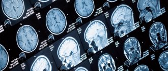
What does a healthy person's brain look like in an image?
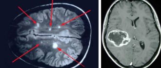
MRI of the head helps to obtain images in which tissues are indicated by darkening and clearing. Brain tissue is gray in color. Leaking cerebral fluid appears as light gray streams. The black cavities in the image are intracerebral sinuses.
If all areas of the brain are developed correctly, then the intensity of the signal received from the tomograph will be the same. In a healthy person, the ventricular system should have normal dimensions. Any expansion or decrease is considered a deviation. Normally, there should be both perivascular and subarachnoid spaces. Pay attention to the condition of the grooves and convolutions. There should be no deviations in them.
The structure of the brain itself should also be within normal limits. It should not be displaced. The eye sockets, ear canals and sinuses should be of normal size. No diffuse or focal changes should be observed in the brain tissue.
During the procedure with contrast, you can carefully examine the condition of the vessels. They must be properly developed. The contrast agent should fill all vessels evenly.
If the MRI of the brain turns out to be inaccurate, that is, the image does not have sufficient clarity, the doctor decides to repeat the study. People may move during the procedure, which makes the picture blurry.
In some cases, the doctor prescribes a procedure with contrast. In this case, a special chemical is injected into the patient’s blood. Thanks to it you can get a clear, high-quality image. In this case, it is much easier to decipher the MRI of the brain.
The types of brain MRI results and the norm are prescribed in medical reference books. To identify abnormalities, the specialist always compares the patient’s images with samples from a healthy person.
What do the diseases look like in the pictures?
Deciphering an MRI of the brain is a complex and lengthy process. Even the patient himself can identify some serious diseases in the images. They are clearly visible in the image. These include:
Stroke
This disease is accompanied by cerebral oxygen starvation. The area where hypoxia is particularly severe is indicated by a light spot on the image. If the procedure was carried out with contrast, you can notice how reduced the blood supply is in this area.
Vascular ruptures help decipher the presence of a hemorrhagic stroke. Such places are displayed as dark cavities, which will have ring-shaped stripes along the periphery. Over time, the thickness of such rings will decrease, therefore, the sooner the patient is examined, the more accurately the diagnosis will be made.
Multiple sclerosis
This disease is accompanied by the appearance of nerve fibers that have lost their myelin layer. Such anomalies will be visible on the image as focal formations. During a procedure with contrast, they will have different shades, since they accumulate chemicals in different quantities.
Such lesions can be located in various areas of the white matter. At the initial stage of the disease, as a rule, one or two lesions are detected. As the disease progresses, the number of lesions can number in the dozens.
Neoplasms
When describing MRI of the brain, it is easiest to detect the presence of tumors. They look like light spots that have an asymmetrical shape and uneven edges.
The neoplasm can impair the functioning of surrounding tissues. If the tumor grows quickly enough, the formation of new blood vessels is observed in this area. If cancer is suspected, experts recommend conducting a contrast study. This will help more accurately identify the location of the tumor and the possibility of surgical removal.
Other pathologies
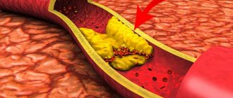
- Vascular atherosclerosis. Determination of diseases of the vascular system is carried out only during studies with contrast. With atherosclerosis, the images will clearly show a decrease in the lumen of blood vessels and the presence of atherosclerotic plaques.
- Aneurysm. The image will show that the walls of the vessels have become thin and expanded.
- Hypertensive angiopathy. The images will show small round cavities located in close proximity to the vessels.
- Malformation. An MRI will show radially arranged vessels that connect closer to the center.
- Hydrocephalus. The ventricular cavities are significantly expanded. The perivascular and subarachnoid spaces are changed.
- Congenital anomalies. Determined by comparing patient images with reference images. If the detected anomalies do not pose a threat to human health, then treatment will not be required.
Deviations from the norm in photographs may appear differently in different people. Therefore, deciphering the results should be trusted only to a practicing specialist with extensive experience.
What can make deciphering an MRI difficult?
The results of MRI of the brain in most cases are accurate and reliable. The contrast procedure always helps to get a clear picture. Therefore, in the absence of any contraindications, it is better to carry it out.
A deteriorated image is obtained in patients with endoprostheses. The presence of metal in them distorts the image. In addition, any metal object left in a pocket may invalidate the test results. Therefore, before the procedure, it is extremely important to remove all jewelry and check the contents of your pockets.
Metal particles may be contained in the ink used to apply tattoos. Therefore, the presence of drawings on the body becomes a contraindication to MRI. They not only worsen the results, but also cause pain during the examination.
The presence of braces can also ruin the picture. In this regard, it is imperative to notify the radiologist about them. If possible, it is better to remove them during the study.
Is it possible to interpret MRI results yourself?
The conclusion of MRI of the brain should be made only by a specialist with extensive experience. In order to correctly determine the presence of anomalies, it is not enough to have samples; it is necessary to have an excellent understanding of the anatomy of the human body. In addition, the doctor must not only analyze the images themselves, but also correlate them with the results of the preliminary examination and tests performed. This is the only way to get a complete picture of the disease and subsequently develop the correct treatment method.
If the deciphered MRI of the brain gives you doubts, you can always get the pictures in hand. The radiologist will give them to you in printed form and record them on any electronic media. You can then ask another specialist to analyze them.
Today it is possible to receive online consultations with specialists. All you have to do is post your photos on a specialized resource. You will receive not only a transcript of the results, but also an explanation of them in simple accessible language. But such consultations should not be taken as final. It is necessary to gather all opinions and consult a doctor you trust.
Remember that you need to do an MRI of the brain and get a transcript only from a specialist with extensive experience in practice.
Syrchin Vladislav Leonidovich Doctor Radiologist of the Highest category Experience - 16 years
Fadeev Stanislav Viktorovich Doctor Radiologist Candidate of Medical Sciences Experience - 15 years.
MRI of vessels and arteries of the neck and MRI of brain vessels: differences
When performing MRI of cerebral vessels, there is no direct possibility of assessing vascular structures. If a patient has a large aneurysm, most likely this will be visible during a routine native examination of the head.
As for the need to visualize vascular structures, for example, the demanded analysis of blood flow through the vascular system, excluding possible narrowings or other defective variants of the structure of the vascular bed, it is necessary to conduct an examination of the head. Also, if the patient has any ischemic conditions or strokes, with the help of additional brain diagnostics, it is possible to determine at the level of which of the arteries the “catastrophe” occurred.
When is magnetic resonance imaging of the cerebral vasculature indicated?
There are a number of diseases and conditions on the basis of which the attending physician gives a referral for such an examination:
- Pathologies and diseases of the cerebral vessels - strokes of various natures, intracranial hemorrhages, etc.;
- Previous injuries and bruises to the head area, injuries to the cervical spine;
- Diagnosis of tumor processes in the brain or cerebellopontine space;
- Impaired or decreased hearing and speech;
- Frequently recurring headaches;
- Feast for the diagnosis of inflammatory processes occurring in the intracranial space;
- Paroxysmal syndrome;
- Suspicion of thrombosis or aneurysm;
- Epileptic seizures;
- Pathologies in the development of the pituitary region and sellar;
- Multiple sclerosis ;
- Neurodegenerative disorders.
Decoding of received images
MRI of the head and neck vessels is a complex diagnostic method and requires special skills that only a radiologist and a neurologist can have. A few hours after MRI of the cerebral vessels, you can get a detailed answer from a specialist about the current situation. Also receive recommendations regarding treatment or further hospitalization of the patient.
The examination is carried out using modern equipment, so in addition to the conclusion, as well as photographs, the patient receives a CD with a recording of the examination. It is the use of the latter medium that makes it possible to find abnormal channels of the vascular system of small sizes.
Types of MRI of the head
Most types of magnetic resonance scanning of the head are available at our medical center, including:
- MRI of the brain;
- MRI of the eye orbits and optic nerves;
- MRI of the sinuses;
- MRI of the pituitary gland;
- MR angiography of cerebral vessels.
What does an MRI of the brain include? During the examination, the doctor assesses the state of blood circulation in this important organ, analyzes the structural state of the nerve nodes and identifies focal formations. If a brain scan reveals pathology of the blood vessels, pituitary gland or sinuses, then all information will be included in the description and conclusion of the MRI, with a recommendation for further further examination.
This month, our medical center is running promotions for diagnostic events and discounts on tomography, including additional bonuses for our patients. This information can be found on the medical center’s website or during a telephone consultation, which can be obtained by number.
Thanks to our 24/7 service, our consultants will help you choose the most appropriate time of day and day of the week for scanning.
Examination with contrast
An MRI examination of the vessels and arteries of the neck using contrast is carried out exclusively according to the doctor’s indications. As a rule, injection of a dye into a vein is not required. At the time of examination, attention is first paid to the native part of the procedure. If at this stage any pathology or mass formation is detected, the radiologist may decide to carry out such a manipulation.
Despite such a simple procedure, there are a number of diseases that require the introduction of a contrast agent for better diagnosis. These include multiple sclerosis.
Benefits and risks of the study
Advantages:
- MRI is a non-invasive imaging technique in which the patient's body is not exposed to ionizing radiation.
- Compared to other imaging techniques, MRI provides clearer, more detailed images of the brain and other structures of the nervous system. This property makes MRI an invaluable tool for early diagnosis and assessment of the status of many diseases, including tumors.
- MRI helps doctors evaluate structural abnormalities of the brain while analyzing its function (functional MRI).
- MRI can detect pathological lesions hidden by bone formations and therefore invisible to other imaging methods.
- The contrast material used in MRI is much less likely to cause allergic reactions than the iodine-based contrast used in traditional x-rays and CT scans.
- MRI is the most sensitive diagnostic tool for detecting brain tumors.
- A type of MRI called magnetic resonance angiography provides detailed images of the brain's blood vessels without the need for contrast material.
- New MRI systems are able to reflect the functioning of the brain and are therefore used to diagnose stroke at the earliest stages.
Risks:
- If appropriate safety guidelines are followed, MRI carries virtually no risks for the average patient.
- When using sedatives, there is a risk of overdose. This is why the radiologist assistant carefully monitors the patient's vital signs.
- Despite the fact that the powerful magnet in the scanner itself is harmless, problems during MRI can arise if there are devices implanted into the patient's body that contain metals.
- When contrast material is injected, there is a very small risk of developing an allergic reaction. Such reactions are usually very mild and go away quickly when appropriate medications are prescribed. If allergy symptoms occur, a radiologist or nurse will immediately provide the necessary assistance.
- One recently described, but extremely rare, complication of MRI is nephrogenic systemic fibrosis, which develops when large doses of gadolinium-based contrast material are administered to patients with impaired renal function.
Up
How often should the examination be carried out?
MRI of the vessels and arteries of the neck is a completely safe diagnostic method and does not leave any traces in the body. It can be carried out countless times, namely as many times as the patient’s condition requires until a complete diagnosis is completed. For some, one study without follow-up is enough. A patient who needs a follow-up examination, for example, after removal of large tumors, undergoes MRI of the vessels and arteries of the neck after 6-12 months.
In what situations should the examination not be carried out?
Earlier we talked about the most important advantages of this scan, but despite their large number, there are also contraindications for MRI. There are absolute and partial bans. Let's consider when you should never do an MRI of the vessels of the neck. So:
- If the body contains inserts, implants or other devices consisting of metal.
- If the patient is overweight, standard tomographs are designed for people weighing up to 130 kg and waist size up to 150 cm.
Doctors include the following as relative indicators:
- Heart failure.
- First trimester of pregnancy.
- Mental and psychological disorders.
- Fear of closed spaces.
- Severe conditions of a person in which he is unconscious (coma, connection to a ventilator, etc.).
Also, it is worth saying that relative restrictions can in some cases be eliminated if scanning is done under anesthesia. Quite often, doctors use clinical sleep so that the patient can remain completely still for some time. This method is used for patients who are unable to control their movements, children and people with mental disorders.
Contraindications
MRI of the head and neck vessels has virtually no contraindications. The only exception is the presence of metals of a certain group in the body. These include products such as pacemakers or other electronics in a metal case. A complete exception may be the presence of titanium structures after osteosynthesis, fractures or joint surgeries. Each situation is considered individually. As a result, each patient can learn about the presence of contraindications.
Is brain imaging dangerous?
Not all patients understand the difference between magnetic and computed tomography, so in the comments you can find opinions about the dangers of both procedures. In fact, CT is a type of X-ray, and MRI is based on the properties of the magnetic field. Due to the absence of ionizing radiation, the second is considered safer and, according to indications, is prescribed even to pregnant women and small children.
Contraindications for MRI
Like any medical procedure, MRI has limitations. This method is absolutely prohibited for use if there are metal and electronic objects in (or on) the human body. Relative contraindications allow the appointment of MRI if symptoms require it, but subject to certain conditions. Among the restrictions are:
- weight above 120 kg;
- claustrophobia;
- pregnancy in the first trimester;
- conditions that prevent you from remaining still during scanning;
- heart disease during exacerbation;
- the presence of an insulin pump, neurostimulator, dental crowns, braces, infusion port system, artificial heart valve, analog hearing aid.
With magnetic resonance imaging with contrast, the risk may be associated with an allergic reaction to the substance injected. Although gadolinium medications rarely cause side effects, for patients with a history of dye allergy symptoms, diagnosis should be made after a preliminary test.
Also, MRI with contrast is not prescribed for people with severe renal and liver failure, pregnant women and after liver transplant surgery. Lactation is not an absolute contraindication, but since gadolinium passes into breast milk, you will have to wait until it is completely eliminated from the body for the next feeding. The mother should worry about the supply of food for the baby in advance.
What is more informative: MRI of head and neck vessels or computed tomography
CT angiography with the introduction of a contrast agent still remains a more informative procedure. This level of detail also has one significant drawback - the absorption of high load rays by the body. The latter can aggravate the development of cancer and completely excludes dynamic monitoring of certain pathological conditions.
The iodine-containing contrast agent administered during CT angiography is quite allergenic. Therefore, not everyone can conduct computer research. The examination itself is variable. At the time of assessing the clinical picture, the neurologist already understands which study will be more relevant for the patient: MRI of the vessels and arteries of the neck or CT angiography.
Choosing a clinic, specialist and price of service
The mrt-mozga service is a full-fledged resource with a database of diagnostic clinics in Moscow (about 300 centers). The resource’s simple and intuitive interface will suggest the best offer. The patient can sort clinics according to various parameters: geolocation of the clinic, price, type of tomograph used, availability of contrast agent, operating hours, promotional offers. The information on the site is constantly updated and expanded.
You can make an appointment by calling 8 (495) 032-07-59 from 09:00 to 00:00. By taking advantage of the site's offer, you can not only make an appointment for free, but also get the best price offer.