Oculomotor nerve (n. oculomotorius, III pair of cranial nerves)
Motor neurons of the oculomotor nerves (n. oculomotorius, III pair of cranial nerves) are located on both sides of the midline in the rostral part of the midbrain. These nuclei of the oculomotor nerve innervate the five extrinsic muscles of the eyeball, including the levator palpebral muscle. The nuclei of the oculomotor nerve also contain parasympathetic neurons (Edinger-Westphal nucleus), which are involved in the processes of pupil constriction and accommodation.
The normal position of the eyeballs and divergent strabismus are shown with weakness of the medial (internal) rectus muscle of the eye on the right (n. oculomotorius, III pair of cranial nerves).
There is a division of supranuclear groups of motor neurons for each individual eye muscle. The fibers of the oculomotor nerve innervating the medial rectus, inferior oblique and inferior rectus muscles of the eye are located on the side of the same name. The subnucleus of the oculomotor nerve for the superior rectus muscle is located on the contralateral side. The levator palpebrae superioris muscle is innervated by the central group of cells of the oculomotor nerve.
Trochlear nerve (n. trochlearis, IV pair of cranial nerves)
The motor neurons of the trochlear nerve (n. trochlearis, IV pair of cranial nerves) are closely adjacent to the main part of the complex of nuclei of the oculomotor nerve. The left nucleus of the trochlear nerve innervates the right superior oblique muscle of the eye, the right nucleus innervates the left superior oblique muscle of the eye.
Abducens nerve (n. abducens, VI pair of cranial nerves)
Motor neurons of the abducens nerve (n. abducens, VI pair of cranial nerves), innervating the lateral (external) rectus muscle of the eye on the side of the same name, are located in the nucleus of the abducens nerve in the caudal part of the pons. All three oculomotor nerves, leaving the brain stem, pass through the cavernous sinus and enter the orbit through the superior orbital fissure.
Clear binocular vision is ensured precisely by the joint activity of individual muscles of the eye (oculomotor muscles). Conjugate movements of the eyeballs are controlled by the supranuclear gaze centers and their connections. Functionally, there are five different supranuclear systems. These systems provide various types of eyeball movements. Among them there are centers that control:
- saccadic (rapid) eye movements
- purposeful eye movements
- convergent eye movements
- holding the gaze in a certain position
- vestibular centers
Weakness of the lateral (external) rectus muscle of the right eye (n. abducens, VI pair of cranial nerves) was shown before and after treatment.
Saccadic (rapid) eye movements
Saccadic (fast) movements of the eyeball occur as a command in the opposite visual field of the cortex of the frontal region of the brain (field 8). The exception is fast (saccadic) movements that occur when the central fovea of the retina is irritated, which originate from the occipital-parietal region of the brain. These frontal and occipital control centers in the brain have projections on both sides in the supranuclear brainstem centers. The activity of these supranuclear brain stem centers of vision is also influenced by the cerebellum and the vestibular nuclei complex. The paracentral sections of the reticular formation of the bridge are the stem center, providing friendly rapid (saccadic) movements of the eyeballs. Simultaneous innervation of the internal (medial) rectus and opposite external (lateral) rectus muscles when moving the eyeballs horizontally is provided by the medial longitudinal fasciculus. This medial longitudinal fasciculus connects the nucleus of the abducens nerve with the subnucleus of the complex of oculomotor nuclei, which are responsible for innervation of the opposite internal (medial) rectus muscle of the eye. To initiate vertical rapid (saccadic) eye movements, bilateral stimulation of the paracentral sections of the pontine reticular formation is required from the cortical structures of the brain. The paracentral sections of the pontine reticular formation transmit signals from the brain stem to the supranuclear centers that control the vertical movements of the eyeballs. This supranuclear eye movement center includes the rostral interstitial nucleus of the medial longitudinal fasciculus, located in the midbrain.
Symptoms of facial paresis
The branches of the facial nerve are responsible for motor function. They provide muscle mobility and make facial expressions lively. If the nerve impulse does not pass, facial movements become impossible. There are three stages of facial nerve paresis:
- Acute (lasts up to two weeks).
- Subacute (lasts up to one month).
- Chronic (illness lasts more than a month).
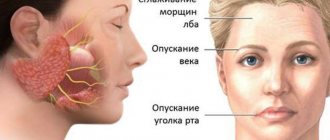
On the affected side of the face there are:
- drooping corner of the mouth;
- smoothing the nasolabial fold;
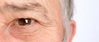
- a wide eyelid, when closing the eye, lagophthalmos is observed (a visible strip of light-colored sclera remains noticeable);
- loss or deterioration of taste sensations on the first third of the surface of the tongue;
- inability to stretch lips;
- leakage of food from the half-open half of the mouth;
- lacrimation (tears begin to flow while eating);
- worsening of hearing in the first days of illness (discomfort with loud sounds);
- pain behind the ear;
- inability to wrinkle the forehead much.
Paresis can occur through three stages:
- Mild degree. Facial asymmetry is slightly expressed. There is a slight distortion of the lips due to inflammation, difficulty when trying to completely close the eyes and frown the eyebrows.
- Moderate severity. The eyelid does not close completely (lagophthalmos); only minor movements are possible in the upper part of the face. The patient cannot puff out his cheek or perform various movements with his lips.
- The degree is difficult. Facial asymmetry is very pronounced. The mouth is distorted, the eye on the side of the paresis almost does not close. The patient cannot perform even the simplest movements that require the participation of facial muscles.
If you experience similar symptoms,
consult your doctor . It is easier to prevent a disease than to deal with the consequences.
Purposeful eye movements
The cortical center for smooth targeted or tracking movements of the eyeballs is located in the occipital-parietal region of the brain. Control is carried out from the side of the same name, i.e. the right occipital-parietal region of the brain controls smooth, targeted eye movements to the right.
Testing of targeted eye movements is carried out by tracking an object from the center to the periphery with the patient's head motionless.
Treatment of convergent strabismus in the MGK
At the Moscow Eye Clinic, patients with convergent strabismus are provided with a wide range of diagnostic and therapeutic procedures. Diagnostics are performed by qualified specialists with unique clinical experience. When prescribing treatment, a strictly individual approach is used. The clinic has an extensive methodological and hardware base for the treatment of such pathologies. These techniques are suitable for both children and adults:
- Infrared laser therapy (exposure to infrared radiation on the organ of vision at close range), the procedures of which make it possible to improve the hemodynamics of eye tissue and relieve spasm of accommodation.
- Laser therapy sessions designed to improve spatial vision and accommodation functions.
- Exercises on the device “Synoptophore Sinf-1”. This device is the main one for stimulating vision, necessary for strabismus and other types of binocular vision disorders. It allows you to carry out sets of special exercises aimed at merging images, which makes it possible to train vision impaired by strabismus. In addition, such training significantly increases visual reserves and improves eye mobility.
- Treatment with the Spekl-M device, using two types of laser, which is indicated for clouding of optical media, amblyopia and strabismus of various origins.
- Especially for young patients with strabismus, MGK offers training with the Rainbow BZR-1 kit. This kit, in a playful way, helps restore central fixation and other functions of binocular vision.
Doctor of Medical Sciences Svetlana Gavrilovna Chernysheva deals with the problems of all types of strabismus at the Moscow Eye Clinic. Svetlana Gavrilovna is an ophthalmic surgeon, professor, the best specialist in Moscow in the field of diagnosing binocular oculomotor pathology in adults and children, as well as its functional and surgical treatment. Proficient in various methods of surgical treatment of all types of strabismus.
You can find out the cost of a particular procedure or make an appointment at the Moscow Eye Clinic by calling 8 8 (499) 322-36-36 (daily from 9:00 to 21:00) or using the online registration form.
Eye movement with changes in gravity and acceleration
Coordination of the movements of the eyeballs in response to changes in gravity and acceleration is carried out by the vestibular system (vestibular-ocular reflex). When the coordination of movements of both eyes is disturbed, double vision develops, since images are projected onto disparate (inappropriate) areas of the retina. In congenital strabismus, or strabismus, a muscle imbalance that causes the eyeballs to be misaligned (nonparalytic strabismus) may cause the brain to suppress one of the images. This decrease in visual acuity in the non-fixing eye is called amblyopia without anopia. In paralytic strabismus, double vision occurs as a result of paralysis of the muscles of the eyeball, usually due to damage to the oculomotor (III), trochlear (IV) or abducens (VI) cranial nerves.
Causes
The most common causes of gaze paresis in adults are:
- diabetes
- arterial hypertension
- atherosclerosis
- injury
- idiopathy.
Less common reasons:
- increased intracranial pressure
- giant cell arteritis
- presence of tumor inclusions
- multiple sclerosis
- stroke
- Chiari malformation
- hydrocephalus
- intracranial hypertension
- previous meningitis.
Among other things, paresis can also occur due to viral diseases such as diphtheria, syphilis, encephalitis, or as a result of complications after the flu.
In some cases, intoxication with ethyl alcohol, heavy metals or combustion products can provoke the appearance of pathology.
In pediatric practice, the most common causes are tumor diseases, injuries and idiopathies.
Reference! Children sometimes develop a benign and quickly recovering isolated abducens nerve palsy, which in some cases is a consequence of previous infections of the ear, nose or throat.
Eyeball muscles and gaze palsies
There are three types of paralysis of the external muscles of the eyeball:
- paralysis of individual eye muscles
- paralysis of friendly movements (gaze)
- mixed paralysis
Paralysis of individual eye muscles
Characteristic clinical manifestations occur with isolated damage to the oculomotor (III), trochlear (IV) or abducens (VI) nerve.
Complete damage to the oculomotor (III) nerve leads to ptosis. Ptosis manifests itself in the form of weakening (paresis) of the muscle that lifts the upper eyelid and disruption of voluntary movements of the eyeball upward, downward and inward, as well as divergent strabismus due to the preservation of the functions of the lateral (lateral) rectus muscle. When the oculomotor (III) nerve is damaged, pupil dilation and lack of reaction to light (iridoplegia) and paralysis of accommodation (cycloplegia) also occur. Isolated paralysis of the muscles of the iris and ciliary body is called internal ophthalmoplegia.
The normal position of the eyeballs and convergent strabismus are shown with weakness of the lateral (external) rectus muscle of the eye on the right (n. abducens, VI pair of cranial nerves).
Injuries to the trochlear (IV) nerve cause paralysis of the superior oblique muscle of the eye. Such damage to the trochlear (IV) nerve leads to outward deviation of the eyeball and difficulty moving (paresis) downward gaze. Paresis of downward gaze is most clearly manifested when turning the eyes inward. Diplopia (double vision) disappears when the head is tilted to the opposite shoulder, which causes a compensatory inward deviation of the intact eyeball.
Damage to the abducens (VI) nerve leads to paralysis of the muscles that abduct the eyeball to the side. When the abducens (VI) nerve is damaged, convergent strabismus develops due to the predominance of the influence of the tone of the normally working internal (medial) rectus muscle of the eye. With incomplete paralysis of the abducens (VI) nerve, the patient can turn his head towards the affected abductor muscle of the eye in order to eliminate the existing double vision using a compensatory effect on the weakened lateral rectus muscle of the eye.
The severity of the above symptoms in cases of damage to the oculomotor (III), trochlear (IV) or abducens (VI) nerve will depend on the severity of the lesion and its location in the patient.
Friendly gaze paralysis
Companionate gaze is the simultaneous movement of both eyes in the same direction. Acute damage to one of the frontal lobes, for example, during cerebral infarction (ischemic stroke), can lead to transient paralysis of voluntary conjugate movements of the eyeballs in the horizontal direction. At the same time, independent eye movements in all directions will be completely preserved. Paralysis of voluntary conjugate movements of the eyeballs in the horizontal direction is detected using the doll eye phenomenon when passively turning the head of a horizontally lying person or using caloric stimulation (infusion of cold water into the external auditory canal).
Unilateral damage to the inferiorly located paracentral section of the reticular formation of the pons at the level of the nucleus of the abducens nerve causes persistent paralysis of gaze in the direction of the lesion and loss of the oculocephalic reflex. The oculocephalic reflex is a motor reaction of the eyes to irritation of the vestibular apparatus, as with the phenomenon of the head and eyes of a doll or caloric stimulation of the walls of the external auditory canal with cold water.
Damage to the rostral interstitial nucleus of the medial longitudinal fasciculus in the anterior midbrain and/or damage to the posterior commissure causes supranuclear upward gaze palsy. Added to this focal neurological symptom is the dissociated reaction of the patient’s pupils to light:
- sluggish pupil reaction to light
- rapid reaction of the pupils to accommodation (changing the focal length of the eye) and looking at nearby objects
In some cases, the patient also develops convergence paralysis (movement of the eyes towards each other, in which the gaze will focus on the bridge of the nose). This symptom complex is called Parinaud's syndrome. Parinaud's syndrome occurs with tumors in the pineal gland, in some cases with cerebral infarction (ischemic stroke), multiple sclerosis and hydrocephalus.
Isolated downward gaze palsy is rare in patients. When this occurs, the cause is most often blockage (occlusion) of the penetrating arteries in the midline and bilateral infarctions (ischemic strokes) of the midbrain. Some hereditary extrapyramidal diseases (Huntington's chorea, progressive supranuclear palsy) can cause restrictions in the movement of the eyeballs in all directions, especially upward.
Mixed paralysis of gaze and individual muscles of the eyeball
The simultaneous combination of gaze paralysis and paralysis of individual muscles that move the eyeball in a patient is usually a sign of damage to the midbrain or pons. Damage to the lower parts of the pons with destruction of the abducens nerve nucleus located there can lead to paralysis of rapid (saccadic) horizontal movements of the eyeballs and paralysis of the lateral (external) rectus muscle of the eye (abducens nerve, VI) on the affected side.
With lesions of the medial longitudinal fasciculus, various gaze disturbances occur in the horizontal direction (internuclear ophthalmoplegia).
Unilateral damage to the medial longitudinal fasciculus caused by infarction (ischemic stroke) or demyelination leads to disruption of the inward adduction of the eyeball (to the bridge of the nose). This can manifest clinically as complete paralysis with the inability to move the eyeball inward from the midline, or as a moderate paresis, which will manifest itself as a decrease in the speed of adducting rapid (saccadic) eye movements to the bridge of the nose (adductive delay). On the side opposite to the lesion of the medial longitudinal fasciculus, as a rule, abduction nystagmus is observed: nystagmus that occurs when the eyeballs are abducted outward with a slow phase directed towards the midline and fast horizontal saccadic movements. An asymmetrical arrangement of the eyeballs relative to the vertical line often develops with unilateral internuclear ophthalmoplegia. On the affected side, the eye will be positioned higher (hypertropia).
Bilateral internuclear ophthalmoplegia occurs with demyelinating processes, tumors, infarction, or arteriovenous malformations. Bilateral internuclear ophthalmoplegia leads to a more complete syndrome of eyeball movement disorders, which are manifested by bilateral paresis of the muscles that lead the eyeball to the bridge of the nose, impaired vertical movements, purposeful tracking movements and movements caused by the influence of the vestibular system. There is a disturbance of gaze along a vertical line, upward nystagmus when looking up and downward nystagmus when looking down. Lesions of the medial longitudinal fasciculus in the overlying (rostral) parts of the midbrain are accompanied by a violation of convergence (convergent movement of the eyes towards each other, towards the bridge of the nose).
Diagnostics
In order to clarify the nature of the pathology and its causes, patients should be examined by an ophthalmologist, endocrinologist, neurologist, cardiologist, rheumatologist and hematologist.
An ophthalmological examination includes examination of the ocular structures, performing functional tests, as well as other studies - x-ray, ultrasound, electrophysiological.
When checking visual acuity, it is revealed that it decreases from insignificant to the level of light perception. Examination of visual fields reveals their defects, which corresponds to damage to various parts of the optic nerve.
Ophthalmoscopy reveals pallor, swelling due to ischemia and an increase in the size of the optic nerve head, its protrusion into the vitreous body. Swelling of the retina around the optic disc is observed; the macular area is characterized by a “star shape”. In the compression zone, narrow veins are identified, in contrast to the periphery, where they are full-blooded. Sometimes exudation and focal hemorrhages are detected.
With angiography of retinal vessels, ischemic optic neuropathy is manifested by retinal angiosclerosis, age-related fibrosis, unequal size of arteries and veins, occlusion of cilioretinal arteries. Ultrasound examinations of the carotid, supratrochlear and vertebral arteries often determine blood flow disturbances.
Electrophysiological studies show decreased function of optic nerve thresholds. A coagulogram reveals hypercoagulability. Determination of cholesterol and lipoproteins shows hyperlipoproteinemia.
Differential diagnosis of ischemic optic neuropathy is carried out with retrobulbar neuritis, space-occupying formations of the orbit and central nervous system.