Causes
Hemorrhage usually occurs after injury. This could be a bruise of the skin, internal organs, a concussion or bruise of the brain, an injection with thin (sharp) objects. Sometimes blood leaves the vessels and pours into the skin and internal organs as a result of infections, autoimmune diseases, and poisoning. The occurrence of hemorrhages and bruises is promoted by increased fragility of blood vessels, fasting, lack of vitamins in food, high blood pressure, and congenital bleeding disorders.
At CELT you can get a consultation with a traumatologist-orthopedic specialist.
- Initial consultation – 3,000
- Repeated consultation – 2,000
Make an appointment
Symptoms
A bruise on the skin is always clearly visible. At first it has a purplish-blue color, and then begins to “bloom”, acquiring yellow and green colors. If a sufficiently large amount of blood accumulates under the skin, a protruding lump forms. At first it is very painful to feel, but later the pain goes away.
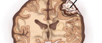
The outpouring of blood into the internal organs and into the substance of the brain is preceded by trauma. The main symptom is pain. In this case, the hematoma is not visible externally. If bleeding continues, the victim becomes pale, weak, and dizzy. With chronic internal bleeding, anemia comes to the fore. Bleeding in the brain is especially dangerous. Compression of brain structures may occur, sometimes leading to death.
Predisposing factors
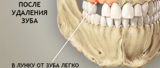
The formation of hematomas occurs after injuries, including pinching, blows, squeezing, and bruises. Subarachnoid hemorrhage does not fall into this category, since it does not appear due to trauma, but due to damage to an unchanged vessel. Often small hematomas appear due to eating large quantities of food or drinking alcoholic beverages. This is due to stretching of the gastrointestinal tract and the appearance of cracks.
The development of pathology is influenced by vascular weakness and problems with blood clotting. Often due to a weakened immune system due to infections or age-related changes, the likelihood of pus accumulating in the affected area increases.
Pathogenesis
The process of cavity formation is based on the process of traumatic injury to a blood vessel (rupture) without the presence of an open wound or with a minor injury.
The reasons for violating the integrity of blood vessels are:
sharp intense local impact from impacts, falls and bruises;- damage caused by bone fragments during fractures or confusion of one anatomical structure relative to another during dislocations.
Under pressure, blood flows out, forming cavities. Some of the leaked blood seeps through the surrounding tissue, causing specific changes in the color of nearby skin. Subsequently, under the influence of the breakdown of red blood cells, the color of the tissue may change (with deep hematomas, the color of the skin does not change).
Over time, the hematoma either resolves or undergoes a series of successive changes, ending with its complete scarring. When opening “fresh” formations, no change in the color and structure of the blood is observed (in rare cases, under the influence of platelets, the blood may thicken).
When old formations are drained, a change in the color and thickness of the blood is observed (it becomes dark burgundy or black). When a cavity is opened and the scarring process has begun, the blood takes the form of a dark, hard and difficult-to-separate substance, which is associated with the beginning of the process of degeneration of injured tissue into fibrous tissue. If the formation becomes infected, its cavity is filled with purulent exudate.
Classification of hematomas
In modern medicine, when classifying hematomas, the following are taken into account:
- Relation to the vessel - pulsating and non-pulsating hematomas.
- Localization - in the cranial cavity, internal organs, under the skin or mucous membrane.
- The state of the blood in the affected area is suppurated, clotted, fresh, infected.
- Symptoms – limited, encysted, diffuse.
There are hematomas that do not fall into this classification. For example, intracerebral, intracranial, intraventricular. They are of the epidural or subdural type and cause serious complications.
Drainage of hematoma and other methods of its treatment
The human body is riddled with blood vessels.
During domestic, industrial or sports injuries, the integrity of the wall of a particular vessel is compromised. This can lead to bleeding or hematoma formation. Hematoma is an accumulation of liquid or coagulated blood in the thickness of soft tissue. This is often a harmless phenomenon, but in some cases doctors are often faced with the need to remove a large hematoma, which can interfere with the functioning of tissues and neighboring organs. Hematomas can form:
- for all types of injuries – closed and open;
- in any part of the human body;
- for injuries of any degree - from minor (in this case, the role is played not by physical effort, but by the fragility of the vascular wall) to severe.
Hematomas can occur not only when physical force is applied to the tissues, but also several hours and days after that. An example is an internal hematoma after surgery - when sutures placed on blood vessels damaged during surgery fail. The possibility of such tissue hemorrhages must be constantly remembered. The timeliness of diagnosis, and therefore the success of hematoma treatment, depends on this.
Soft tissue hematomas
Soft tissue hematomas are divided into 3 types:
- Lungs - appear 24 hours after injury and are accompanied by mild pain. No special treatment is required.
- Medium - appear within 5-6 hours and are accompanied by pain and swelling. The motor function of the limb deteriorates. Consultation with a traumatologist is required.
- Heavy - formed within 2 hours after tissue damage. The function of the limb is impaired, acute pain and diffuse swelling are observed. You should immediately consult a doctor to determine a treatment strategy.
Immediately after the injury, swelling appears, and the skin acquires a purplish-bluish tint. After 5 days, the skin takes on a green tint as hemoglobin breaks down. Gradually, the hematoma resolves and “flows” down.
If there are no complications, the hematoma will resolve on its own. In the worst case, a hard area appears that causes discomfort and impairs motor function. When an intramuscular lump forms, external symptoms are rarely observed, but the limb swells significantly and an area forms inside, the touch of which causes severe pain.
Note! For chronic intramuscular hematomas, an MRI is prescribed to determine the location and extent of tissue damage.
When large lumps form, surgical intervention is required. Treatment is carried out by a traumatologist. The opening of infected seals is performed by a surgeon after a comprehensive diagnosis. The operation is performed on an outpatient basis, but for large hematomas hospitalization is required. An autopsy is performed, during which blood clots are removed and washing is carried out. Drainage and suturing are required. Sutures are not applied only for infected hematomas. Antibiotics are often prescribed in combination to eliminate the infection.
Causes of hematoma
In a healthy person, hematomas resolve within three weeks. The period may increase slightly if a person takes blood thinning medications:
- Eliquis;
- Warfarin;
- Clopidogrel;
- Prasugrel (Effient);
- Xalerto.
Permanent non-healing hematomas that occur even after minor exposure to the skin are a sign of hepatitis C, HIV, and parvovirus. In such patients, bruises may not heal for months. It is recommended to consult a hematologist, take blood tests and find out the true cause of the problem. Sometimes long healing of hematomas is provoked by:
- thrombocytopenia (low platelet levels in the blood);
- aplastic anemia (diagnosed when the bone marrow stops producing certain blood cells);
- regular consumption of large amounts of alcohol;
- vitamin D deficiency.
But don’t worry, because for the vast majority of people, bruises go away quickly enough without the help of doctors.
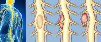
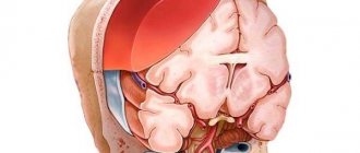
Intracranial hematomas
Intracranial hematomas are divided into the following types:
- Epidural.
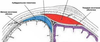
- Subdural.
- Intracerebral.
- Intraventricular.
Epidurals appear in 1-3% of cases and are due to injury to the middle meningeal artery. Pathology is often observed with skull fractures or depressed fractures. A hematoma develops in 2-3 hours or within 24 hours. Lack of treatment leads to coma. The first symptoms are confusion and weakness. Children rarely lose consciousness after a severe blow. Significant swelling of the brain does not lead to the detection of a light gap (which is rare in adults).
Subdurals appear in 1-7% of cases and pose a threat to human life, since death occurs in 60% of cases. There is an acute, subacute and chronic form of the pathology. Bleeding occurs due to a rupture of a vein or artery in the damaged area. People report nausea and severe headaches. Symptoms characteristic of compression of the brain stem are often observed. Lack of treatment and worsening symptoms lead to coma.
Intracerebral are observed extremely rarely with severe traumatic brain injuries. The light gap is not visible, the development of pathology occurs quickly. Hemiplegia or hemiparesis often occurs, as well as extrapyramidal symptoms.
Intraventricular diseases are rarely diagnosed due to the serious condition of patients. There are acute disturbances of consciousness, an increase in body temperature, a decrease in heart rate, and an increase in blood pressure. To establish a diagnosis, a survey of close people is carried out, since the patient is unconscious. To establish the location of the hematoma, MRI is used. In the most severe cases, lombal puncture is used.
Treatment of hematoma
A healthy body has sufficient resources to reabsorb blood accumulated subcutaneously. But if possible, then within 48 hours after injury it is worth applying a cold compress. Cooling the hematoma for 20-30 minutes reduces the risk of developing edema several times!
Also recommended:
- treat the bruise with arnica ointment;
- take acetaminophen-based pain relievers (if throbbing pain occurs);
- If subcutaneous bumps occur, use heparin ointment.
During home treatment, you should avoid squeezing or rubbing the area with the hematoma. Anti-inflammatory drugs containing aspirin and ibuprofen are also prohibited. The fact is that such medications can thin the blood and slow down the healing process.
Diagnostics
A hematoma is diagnosed by visual examination. If the hemorrhage is located deep under the skin, in internal organs, or in a joint, it is often very difficult to assess its size and possible consequences.
Patients are prescribed an examination, which may include:
- Ultrasound of internal organs, joints;
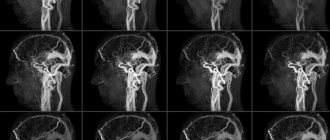
- computed tomography and magnetic resonance imaging;
- puncture (puncture with a needle): for example, a puncture of the knee joint is often done if there is a suspicion that blood has accumulated in it after an injury.
Damage diagnostics
A visual inspection of the damaged area and collection of anamnesis is the first step in providing medical care. If the injury is severe, an x-ray may be ordered.
If damage to internal organs is suspected, an ultrasound examination or magnetic resonance imaging is performed. Blood and urine tests may also be needed.
Treatment of bruises
First aid for bruised limbs begins with a thorough examination of the bruised area (flexion and extension of the arms and legs). In case of severe injury (before examination by a doctor), the injured limb must be immobilized.
If there are bruises to the chest, head or abdomen, you need to lay the victim on a flat surface, apply a cold compress and urgently call an ambulance. It is not advisable to take painkillers on your own so as not to miss the moment when your health worsens.
You need to pay attention to the following symptoms:
- the appearance of weakness and loss of consciousness (in case of a head injury, it is important to exclude intracranial hematoma);
- severe pain (this may indicate internal organ damage or a fracture),
- decreased breathing, blood in the urine (possible internal bleeding).
Treatment of hematomas
As a rule, the main part of the treatment of bruises comes down to the treatment of hematomas. In order to stop the bleeding, a cold compress is applied to the site of the bruise. Under the influence of cold, the blood vessels narrow and the hemorrhage stops.
It should be noted that cold is effective only for the first 12 hours after injury. On the second day, the best remedy for bruises (and a remedy for hematomas) is a warm compress, which, along with physical therapy, helps resolve hemorrhages.
Treatment of a hematoma in a hospital may consist of puncturing the skin on the damaged area and removing the accumulated blood.
Also, as prescribed by a doctor, medicinal treatment of bruises can be carried out, including the administration of anti-inflammatory, absorbable and vasodilating agents.
The MedicCity clinic operates a paid emergency room every day from 9.00 to 21.00. Day and night, our professional traumatologists are ready to provide you with the necessary assistance in treating bruises and any other injuries!
Treatment
Minor bruises can be treated conservatively: physiotherapeutic procedures and medications are prescribed.
In case of large accumulations of blood, the hematoma is treated surgically: it is opened, the blood or pus is evacuated, washed with antiseptics and drainage is installed. Antibiotics are prescribed if necessary.
When hemorrhaging into internal organs, it is often necessary to perform surgical intervention, during which it is necessary not only to remove the spilled blood, but also to stop the bleeding.
The multidisciplinary CELT clinic employs experienced traumatologists and surgeons who perform operations on hematomas of various locations. Modern techniques used in our clinic help provide effective treatment and minimize the risk of complications.
Hematoma puncture
Light hematomas are treated by applying aseptic dressings, ointments, compresses, and, if necessary, prescribing painkillers. Physiotherapeutic procedures will help speed up the resorption of the hematoma. But for extensive, poorly healing lesions, these therapeutic measures are not enough; puncture of the hematoma is recommended. Puncture of accumulated blood is one of the most complex surgical procedures.
Before the procedure, the state of the hematoma is determined by palpation. The skin is treated with an antiseptic, and if necessary, the patient is given an anesthetic.
After preparatory manipulations, blood is sucked out using a thin puncture needle. To speed up the removal of the contents, lightly press on the tissue surrounding the hematoma. After completing the procedure, antibiotics are injected with another syringe and an antiseptic pressure bandage is applied.
In case of extensive damage, the puncture may be performed several times. Thermal procedures are recommended to speed up the healing process.
Opening a hematoma
In case of extensive hematomas or in cases where the patient did not immediately seek medical help, the lesion on the skin is opened. Opening an extensive hematoma involves cleaning and disinfecting the wound and removing blood clots.
An accumulation of blood in tissues that forms a cavity is called an encysted hematoma. It will not be able to resolve on its own, without treatment.
Opening of the encysted hematoma is carried out under local anesthesia. An incision is made in the center of the cavity through which blood clots are removed. The wound is washed with Chlorhexedine or hydrogen peroxide to prevent infection. Drainage of the wound is carried out using a tube or rubber outlet.
If there is no pus, the wound is sutured and a bandage is applied. Otherwise, in the presence of purulent discharge, antibiotic therapy is continued.
Opening a hematoma on the leg is also indicated when there is extensive damage caused by trauma or when there is a significant risk of infection. With the help of ice, swelling is eliminated and pain is relieved, and then an autopsy is performed to cleanse the blood clots.
After surgery, the patient needs complete rest. External medications are prescribed to eliminate pain and inflammation, and physiotherapeutic procedures are performed.
Postoperative care Further treatment involves periodic treatment of the wound with antiseptic solutions and regular change of dressings. Tubular drainage is not removed. If the postoperative period is favorable, the sutures are removed seven days after the autopsy.
Postoperative care includes the following procedures:
- taking antibiotics;
- treating the hematoma site with antiseptic solutions or water-soluble ointments;
- physiotherapy;
- taking painkillers, anti-inflammatory, immunomodulating drugs (according to indications).
Provided that all medical recommendations are followed, the prognosis for healing of the hematoma site is favorable. In the absence of proper treatment, there is a risk of infection and deterioration of the patient's condition.
Prevention of hematomas involves following precautions and rules of behavior on the roads and in public places to avoid falling from a height or other similar injuries.
Orthopedics and traumatology services at CELT
The administration of CELT JSC regularly updates the price list posted on the clinic’s website. However, in order to avoid possible misunderstandings, we ask you to clarify the cost of services by phone: +7
| Service name | Price in rubles |
| Appointment with a surgical doctor (primary, for complex programs) | 3 000 |
| X-ray of the chest organs (survey) | 2 500 |
| Ultrasound of soft tissues, lymph nodes (one anatomical zone) | 2 300 |
All services
Make an appointment through the application or by calling +7 +7 We work every day:
- Monday—Friday: 8.00—20.00
- Saturday: 8.00–18.00
- Sunday is a day off
The nearest metro and MCC stations to the clinic:
- Highway of Enthusiasts or Perovo
- Partisan
- Enthusiast Highway
Driving directions