Magnetic resonance imaging is a hardware diagnostic method that is used in medical centers to obtain the most anatomically accurate images of the patient’s internal organs and tissues. Diagnostics is carried out in a non-invasive way, that is, no instruments are introduced into the patient’s body, and no punctures are made. From the point of view of the subject, the main advantage of MRI is the comfort and safety of the procedure. Doctors value magnetic resonance imaging for its:
- high tissue contrast;
- informative and comprehensive assessment of all body tissues;
- high sensitivity during oncological search.
Since the examination method does not involve any radiation exposure, it is available to a wide range of patients, including young children, pregnant women, cancer patients and the elderly.
Free diagnostic consultation
If in doubt, sign up for a free consultation. Or consult by phone
+7
All about diagnostics
How does an MRI machine work?
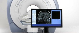
The operating principle of a magnetic resonance imaging scanner is based on the effect of nuclear magnetic resonance. The installation itself consists of a scanner with a powerful magnet inside and a computer installation that analyzes the data and displays it on the monitor in the form of three-dimensional three-dimensional images. When the patient's body enters the scanning device, it is exposed to a strong magnetic field and radio frequency pulses. This causes vibrational movements in the protons of the hydrogen atoms of the body's cells. The computer reads these resonant pulses and builds tomograms - three-dimensional images - based on them.
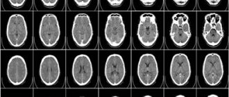
The more water is contained in the tissues of the body, the better the resonance effect will be, and the organs will appear more accurately and in more contrast in the photographs. Since the human body is 70% water—blood is 98% water, muscle is about 75%, and bone is about 28%—most tissue will show up very well on an MRI. The only exceptions would be the chest organs: lungs, bronchi, since they contain a lot of air and little water. Bone structures are also difficult to visualize on MRI. The water content in bones is minimal, so they are displayed on tomograms, but not clearly enough to fully judge the quality of bone tissue. The optimal method for examining the chest organs and bones will be x-ray diagnostics - computed tomography and radiography.
| OPEN TYPE MRI | CLOSED MRI |
In what subjects can you write a PT and where can you take it?
RIKZ conducts rehearsal testing in 15 subjects: Belarusian language, Russian language, mathematics, physics, chemistry, biology, history of Belarus, world history (modern times), geography, foreign language (English, German, French, Spanish, Chinese), social studies.
You will find a list of educational institutions where you can register and take RT in your personal account on the RIKZ website. By filling out the registration form, you will see a proposed list of cities (or regions), as well as schools (gymnasiums, lyceums or colleges). All you have to do is decide which option is convenient for you.
Go not to three rehearsal tests, as advised, but to six. The more RTs you write, the better your scores will be. After all, RT is absolutely no different from CT in terms of its atmosphere. The same thing, only my hands are shaking very much from excitement.
Yakov Malyonkin, graduate of training courses for CT Adukar
Magnetic resonance imaging and its varieties
In MRI centers, tomography can be performed using several basic protocols:
- Native MRI;
- MRI with contrast
- Magnetic resonance angiography.
The basic MRI procedure is called non-contrast or native tomography. This is what is used in most cases. During this examination, the focus of the scan is soft and bone tissues, and the patient’s internal organs.
The procedure for magnetic resonance imaging with contrast is a special protocol during which, in addition to the native scan, an examination is performed when a special contrast compound is injected into the patient’s body. In most cases, a drug based on salts of the rare earth metal gadolinium is used for contrast enhancement. The purpose of a contrast agent is to maximize tissue contrast. Due to this, doctors have the opportunity to more clearly visualize all small formations, search for metastases and very small foci of inflammation. Based on the type of contrast accumulation and release, the radiologist can judge whether the tumor is benign or malignant.
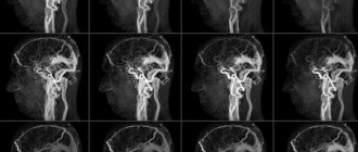
If the subject of examination is the human vascular system, an MR angiography protocol is used. During it, the computer program is set in such a way that only the vascular bed is visualized in the images. Using the angioprogram, doctors can assess the condition of arteries and veins and identify most vascular pathologies, for example, occlusions, malformations, kinks, aneurysms, bifurcations, ruptures and intimal tumors.
How is an MRI scan done?
- The patient is positioned on the table in a supine position.
- The main condition is complete immobility. To achieve this, the patient's limbs are secured with straps and bolsters. If the procedure is psychologically difficult to tolerate, sedatives may be taken.
- After fixing the patient, the examination begins. During operation of the device, characteristic sounds may appear. They are not too loud and do not cause discomfort, but earplugs or headphones can be used if desired. The second option is preferable, as it will allow you to communicate with the doctor and follow his instructions.
If a contrast agent is used, the procedure is carried out according to a similar plan. The only difference is that before the MRI begins, the patient is injected with a contrast agent.
Indications
Magnetic resonance imaging can be done in MRI centers either by a doctor’s referral or on the patient’s personal initiative. Reasons for scheduling an MRI may include:
- Frequent pain of unknown etiology;
- Head and body injuries;
- Dizziness, fainting, convulsions;
- Disorders of the musculoskeletal system;
- Deterioration of sensitivity, vision, speech, hearing;
- Decreased mental activity, memory, concentration;
- Pain in the abdomen and groin area;
- Chronic diseases.
Contraindications
Magnetic resonance imaging has a number of contraindications dictated by the physics of image acquisition. Since the MRI machine is a large magnet, it tends to attract or heat any metal objects. Therefore, MRI scans are not performed on patients who have ferromagnetic metal in their body. In modern medicine, most implants are made of safe non-ferromagnetic alloys, such as titanium or medical plastic. It is absolutely safe to perform MRI with them. However, if the patient has steel prostheses, iron bullets or shrapnel in his body, magnetic resonance imaging will be contraindicated. The machine's strong magnetic field can cause metal inclusions to move or become very hot, causing either internal bleeding or burns.
Magnetic resonance imaging should be performed with caution in people who have artificial pacemakers, such as pacemakers, neurostimulators, or hearing aids. A strong magnetic field can damage any electronic device. Therefore, if the product passport does not indicate that it is MRI compatible, the study cannot be done.
The remaining limitations to MRI are relative. For example, due to their heavy weight or size, overweight and large people may have difficulty finding the right model of magnetic resonance imaging scanner. Most MRI machines are designed for a body weight of no more than 120 kg and a circumference of no more than 120 cm. However, there are installations with a load capacity of up to 200 kg and an aperture of 140 cm.
People with severe claustrophobia sometimes find it difficult to endure a full scanning session in closed machines. Open-circuit tomographs were specially created for patients prone to panic attacks.
If you belong to the category of patients for whom MRI is contraindicated, alternative forms of scanning for you would be computed tomography, quadrilateral radiography and ultrasound examination using an expert-class device.
You can read in more detail about all the absolute and relative contraindications to MRI here.
Reliability of the result
The reliability of the result and consequences is influenced by proper preparation. To do this, the patient must think about the clothes he will wear during the MRI. There should not be any metal parts on it. The same goes for jewelry - they all need to be removed. It is also worth considering that burns may occur at the site of tattoos made with the use of dyes containing metal particles, and the operation of implants and stimulators may be impaired. Therefore, all tattoos, ECS, metal structures in joints and teeth should be reported to the doctor.
Preparation
Most MRI examinations do not require any preparation from the patient. The exceptions are MRI of the pelvic organs, MRI of the abdominal organs and MRI of the gastrointestinal tract.
- A reminder to the patient about preparing for MRI of the pelvic organs.
- A reminder to the patient about preparing for an MRI of the abdominal organs.
Before undergoing the procedure, you should remember that the tomograph scans a person using a magnetic field, which is created during the procedure in a tomographic machine. It follows that everything metallic: jewelry, watches, hairpins, clothes with a zipper should be left outside the MRI room. You should also remove all electronic devices from your pockets.
If you suffer from claustrophobia or cannot lie still, doctors recommend taking sedatives before the diagnosis.
If you have an MRI with contrast, tell your healthcare provider about any allergies you have and any chronic kidney disease you have. In the case of contrast-enhanced tomography, eating is not recommended 2 hours before the diagnosis.
How to pay for participation in rehearsal testing?
Payment is confirmed in your personal account on the RIKZ website using the transaction number in the ERIP. This year you need to pay 7.60 BYN for one item.
To pay for RT,
1) select:
— item “System “Calculation”” (ERIP);
— education and development;
— RIKZ;
— RT, on-line registration;
2) enter the personal account you received in the rehearsal testing participant registration system;
3) make sure that the full name transmitted by the “Raschet” system and displayed on the screen of the payment terminal, computer or mobile phone (depending on how you pay) corresponds to your last name, first name and patronymic. In case of discrepancy (this is possible if you made a mistake when entering your personal account), cancel the current action and repeat the process from the beginning;
4) pay.
This procedure can be done at a bank (then a commission will be charged from you), an ATM, an information kiosk, or cash settlement centers.
Sign up for an intensive online training course for the CT! A month before the CT, we will repeat and systematize the knowledge, solve typical CT tasks and gain confidence in our abilities! Find out more and sign up
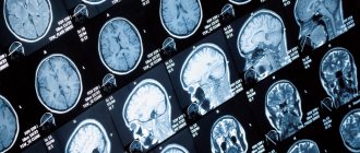
results
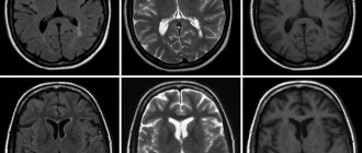
The results of the magnetic resonance examination will be a series of three-dimensional black and white images. On average, an MRI machine produces 1,000 to 1,500 high-resolution images per scan. Their interpretation is carried out by a radiologist. He evaluates the anatomical structures and in his conclusion notes the main characteristics: size, location, degree of invasion into neighboring organs. If a contrast-enhanced examination was carried out, in addition to these data, the diagnostician indicates the behavior of the tissues after the administration of contrast. If any anomalies are detected, the doctor gives their initial interpretation.
Based on the results of the magnetic resonance examination, the patient will be given a written doctor’s report and a series of tomograms recorded on electronic or film media. The X-ray report is not a final diagnosis. The main task of the radiologist is only to describe all detected abnormalities. With this data, the patient should be referred to a specialized specialist who, taking into account all the results of the examination, medical history, tests and diagnostics, will be able to make a final diagnosis and propose a treatment strategy.
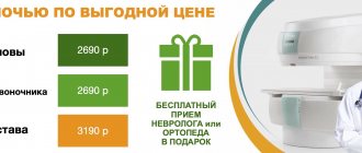
In some clinics, MRI results include a free consultation with a specialist with extensive diagnostic experience, who will talk in detail about the results of the study and give recommendations on further actions. It could be:
- free consultation with a radiologist;
- free consultation with a neurologist based on the results of the tomography;
- free consultation with an orthopedist based on the results of the tomography.
EXAMPLE OF MRI CONCLUSION - spine
MR signs of degenerative-dystrophic changes are revealed in the form of a decrease in the intensity of the MR signal from the intervertebral discs in the L1-S1 segments on T2-WI due to dehydration of the nucleus pulposus of the discs, with a decrease in height in the L4-S1 segments, small anterior and posterolateral marginal bones growths in segments L1-S1, with calcification of the anterior longitudinal ligament at the points of attachment to the vertebral bodies. In the intervertebral joints, degenerative changes are visualized in the form of compaction, unevenness of the articular surfaces, and hypertrophy of the yellow ligaments. In the adjacent sections of the vertebrae L4-L5, L5-S1, zones of heterogeneous increase in the MR signal on T1, T2-WI and hypointense on STIR-IP (due to fatty degeneration) are determined. Irregularities of adjacent endplates of the vertebral bodies Th12, L1, L2, L3, L4, L5, S1 due to Schmorl’s cartilaginous nodes.
MRI of the spine
| Name of service | Price | Discount |
| MRI of the cervical spine | from 2100 rub. | At night |
| MRI of the thoracic spine | from 2100 rub. | At night |
| MRI of the lumbosacral spine | from 2100 rub. | At night |
| MRI of the sacrococcygeal spine | from 1500 rub. | At night |
| MRI of the cervical spine and craniovertebral junction | from 2100 rub. | At night |
| MRI of the sacroiliac joints | from 2100 rub. | At night |
| MRI of the entire spine | from 6500 rub. | At night |
| MRI of the central nervous system (all parts of the spine + brain) | from 9000 rub. | At night |
| Cost of contrast administration | from 2100 rub. | At night |
MRI of joints
| Name of service | Price | Discount |
| MRI of the knee joint | from 2500 rub. | At night |
| MRI of the hip joints | from 2500 rub. | At night |
| Shoulder MRI | from 2500 rub. | At night |
| MRI of the elbow joint | from 2500 rub. | At night |
| MRI of hands | from 4000 rub. | At night |
| MRI of the foot | from 4000 rub. | At night |
| MRI of the ankle | from 2500 rub. | At night |
| MRI of the wrist joint | from 4200 rub. | At night |
| MRI of the temporomandibular joints (pair) | from 3800 rub. | At night |
| Cost of contrast administration | from 2100 rub. | At night |
Vascular MRI
| Name of service | Price | Discount |
| MRI of cerebral vessels (MR angiography of the brain) | from 2100 rub. | At night |
| MRI of neck vessels (MR angiography of the neck) | from 2100 rub. | At night |
| MRI of spinal vessels (MR angiography of the spine) | from 3200 rub. | At night |
| MRI of the abdominal or thoracic aorta | from 4100 rub. | At night |
| MRI of extremity vessels | from 4000 rub. | At night |
| MRI of renal vessels | from 3800 rub. | At night |
| MRI of pulmonary vessels | from 3800 rub. | At night |
| MRI of pelvic vessels | from 3800 rub. | At night |
| MRI of the coronary vessels of the heart | from 6500 rub. | At night |
| MRI of the brain and MR angiography (comprehensive examination of the head) | from 4800 rub. | At night |
| MRI of the cervical spine and MR angiography of the neck (comprehensive examination of the neck) | from 4800 rub. | At night |
| MR venography or MR arteriography | from 3200 rub. | At night |
| Cost of contrast administration | from 2100 rub. | At night |
Tissue MRI
| Name of service | Price | Discount |
| MRI of soft tissues of the face | from 4200 rub. | At night |
| MRI of soft tissues of the buttocks | from 4200 rub. | At night |
| MRI of soft tissues of the neck | from 4200 rub. | At night |
| MRI of soft tissues of the extremities | from 4200 rub. | At night |
| MRI of the tongue and soft tissues of the throat and larynx | from 4000 rub. | At night |
| Cost of contrast administration | from 2100 rub. | At night |
MRI of organs
| Name of service | Price | Discount |
| MRI of the lungs and mediastinal organs | from 3200 rub. | At night |
| MRI of the pelvis in women | from 3200 rub. | At night |
| MRI of the pelvis in men | from 3200 rub. | At night |
| MRI of the external genitalia | from 3000 rub. | At night |
| Abdominal MRI | from 3200 rub. | At night |
| Abdominal MRI with MR cholangiography | from 4000 rub. | At night |
| MRI of the retroperitoneum | from 3200 rub. | At night |
| MRI of the gallbladder and bile ducts (MR cholangiopancreaticography) | from 3200 rub. | At night |
| MRI of the urinary tract (MR urography) | from 3200 rub. | At night |
| MRI of the intestine (MR colonoscopy) (MR enterography) | from 3200 rub. | At night |
| Hydro MRI of the intestine | from 6000 rub. | At night |
| MRI heart | from 6000 rub. | At night |
| Whole body MRI/Whole body diffusion MRI | from 11,000 rub. | At night |
| MRI oncology search | from 8000 rub. | At night |
| Fetal MRI | from 5500 rub. | At night |
| Cost of contrast administration | from 2100 rub. | At night |
MRI of glands
| Name of service | Price | Discount |
| MRI of the thyroid gland | from 4000 rub. | At night |
| MRI of the mammary glands (MR mammography) | from 4000 rub. | At night |
| Cost of contrast administration | from 2100 rub. | At night |
MRI of the head
| Name of service | Price | Discount |
| MRI of the brain | from 2100 rub. | At night |
| Brain MRI with contrast | from 5100 rub. | At night |
| MRI of cerebral vessels | from 2100 rub. | At night |
| MRI of the pituitary gland | from 2100 rub. | At night |
| MRI of the pituitary gland with contrast | from 5000 rub. | At night |
| MRI of the sinuses | from 2050 rub. | At night |
| MRI of the eye orbits and optic nerves | from 2500 rub. | At night |
| MRI of cranial nerves | from 3200 rub. | At night |
| Cost of contrast administration | from 2100 rub. | At night |