Do you need preparation for MRI examinations?
Magnetic resonance imaging, as a reliable and accurate diagnostic method that is not affected by harmful X-ray radiation, has virtually no health contraindications. But there are a number of factors that cannot be ignored:
- the presence of a pacemaker located under the skin;
- metal clamps on vessels;
- the presence in the body of foreign metal objects of a fragmentary nature;
- metal crowns or pins in the mouth;
- presence of an implanted cochlear prosthesis;
- internal central nervous system stimulants;
- the presence of a compression-distraction device;
- built-in metal pins;
- the presence of a stationary prosthetic limb.
The above factors prohibit you from undergoing tomography. There are also conditional rules that you just shouldn’t forget about:
- When choosing comfortable clothes for the upcoming procedure, carefully inspect the items for the absence of metal buttons, zippers, hooks, buckles and decorations.
- When going for an MRI diagnosis, remove all jewelry from your body in advance.
- Check your pockets first so as not to miss even a small thing that could interfere with the diagnosis.
In addition to external reasons that prevent MR examination, there are also psychological factors that need to be taken into account.
It is important to warn the patient that he will be immobilized for a long time. The duration of the process can be 30-60 minutes.
Prices for magnetic resonance imaging of the brain in Moscow
Various factors influence how much an MRI costs in each case. The price may change depending on whether the specialist will use a contrast agent, whether the results will need to be recorded on disk, etc. The approximate cost of magnetic resonance imaging is indicated on the page.
You can make an appointment with a doctor at the Diagnostic and Treatment Center on Vernadsky. To do this, call us by phone at any time convenient for you. You can also leave an online application through a special form presented on the website.
Preparing for an MRI with contrast
Contrast is a special dye solution administered intravenously. Before performing an MRI using a contrast agent, specialists need to make sure that the patient does not have problems with the kidneys or liver. A preliminary opinion from a urologist may be required.
It is important to exclude the presence of allergic reactions to the contrast agent. To do this, tests are carried out in advance for rejection of the composition.
Modern drugs are produced based on gadolinium salts. They do not cause allergies, but are excreted through the liver and kidneys. If you have chronic diseases of these organs, problems may arise. Therefore, preliminary consultation with a specialist is necessary.
What does a brain MRI with contrast show?
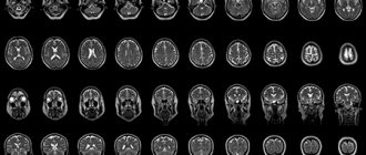
In the first series of images of the brain in the form of detailed layer-by-layer thin sections, the cerebral cortex, white and gray matter, meninges, corpus callosum and other parts of the central nervous system are easily identified. An MRI diagnostic doctor sequentially examines all obtained images, paying attention to any deviations from the norm. Particular emphasis is placed on the presence of focal changes, cysts, and space-occupying formations. The condition of the cerebrospinal fluid pathways through which cerebrospinal fluid circulates is studied. The course of blood vessels and cranial nerves is traced.
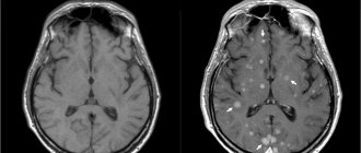
When abnormalities are detected that cannot be identified by conventional methods, contrast is introduced. Then the doctor, studying the images with contrast, receives additional information - MRI with contrast allows you to assess the rate at which pathologically altered tissues accumulate and release contrast. By studying the shape and boundaries of the lesion, its relationship with other anatomical structures and other features, doctors can more accurately make a diagnosis. This is possible due to the fact that any disease has unique characteristics of interaction with contrast agents.
Preparing for an abdominal MRI
To be prepared for diagnosing internal organs, you need to familiarize yourself with some rules:
- the procedure is carried out on an empty stomach, so it is advisable to refrain from eating 6-12 hours before the session;
- several days before the study you need to follow a diet that excludes raw fruits and vegetables, sweets, drinks containing gas, dairy products, legumes and mushrooms;
- two to three days before the tomography, take medications that reduce flatulence (Mezim or Espumisan);
- 30 minutes before the procedure you need to take an antispasmodic agent, for example No-Shpa.
MRI with contrast: how the procedure works
Bolus MRI
There are 2 ways to do enhanced magnetic resonance imaging.
In MRI with dynamic administration of contrast (bolus magnetic resonance imaging), the drug is administered intravenously during the entire diagnostic procedure using a special device - an automatic dispenser, at a given speed. This helps to synchronize the progress of the study with the accumulation of the indicator in the bloodstream and allows you to select the desired phase of contrasting.
Diagnostics takes place in real time, so tomograms are more informative and easier to interpret compared to those obtained with the simultaneous administration of the working substance.
Bolus MRI is prescribed if you need to look at dense parenchymal organs - liver, kidneys, etc. or differentiate the structure of tumors.
MRI without contrast is ineffective for assessing the size of a tumor and its dynamics during treatment.
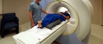
The patient must lie motionless on the tomograph table during the procedure; any activity can lead to the appearance of artifacts in the images.
There is no pain; if necessary, you can contact the medical staff using a special button.
During contrasting there may be:
- feeling hot or cold;
- drowsiness;
- taste in mouth;
- sudden urge to urinate;
- dizziness;
- nausea.
There is no need to worry; after a while these phenomena will disappear on their own.
To reduce vegetative symptoms, you need to have a light snack before diagnosis.
Manifestations to be reported immediately:
- labored breathing;
- pain and burning at the injection site, feeling of fullness;
- the appearance of an itchy rash on the skin.
The contrast does not affect the ability to drive a vehicle or mental activity.
Some people feel uncomfortable (panic) in a confined space. This disorder is called “claustrophobia,” and in these cases sedation may be used. The administration of sedatives before MRI is also performed in patients with psychiatric symptoms, because such clinical manifestations may be a consequence of a brain tumor. But such patients are diagnosed according to indications only in public clinics.
Preparing for an MRI of the pelvis
This examination includes the study of organs such as the prostate gland, bladder and adjacent organs, the uterus and its appendages. To prepare for an MRI of the pelvic organs, perform the following procedures:
- cleaning the gastrointestinal tract with an enema;
- fasting for six hours;
- for the examination to be effective, it is necessary that the bladder is partially filled; for this, it is recommended to refrain from going to the toilet two to three hours before the examination and drink half a liter of clean water without gas an hour before the session;
- take the antispasmodic within 30-40 minutes.
Contraindications for magnetic resonance imaging of the vascular system of the head and neck
The ban on MRI of head and neck vessels is due to the peculiarities of the interaction of electromagnetic radiation with certain metals and magnets. The examination is prescribed with caution in certain patient health conditions.
MRI of cerebral vessels is contraindicated if the patient has the following devices in the body:
- pacemaker and other stimulating implants and pacemakers, cochlear implant - implants may fail under the influence of a magnet;
- metal intravascular stents, artificial heart valves;
- endoprostheses, foreign metal bodies in the body;
- metal clips installed in the vessels of the brain after surgery - under the influence of a magnetic field they can move and cause bleeding.
Not every material prohibits MRI of vessels and arteries of the neck and head. Metals that react to a magnetic field by displacement and heating: stainless steel, iron, nickel, cobalt. In other cases, the procedure is not prohibited.
In some patient conditions, examination is allowed by the doctor’s decision on an individual basis:
- pregnancy up to 12 weeks - the fetus is especially sensitive to external factors;
- serious condition of the patient, severe pain;
- epilepsy, seizures, mental illness;
- body weight should not be more than 130 kg - the devices have restrictions on the patient’s weight and waist circumference.
The procedure can be performed in patients who find it difficult to maintain a stationary body position under general anesthesia or in an open-type apparatus. During the entire period of using MRI, there have been no cases of fetal pathology developing after the procedure in the first trimester of pregnancy, however, doctors are careful. If the patient suffers from a mild form of claustrophobia, he may take sedatives before the examination.
MRI of the vessels of the head and neck with the use of contrast agents is contraindicated in pregnant women at any stage, as well as in patients with renal failure.
Preparing for MRI in women
Additional rules exist for conducting research in women:
- If a woman is pregnant or suspects pregnancy, it is necessary to warn the doctor about this.
- If possible, avoid undergoing tomography during the menstrual cycle, when internal organs and arteries change in size and contours. Tomography results may be incorrect.
- If a session was carried out using a contrast agent, women who are breastfeeding must interrupt breastfeeding for two days until the dye is completely removed from the body.
When is the examination carried out?
Before undergoing an MRI of the head and neck vessels, it is advisable to consult a neurologist. The doctor will assess the patient’s symptoms and condition and refer him for the most appropriate study.
The examination is usually carried out if the patient observes the following phenomena for a long time:
- prolonged headaches of unknown cause that cannot be treated with known drugs;
- periodic dizziness, loss of consciousness, fainting, darkening of the eyes, difficulty in spatial orientation;
- unstable blood pressure;
- decreased vision;
- numbness of the arms, shoulder girdle;
- actively developing osteochondrosis of the cervical spine with corresponding symptoms;
- herniated disc in the cervical region;
- subluxation, instability of the cervical vertebrae;
- traumatic brain injury, spinal injury;
- suspicion of cancer or mass formations in the brain.
The value of MRI of the head and neck vessels lies in the possibility of making an early diagnosis. The examination helps to start therapy in a timely manner and prevent the progression of the disease. Having received the necessary treatment, the patient can undergo the procedure again an unlimited number of times without risk to health. The doctor monitors the effectiveness of therapy and, if necessary, makes adjustments or changes tactics.
MRI of cerebral vessels is actively used by neurosurgeons to plan operations. Information about the location and structural features of the vascular system allows you to avoid unforeseen situations and make precise interventions. MRI is important in the postoperative period to monitor recovery processes.
How to prepare children for MRI
Before going to the procedure with your child, relieve him of internal fears about an unfamiliar situation. The day before, you need to talk to the baby, tell him what awaits him. You need to convince him that the process is safe and painless.
MRI in children has some peculiarities. Since it is difficult for a child to remain motionless for a long time, the session is carried out with the help of mild sedatives or under general anesthesia. During the entire tomography process, the child’s condition is monitored by an experienced specialist – an anesthesiologist.
Before conducting diagnostics with a contrast agent, the presence or absence of allergic rejection to the drug is determined. A severe form of bronchial asthma in a child’s diagnosis may also be a contraindication.
Basic guidelines that people should follow when preparing for an MRI
When using a contrast agent, you should inform the specialist performing the procedure about the presence of allergies, problems with the heart, liver or kidneys, or other previously identified diseases. The patient must tell the doctor what medications he took in anticipation of the procedure.
If a person experiences fear of closed spaces, this should also be reported to the doctor. In such cases, doctors often prescribe sedatives to help prevent panic attacks during the examination and complete it successfully.
It is not advisable for women in the first trimester of pregnancy to undergo an MRI. This is explained by the fact that the effect of a magnetic field on an embryo during organ formation has not been fully studied. Patients during lactation should avoid breastfeeding for two days after the procedure. This will prevent the penetration of gadolinium salts into the baby’s body during MRI with contrast.
Immediately before the examination, the patient is asked to remove his clothes and change into a more comfortable medical gown. He should not enter the diagnostic room with bank cards, telephone, jewelry or costume jewelry made of metal. You must also remove your watch if you have one.
Having completed all these instructions, the patient lies down on a retractable table and follows all the doctor’s recommendations during the procedure.
Preparing for an MRI of the brain
If a doctor sends a patient for an MRI of the brain, you also need to know that:
- if there are metal crowns, bridges, dentures, or braces in the oral cavity, visit the dentist in advance and temporarily remove these objects;
- all removable electronic and metal devices must be left outside the threshold of the room with the MR installation;
- emotionally prepare yourself for the upcoming procedure in order to avoid immersion in a stressful state, remember that the diagnosis is absolutely safe;
- If you cannot get rid of your anxiety, ask your doctor to give you a sedative.
By following the above rules, the patient will help specialists conduct an effective diagnosis. The MR procedure will be comfortable and high quality.
What will the examination show?
MRI of the vessels of the head and neck is performed in angiography mode - in this case, only areas of the vascular system are visible in the images, without surrounding tissues. The examination makes it possible not only to assess the condition of the arteries and veins, but also to determine the speed of blood flow in the vessels in real time. Based on the scanning results, a three-dimensional 3-D model of the area under study is constructed.
Signs of which diseases will be shown by MRI of cerebral vessels:
- stroke - hemorrhagic (leakage of blood from the vascular system into the brain tissue or meninges) or ischemic (cessation of blood supply to a part of the brain, more common);
- arterial aneurysm - pathological expansion and thinning of the vascular wall with the risk of rupture;
- arterial dissection as a result of congenital weakness of the arterial wall or damage by a pathological process;
- thrombosis, thromboembolism;
- inflammatory diseases of the vascular system – phlebitis, arthritis, systemic vasculitis;
- vascular tumors – hemangioma, hemangioblastoma, angioreticuloma;
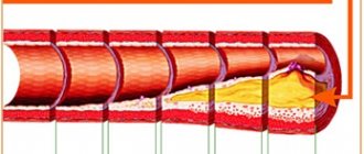
- atherosclerosis;
- narrowing of the lumen of the vessel due to structural features or compression from the outside by a space-occupying formation;
- congenital developmental anomalies and structural features - absence, decrease or increase in diameter, bifurcation of some parts of the vascular system, arteriovenous malformation.
MRI of the vessels and arteries of the neck will show structural defects and pathological processes leading to impaired cerebral circulation:
- dissection of the carotid and vertebral arteries - damage to the artery wall as a result of injury, bending, with further formation of a hematoma between the walls of the vessel, narrowing of the lumen of the artery and disruption of the blood supply to the brain;
- pathological tortuosity of the carotid artery;
- developmental anomalies and acquired lesions of the vertebral artery;
- neoplasms;
- aneurysm of a section of the vascular system;
- pathologies in the area of the carotid sinus.