PET/CT examination - what is it?
The deciphering of the abbreviation sounds like positron emission tomography - computed tomography. This diagnostic method combines the study of the structure and functional characteristics of tissues. The technology is especially widely used in oncology to identify and determine the degree of development of malignant tumors.
Why?
Today, up to 90% of PET/CT studies in the world are performed on people suffering from cancer. The study is important from a number of perspectives:
- The study is important from the point of view of determining the degree of development and spread of the tumor process, detection of metastases, both regional and distant.
- In medical practice, there are situations when doctors are unclear about the nature of the process occurring in a particular organ. The study allows you to differentiate the process, distinguish between benign and malignant.
- The study helps to understand whether the treatment is effective.
- The study can be used to diagnose relapse of the disease.
When it comes to diagnosis, many cancers have the ability to actively accumulate fluorodeoxyglucose. This substance is used during the study. However, some types of cancers have low metabolism.
These types include:
- Highly differentiated cancerous neoplasms of the thyroid gland; Benign tumors of any location;
- Some kidney tumors;
- Some bone tumors;
- Liver tumors;
- Prostate tumors;
- Some types of sarcomas, lymphomas.
Having a low metabolic rate, they are poorly visualized by PET/CT. This means that other diagnostic methods are required.
General contraindications to PET/CT
Absolute:
pregnancy (confirmed or suspected) and breastfeeding. If it is necessary to perform an examination on a patient who is breastfeeding, breastfeeding is stopped for the entire duration of the examination. The re-initiation of breastfeeding is determined by the study physician.
Relative:
diabetes mellitus in the stage of decompensation for patients who are planning a study with the radiopharmaceutical 18F-FDG - consultation with an endocrinologist and additional preparation are required in order to achieve optimal blood glucose levels for PET/CT up to 7.7 mmol/l;
Carefully:
in case of insufficiency of renal function (since PET/CT data may be distorted due to the retention of radiopharmaceuticals in the body).
Description of research technology
The basis of the research technology is the study of tissue characteristics, structural and functional. Functional characteristics can be assessed through metabolism. Let's take the following example. We choose a substance that is necessary for all cells of the body. We mark it with a radioactive label, introduce it into the body and observe the places of its maximum accumulation.
Glucose is a universal substance in the human body. With the help of glucose, almost all tissues and cells are nourished. In malignant tumors, the greatest consumption occurs, since the growth and reproduction of tumor cells requires a lot of energy.
In a PET/CT scan, glucose is labeled with radioactive atoms with a short half-life. Once in the body, glucose actively accumulates in tissues with the most intense metabolism, i.e. in cancerous tumors.
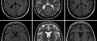
The tag disintegrates, emitting energy in the form of gamma rays. A special device records this process. The data that the doctor receives creates a visual model showing the location of the tumor, its size and metastases.
Radioactive tracers accumulate only at the location of atypical cells; healthy tissues are not visualized.
When the doctor needs to examine both healthy and changed structures, CT comes to the rescue, allowing you to obtain a detailed picture with millimeter accuracy.
After data from both scanning systems is received, they are superimposed on each other, thereby achieving an image that gives a clear picture of the location of tumor foci.
Positron emission tomography is a breakthrough in the diagnosis of cancer
Computed and magnetic resonance imaging, ultrasound and endoscopic research methods... The arsenal of modern medicine is wide and allows us to identify many health- and life-threatening ailments at the earliest stages.
At the same time, science does not stand still. In European countries and the USA, along with the now traditional diagnostics, the positron emission tomography method is actively used. In our country it is not yet so widespread. What sets it apart from other modern medical imaging methods? In terms of diagnostics, what can PET do that CT and MRI cannot?
Let's consider these and other questions.
What is positron emission tomography and what is the essence of this study?
Positron emission tomography (or, otherwise, PET) is a modern method of medical imaging, which is based on its own special principle.
The essence of PET is as follows. Before the study, a pharmaceutical preparation containing a radioactive component is injected into the body - a radiopharmaceutical (this can be, for example, chemically modified glucose). In the body it decays with the release of positrons. The latter begin to interact with electrons of organs and tissues, resulting in special electromagnetic radiation that is recorded by special sensors. Based on its characteristics, a conclusion is drawn about the state of the observed structures at the molecular and cellular level.
PET scan shows not only the structure of the area under study, but also its function. Glucose from the administered radiopharmaceutical is “included” in the metabolism, and with the help of the device it is possible to record how it “behaves” in a particular tissue or organ. For example, it is known that malignant tumor cells grow rapidly. This happens for a reason: rapid growth requires, in particular, energy. Thus, the administered glucose is more intensively consumed by the cells of the tumor tissue, which makes it possible to “see” them on tomograms.
What is PET/CT (or PET-CT)?
This is a study that combines data obtained using both methods, which makes it possible to accurately identify the presence and location of a pathological formation.
What are the indications for positron emission tomography?
First of all, these are many oncological diseases, including those with the formation of metastases. This method will also help with neurological diseases - in particular, with cerebral vascular pathology, epilepsy, memory impairment, Alzheimer's disease and other types of dementia, degenerative brain lesions (Parkinson's disease, Huntington's disease).
What role does MRI play in diagnosing epilepsy? Read here
In addition, PET is used to assess blood circulation and vitality of the heart muscle (myocardium).
Are there any contraindications to PET CT?
Yes, they are.
— The study is not prescribed during pregnancy or its potential. If it is necessary for nursing mothers to carry it out after the procedure, resumption of feeding is allowed no less than 24 hours later;
— Kidney and liver failure;
— The use of the method in patients with diabetes mellitus is limited;
— Allergy to components of the radiopharmaceutical and contrast agent;
— Diseases of the thyroid gland (when planning to use contrast in CT);
— General serious condition of a person and some others.
MRI has its own contraindications, which you can read about here
All details regarding possible contraindications should be discussed with your attending physician and the doctor who will perform the diagnostic procedure. Give them only reliable and as detailed information as possible.
How to prepare for the examination and undergo a PET scan correctly? Is it necessary to follow a diet before the examination?
There are different types of PET, each of which has its own characteristics in preparation. To understand the basic principles, consider the option for diagnosing tumors.
In order for the diagnosis to be as informative as possible, the following recommendations must be followed:
- be sure to inform the doctor who will conduct the study about any allergies or reactions to medications you have, or any existing or suspected pregnancy;
— bring with you a list of medications you are taking;
- 48 hours before the test, stop intense physical activity and avoid massage;
— 36 hours before the test, avoid hypothermia. If the procedure is carried out in the cold season, then dress warmly on the day of the examination;
- 24 hours before the test: follow a very low carbohydrate diet, completely eliminating sugar. Do not use chewing gum or mints;
- 6 hours before the test: stop eating, drink only non-carbonated unsweetened water in large quantities; continue taking prescribed medications unless otherwise advised by your doctor;
— Patients with diabetes should consult their doctor about any changes in diet and medication.
How is positron emission tomography performed?
First, a radiopharmaceutical is injected into the patient's body. In this case, a person usually does not feel anything; occasionally there is a feeling of heat. After administration, large amounts of water should be taken as this will affect the quality of subsequent images.
Within an hour, the product is distributed throughout the organs and tissues, including the pathological focus. All this time you need to lie still and avoid talking. This contributes to the fact that during the study the neoplasm is more clearly identified, where more of the radiopharmaceutical is concentrated.
Then the scanning procedure itself is carried out. First, a CT scan is performed (sometimes, if indicated, with contrast enhancement), and then a PET scan is performed. Both studies take place in the same tomograph and, like MRI, CT and ultrasound, are completely painless.
When are CT, MRI and ultrasound prescribed? Find out here
What to do after undergoing positron emission tomography?
In the absence of contraindications, it is recommended to consume at least 2 liters of fluid per day and limit salty and smoked foods. Avoid contact with pregnant women and children for 24 hours. Breastfeeding women should refrain from feeding for 24 hours. During this time, express all milk produced; Other family members need to care for the baby during this period.
If any health symptoms occur, immediately notify medical personnel. If complaints appear after you have left the clinic, contact the doctor who conducted the study or call an ambulance.
What are the advantages and disadvantages of positron emission tomography?
Among the advantages of PET are the non-invasive and painless procedure, high diagnostic accuracy, detection of pathologies in the early stages, the ability to examine all organs at once, and monitoring the effectiveness of the therapy.
The disadvantages of the method include the presence of a certain radiation exposure provided by a radiopharmaceutical, and when PET is combined with CT, by x-ray radiation. Examination of patients with diabetes mellitus is problematic or impossible. The reliability of diagnosis strongly depends on the quality of preliminary preparation. PET alone is not always sufficient to make a definitive diagnosis, so it is usually combined with CT or MRI. Also, the disadvantages of the method include its relative high cost.
Author: Enver Aliyev
***********************************************************************************
Realizing that people’s awareness of this method is high, for an expert opinion we turned to Andrei Vladimirovich Korobov, candidate of medical sciences, specialist in the field of radiation diagnostics, member of the board of the medical group, director of the Expert Institute.
- Andrey Vladimirovich, when is it necessary to do without PET CT?
Taking into account the unique ability to determine and visualize this combined method of cellular and molecular metabolism, the use of such an expensive method as PET CT can be considered fully justified in the diagnosis of any oncological diseases with expected high metabolic activity. This allows you to monitor the patient’s entire body for the presence of a primary tumor and metastases in one session, measure functional changes in biochemical processes, distinguish benign from malignant formations with high accuracy, promptly determine the effectiveness of the treatment used, and conduct early diagnosis of an impressive list of oncological diseases.
- Is there an alternative to PET CT?
All medical imaging methods are, to one degree or another, alternative to each other. At the same time, if you ask the question: “what is better to do...” and start listing ultrasound, radiography, computed tomography, MRI, SPECT-CT, PET-CT, and other methods, then with a high degree of probability you will get the answer which is better and more correct in a full-fledged diagnostic process, use several different methods. In this case, the choice is largely determined by the doctor based on the expected type of disease. There are various legends extolling certain individual methods of radiation diagnostics. Legends that exaggerate the identification of one or another unique information that is inaccessible to identification by other methods. At the same time, as a rule, the fact is kept silent that in the example given, the patient has already undergone a lot of other studies, which excluded a great many missing changes and significantly narrowed the direction for the search. Of course, in the search for small lesions in suspected cancer with high metabolic activity, PET, and even more PET CT, has undeniable advantages.
A research method such as single photon emission computed tomography (SPECT) has similar properties to PET. Both methods belong to the field of nuclear medicine. A full, correct comparison of these methods in terms of their value for the diagnostic process is difficult to achieve due to both the relative scarcity of both methods and the difficulty of justifying their simultaneous use in the same patients. That is why discussions about the comparative advantages of these diagnostic methods, which are similar in physical meaning, are still ongoing. Authors of scientific publications point out the advantages of PET CT in sensitivity, specificity and diagnostic accuracy. At the same time, these SPECT CT indicators are also quite high, reaching values close to 90%, which, despite the many times higher cost of PET, gives grounds for the wider use of SPECT.
- Where can I get positron emission tomography in our country?
These studies are currently available mainly in large cities. The data varies, but today PET can be performed in Moscow (PET Technology, Sofia Cancer Center), St. Petersburg (LDC MIBS), Voronezh (MMCRDiLOZ). The Federal Network of PET Technology also has its branches in Ufa, Lipetsk, Yekaterinburg, Belgorod, Tambov, Orel, and Kursk. There are also centers in Tyumen, Chelyabinsk, Kazan.
If you need to undergo such research, I recommend checking the relevance of the information, since the list of cities is constantly expanding.
For reference:
Andrey Vladimirovich Korobov
Candidate of Medical Sciences, specialist in the field of radiation diagnostics, member of the board of the medical group, director of the Expert Institute.
Types of drugs used
The potential of a study depends on the arsenal of radiopharmaceuticals used in it. These drugs are labeled with unstable isotopes that make them radioactive.
Today, isotopes of such elements as:
- Nitrogen-13;
- Oxygen-15;
- Carbon-11;
- Fluorine-18.
In oncology, fluorine is most common, since it has the longest half-life and at the same time the lowest radiation energy.
These advantages of fluorine make it possible to obtain high-quality images with high spatial resolution. In addition, the relatively long half-life makes it possible to transport the drug from the site of production to the clinic.
The most common drug used is fluorodeoxyglucose. It is an analogue of glucose. Atypical cells absorb it faster, it actively accumulates, and this is ideal for scanning.
The downside of fluorodeoxyglucose is that it accumulates in brain tissue and nephrons, which in turn can cause these organs to glow, even when they are healthy.
This drawback stimulated the search for other, more advanced drugs, and now such drugs have been created.
An example of a modern drug is 18F-FET. It is intended for the brain and contains the amino acid tyrosine, labeled with the fluorine-18 isotope. Tyrosine has a very high selective accumulation in brain tissue, which is important for imaging neurotumors.
The drug is also used to diagnose oropharyngeal tumors, to detect metastases and to diagnose lesions of the cervical lymph nodes.
How is the procedure performed?
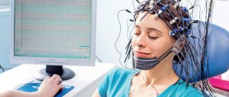
Before starting diagnostics, you must turn off your cell phone. In the waiting room you need to change into comfortable, warm clothes. You will first need to fill out a health questionnaire and medical documentation. After this, you will have a conversation with a diagnostician. He will collect anamnesis and study the medical history. In the waiting room you need to lie down, close your eyes and relax. After this, methionine is administered intravenously to the patient using a thin catheter. The patient returns to the waiting room and rests for 20-30 minutes until the substance spreads throughout the body. At this time, it is not recommended to read, listen to music, or talk.
After this time, a scan is carried out. It takes about half an hour. Positron emission tomography is done in a darkened room. This is necessary to make the patient feel relaxed. The examination is a non-invasive diagnostic method. It does not cause discomfort. Performed in a supine position. Medical staff monitors the progress of the procedure.
The total examination time takes about 90 minutes. Results are usually ready the next day.
Indications for the study
- Search for the primary tumor site when metastases are detected;
- Determination of the stage of the oncological process;
- Differentiated diagnosis of relapse and post-treatment changes;
- Monitoring the course of the disease, detecting relapse;
- Planning of radiotherapy and surgical manipulation;
- Planning a biopsy and finding the most aggressive area of the tumor.
Radiofrequency ablation of liver metastases
Preparation
When preparing for the examination, you must adhere to a number of rules:
- The day before your scheduled procedure, follow a low-carbohydrate diet.
- Come to the examination on an empty stomach.
- On the eve of the study, avoid heavy physical activity.
- On the day of the procedure, drink plain water.
- Stop chewing gum.
- Come to the procedure in comfortable, comfortable and preferably warm clothing.
It is recommended to bring information about the disease, diagnoses, extracts from other hospitals, results of other studies, etc. to the research procedure.
When is it prescribed and what does this study show?
The main area of application of PET CT is the diagnosis of malignant neoplasms. The study may be prescribed if the oncologist needs:
- identify a tumor node at stage zero, that is, when it cannot be detected in any other way;
- clarify the nature of the pathology detected during CT scanning, ultrasound, x-ray, etc.;
- determine the presence and localization of metastases, including distant ones;
- evaluate the results of chemotherapy at the very beginning of the course of treatment;
- differentiate tumor relapse or progression from necrosis that developed as a result of radiation;
- solve other specific problems.
The most important advantage of this method in oncological diagnostics is the ability to determine absolutely all tumor foci, including the smallest metastases up to 1 mm in size, during one examination.
The second direction is the diagnosis of neurological diseases. Based on the examination results, neurologists can:
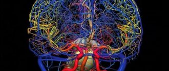
- assess the condition of the arteries supplying the brain and their role in cerebrovascular accidents;
- differentiate between malignant and benign brain tumors;
- establish the cause of dementia, including diagnosing Alzheimer's disease in the early stages;
- accurately identify foci of epilepsy, which is especially important when planning surgical intervention;
- obtain valuable diagnostic information for Parkinson's disease, Huttington's disease, degenerative diseases of the central nervous system (multiple sclerosis, etc.)
Cardiologists rarely resort to PET CT. In this case, the procedure allows:
- diagnose a temporary adaptive decrease in the volume of work of the heart muscle and differentiate it from heart failure;
- accurately determine areas of myocardial ischemia before surgery to restore impaired blood circulation (angioplasty, shunt installation);
- identify areas of post-infarction cardiosclerosis.
Types and modes of research
Depending on the diagnostic purposes, the following may be prescribed:
- full body scan;
- local brain scan;
- examination of the chest area.
Based on the type of radiopharmaceuticals (RP) used, PET is divided into:
- With 18F-fluorodeoxyglucose. Allows you to identify areas with different intensity of glucose accumulation. The cells of most cancers and other malignant tumors take up glucose most quickly. The more the drug accumulates in the tumor tissues, the brighter they glow in the images and the higher the degree of malignancy of the cancer cells.
- With choline, gallium-PSMA, PSMA-7. It is used in the diagnosis of prostate cancer, the tumors of which actively accumulate this substance. Choline is also sometimes used in liver and brain examinations.
- With methionine or tyrosine. Provides high quality images when visualizing gliomas (a type of brain tumor);
- With 13N-ammonium. Used to diagnose changes in the heart muscle.
- With FP-CIT and F-DOPA. Used for Parkinson's disease.
- With F18-FBB. Prescribed for Alzheimer's disease.
If it is necessary to obtain clearer CT images, the patient may need the injection of a contrast agent, and therefore PET/CT is also distinguished without contrast and with contrast.
Limitations, contraindications, complications
In addition to the contraindications common to any diagnostic methods associated with radiation exposure, positron emission tomography is not performed:
- in the first month after completion of chemotherapy;
- in the postoperative period;
- in the presence of severe inflammatory processes or during the height of an infectious disease.
In all of the above cases, the reliability of the results will be insufficient.
In addition, examination by this method is not indicated for patients during radiotherapy treatment and immediately after its completion. The recommended pause after a course of RT is 3 months.
If the prescription rules are followed, the examination does not cause any side effects, and the radiation dose is so small that, if indicated, it can be prescribed to children and nursing mothers. The only possible complication is individual intolerance to radiopharmaceuticals (extremely rare) or contrast agent (higher probability).
Preparation for PET diagnostics
Preparation for the study varies depending on the type of study and the patient’s health characteristics.
General recommendations boil down to limiting physical activity, stopping drinking alcohol and smoking 24 hours before, and stopping carbonated water and food 6 hours before the test.
These recommendations must be followed because intense physical activity, excess glucose, the presence of nicotine and alcohol in the blood lead to changes in the intensity of radiopharmaceutical accumulation, and gases and food make scanning of the abdominal organs difficult.
To increase the accuracy of diagnosis and reduce the risk of side effects from the administration of 18F-fluorodeoxyglucose, patients with diabetes mellitus take drugs that normalize the metabolism of sugars (trimetazidine, etc.) for 8–12 days before the examination. The day before the tomography, coffee is excluded from their diet and taking caffeine-containing medications is prohibited.
Before the choline PET/CT scan, a special diet is also prescribed, excluding protein-rich foods, B vitamins and some vegetables.
How the research is carried out
The patient is administered a radiopharmaceutical, most often intravenously, as well as a diuretic to accelerate the distribution of isotopes throughout the body. The radiopharmaceutical dose depends on the scope of the study and the patient’s weight.
After a certain period of time, during which the maximum accumulation of isotopes occurs in the parts of the body being studied, a scan is performed. In this case, the couch with the subject moves slowly inside or along the module with scanners. The whole process takes from half an hour to an hour or a little more.
After the procedure, it is recommended to consume large amounts of fluid to remove isotope decay products from the body, the lifespan of which is very short - from 2 to 22 hours. Thanks to this, no later than 24 hours later you can communicate with others, including pregnant women and children, without any restrictions.
Decoding the results
Data interpretation is carried out by radiologists with appropriate specialization. As a rule, it takes no more than an hour to evaluate the results and draw up a conclusion.
Carrying out
The procedure itself takes about an hour, but it is necessary to keep in mind the preparation, preparation of the necessary papers and post-procedure rest.
The procedure is carried out in comfortable clothing; it is necessary to remove everything containing metal.
It is necessary to inform your doctor about the pain that may arise from prolonged immobility. Taking this into account, he will be able to select an individual procedure for the procedure.
After the injection, you must lie silent, motionless and relaxed. Immobility has a beneficial effect on the correct distribution of the administered drug. This is important for image quality.
The first stage of the examination is the administration of the drug. It lasts about an hour. The drug is administered intravenously. Sometimes the patient may feel fever, nothing more. The distribution of the drug among the cells lasts about an hour.
The second stage is scanning. Patient in a tomograph. CT is performed first, then PET. If required, an additional contrast agent is administered. The duration of this stage varies from twenty to forty minutes.
Upon completion, the CT and PET images are superimposed.
Preparing the patient for PET/CT
The study is performed strictly on an empty stomach.
Preparation is carried out in accordance with the “Patient Preparation Instruction” corresponding to the study with the prescribed radiopharmaceutical (RP).
The memo is issued:
- when registering a patient for a study at the registry of the PET center (for outpatients);
- the attending physician in the department (for patients hospitalized in departments of healthcare institutions);
- by email (for out-of-town patients booked for PET/CT by doctors from other medical institutions).
On the day of the study, upon registration, the patient provides the following documents:
- statements of previous special treatment (surgeries, radiation therapy, chemotherapy), if any;
- data from the morphological report (cytology, biopsy, final morphological report upon tumor removal), if available;
- results of diagnostic studies for the last 3-6 months (ultrasound, X-ray, angiography, computed tomography (CT), magnetic resonance imaging (MRI), positron emission tomography (PET/CT) on digital media (disks) and in paper form (description and conclusion);
- results of laboratory research methods (biochemical blood test, general blood test, blood test for tumor markers);
- data on the presence of allergies to medications (x-ray contrast agents, etc.);
- data on the presence of concomitant pathology that affects the performance of PET/CT and interpretation of results (diabetes mellitus, renal failure, etc.).
What after?
Afterwards, it is not recommended to be in crowded places for 24 hours, especially to have contact with pregnant women and children. In the family, it is necessary to maintain a social distance of 1 -1.5 from each other.
It is necessary to consume a lot of fluid, two and a half liters or more. This is necessary to remove contrast and radiopharmaceuticals faster and safer.
You can drink any decaffeinated drinks, not just water.
Cost of the study.
Prices for PET/CT can vary significantly depending on the chosen medical center, the complexity of the diagnosis, the tomograph model used, and the experience of the medical staff. Thus, the cost of such a brain study in Russia is in the range of 25,000-85,000 rubles.
If you are a compulsory medical insurance card holder, you can undergo PET/CT completely free of charge. To do this, you just need to take a referral from your attending physician and make an appointment in advance at a radiomedicine center that practices duty-free diagnostics. It is worth considering that the queue for such an examination can last more than one month.
Complications
Complications during the examination procedure are mainly associated with the administration of contrast.
It could be:
- Allergy caused by vein puncturing;
- Damage to blood vessels;
- The appearance of a hematoma;
- Neuritis
A very rare complication is renal dysfunction.
Oral administration of contrast may cause nausea, vomiting, and diarrhea.
After the procedure, dizziness and weakness may occur due to the fact that the stomach is empty.
Radiation exposure to the patient during examination
The choice of radiation dose is influenced by the radiopharmaceutical used and the scope of the study. So scanning the head requires less radiation than scanning the whole body. Minimizing harm from using the method is achieved if the following requirements are followed:
- Use isotopes with a half-life of several minutes or hours;
- Calculate the dose individually for each patient, taking into account his age, height, weight;
- Use special filters on scanners, as well as programs that reduce radiation doses;
- Consume the required amount of fluid, this will allow you to quickly remove the radiopharmaceutical from the body
By contacting the Onco.Rehab integrative oncology clinic, you will find out where it is best to undergo the study that we told you about in this article.
Passing PET/CT diagnostics
The patient must arrive at the clinic exactly at the appointed time. In this case, it is necessary to take all medical documents that are of interest within the framework of the disease - data from previous CT scans, histology data, laboratory tests, a statement of treatment, as well as data on concomitant pathology that may affect the diagnostic results (diabetes mellitus, kidney disease) .
After completing the paperwork, the patient is taken to the treatment room. There they take a blood test for glucose and measure anthropometric data. If everything is in order, the patient is taken to a relaxation room. There he is comfortably positioned, and a peripheral intravenous catheter is installed, through which the radiopharmaceutical is administered. To ensure it is evenly distributed, you need to drink a glass of water (or a contrast agent that the nurse will suggest) every 15 minutes. After administration of the radiopharmaceutical, you must be in a relaxed state and not talk. Any physical activity can lead to uneven distribution of the tracer and distort the results.
After 60–90 minutes, the patient is invited for a scan. First, a CT scan is performed, and then a PET scan. A CT scan takes a matter of minutes, while a PET scan takes up to half an hour. In this case, it is important to lie still so as not to provoke the formation of artifacts in the photographs that can distort the image. Both studies are carried out in the same tomograph and do not cause pain. After the procedure is completed, the patient must remain in the clinic for another hour, as a repeat scan may be necessary to clarify the data.
After a PET examination, the patient should not be in crowded places or have contact with children and pregnant women for 24 hours. In a family circle, you should keep a distance of at least 1 m from each other. Such restrictions apply for 24 hours after administration of the radiopharmaceutical.
In addition, during this period it is necessary to take a sufficient amount of liquid, at least 2.5 liters. This will help remove the contrast and radiopharmaceuticals faster and safer. You can drink more than just water. You can consume any caffeine-free products - juices, sparkling water, fruit drinks, mineral water, etc.