General information
The diencephalon is the lower, most massive part of the brain stem that carries a huge functional load.
On the sides it is limited by the hemispheres (and covered with them on the sides and top, like a cap), in front - by the optic chiasm, on the upper side of the trunk - by the corpus callosum. The most important load is borne by nuclear formations: the thalamus (visual thalamus), hypothalamus (perituberal space), epithalamus and metathalamus.
The hypothalamus and pituitary gland form the hypothalamic-pituitary system.
Structure of the diencephalon
The main topographic structures of this section are the ventricular cavity, thalamus, subtubercular space (hypothalamus), epithalamus (supratubercular space), metathalamus (subtubercular region).
- The third ventricle is a slit-like cavity. On the lateral sides it is limited by the thalamus, behind by the commissure of the epithalamus (through which it communicates with the aqueduct), and in front by the columns of the fornix. The lower wall is formed by the inner side of the hypothalamus, and the upper wall is formed by the vascular weave, over which hangs the fornix of the brain, separating the ventricle from the corpus callosum.
- The thalamus is responsible for the experience of pain. With damage to the thalamus of a mechanical or organic nature, symptoms such as insensitivity of large areas of the body to pain or, conversely, painful hypersensitivity may appear. It includes 40 pairs of thalamic nuclei, which according to functional features are divided into 3 groups. The associative nuclei communicate through the nerve fibers of the tracts with the occipital and parietal temporal regions of the cortex responsible for vision, hearing and speech. Damage to these connections leads to disruption of the corresponding processes. Specific nuclei (for example, the geniculate body) perform the function of switching signals coming from sensory organs, muscles and visceral organs. They contain specific neurons with very long axons and almost no dendrites. The function of nonspecific nuclei is similar to the function of the reticular formation, and disruption of their work leads to confusion or lack of consciousness.
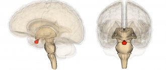
- The hypothalamus is localized in front of the cerebral peduncles and is the main control center for life support functions and (communicating with the pituitary gland) regulation of metabolism. He also manages sexual function, growth processes, and coordinates all the activities of the autonomic nervous system. The blood supply system of this organ has increased permeability to hormones and nutrients. It contains 48 pairs of nuclei. Typically kernels are classified as follows:
- posterior group: mamillary, premamillary and supramamillary;
- anterior group: supraoptic, preoptic, supraoptic, anterior, paraventricular;
- middle group: lateral, ventromedial and dorsomedial.
- The epithalamus is divided into the pineal gland (epiphysis) and the space on its sides, which includes the nuclei of the olfactory analyzer and forms the cover of the third ventricle.
- The metathalamus is the geniculate body located near the thalamic cushion. The lateral body is the subcortical instance of the visual analyzer (its nuclei are connected to the lower pair of colliculi), and the medial body (connected to the upper pair of colliculi) is the auditory one.
Thalamus
The thalamus are paired, small, ovoid structures that occupy almost the entire (80%) diencephalon. The main function of this department is the convergence (unification) of all sensitive pathways, their processing and transmission to the cortex. It also prevents unnecessary or low-value signals from entering the brain, which reduces the load on the cortex. The thalamus contains approximately 40 nuclei - clusters of neurons with specialized functions. They are divided into three groups:
- Specific (projection) switch sensory information to the cerebral cortex, modulate a specific signal by which the brain determines where the irritation came from and perceives it. They also process pain information (the highest center of pain sensitivity is located here), so if the thalamus is damaged, it is possible to either reduce the pain threshold or increase it. With the help of specific signals, the thalamus coordinates the actions of the higher lying parts of the central nervous system;
- Nonspecific are associated with the reticular formation, their function is associated with the creation of background excitation. They modulate nonspecific signals that support the excitation of cortical neurons, and also take part in the formation of emotions and facial expressions;
- Associative lobes connect different lobes of the cerebral cortex: temporal, parietal, occipital.
The metathalamus is the medial and lateral geniculate bodies, which constitute the subcortical center of hearing and vision, and are responsible for orientation reflexes. They are connected to the quadrigeminal midbrain (which is an ancient visual center). Their damage threatens complete loss of vision or hearing (while maintaining the integrity of the optic and auditory nerves).
If we talk about the structure of the diencephalon, we also need to highlight the subthalamus, which is the Lewis nucleus. It is strongly connected to the extrapyramidal system and is involved in the system of muscle control and coordination of fine movements. There is also an undefined zone, the functions of which are unknown.
Functions of the diencephalon
There are several groups of processes that are regulated by the diencephalon:
- the functioning of the senses, processing sensory signals, interpreting them in terms of significance for the body. The hypothalamus has centers of vision and hearing in the thickness of its geniculate bodies, and the thalamus functions as a regulator of visual, skin, and auditory sensitivity. Some of its processes extend to the cortex (thalamocortical pathways), the other part to the striatum;
- control of vegetative processes. Numerous centers responsible for regulating life support and metabolic processes are localized in the subcortex of the hypothalamus. There are sensations of hunger, thirst, and physical discomfort. The hypothalamus also controls the body's thermoregulation;
- regulation of biorhythms and daily activity by the pineal gland;
- participation in the regulation of emotions and voluntary movements;
- hormonal function of the pituitary gland (regulates the production of thyroid hormones, numerous sex hormones, growth hormone, follicle-stimulating hormone).
Thalamic brain
Many students make the mistake of saying that the diencephalon consists of the thalamus and hypothalamus. This is incorrect, because with such a classification we lose many important structures that are not the thalamus, but are part of the thalamic brain. Let's look at this in detail.
Thalamus
Wikipedia has a very good picture of the projection of the thalamus in relation to other parts of the brain:
Also, the thalamus has always reminded me in shape of the spaceship of the inhabitants of Cloud City on the planet Bespin. It was there that the most dramatic events of the 5th episode of Star Wars took place. So, the residents of Cloud City traveled on small spaceships that looked like two cabins connected by a small isthmus:
In this illustration from the atlas of Yu.L. Zolotko you can see both thalami and partially the caudate nucleus, which is located directly in front of the thalamus. Here the thalami are sagittally truncated by approximately half, but the absence of the cerebral hemispheres gives us the opportunity to see the thalami themselves and other components of the thalamic brain, about which we will see below.
So what is the thalamus? The thalamus is a paired egg-shaped formation with a pronounced sharpening in the front and a pronounced thickening in the back. The anterior point is called the anterior tubercle of the thalamus (tuberculum anterius thalami), and the posterior thickening is called the pulvinar thalami. The two thalami are connected to each other by a thin isthmus called the interthalamic fusion (adhesio interthalamica).
As I said before, the thalamus has the old school name “optic thalamus”. This is due to the fact that the thalamus actually processes and conducts visual signals, being one of the subcortical centers of vision. However, the thalamus processes not only visual signals, but in general all signals of all types of sensitivity.
According to the apt expression of Professor V.A. Dubynin, the thalamus is the “secretary” of the cerebral cortex. This is a very accurate definition, because the thalamus not only duplicates the functions of the cortex, it processes a huge amount of visual, auditory, tactile, proprioceptive information and decides which of these information flows to send to the cortex and what to slow down or completely block.
Indeed, the thalamus actively blocks unnecessary flows of information that tend to get into the cortex - you can appreciate this effect while observing a small, restless child who cannot concentrate on any one task and is constantly distracted. This is a consequence of imperfect development of the central nervous system, including the thalamus, which is only learning to stop numerous impulses and impulses that interfere with performing the same work, for example, eating.
To summarize, let's remember that the thalamus is the main subcortical center of almost all types of sensitivity. All sensory pathways going to the cortex are somehow connected to the thalamus.
Nuclei and pathways of the thalamus
In this lesson we will study the material a little differently than in previous neuroanatomy guides. Today we will deviate from our usual scheme, in which we studied the nuclei separately and the pathways separately (if you don’t understand what these are, look here). The thalamus has a lot of nuclei interspersed with relatively little white matter, so I decided to look at both nuclei and tracts at the same time.
In order to outline the approximate location of the thalamic nuclei on the diagram, we will need to draw the following marking in the form of something similar to a diverging lens:
Anterior nuclei of the thalamus
Having thus divided the thalamus into several fields, we can mark the nuclei. First of all, we will note a group of nuclei called the anterior nuclei of the thalamus (nuclei anteriores thalami). The subcortical sense of smell[/anchor] is located here.
The conducting path of the olfactory analyzer is interesting in that here the impulse first enters the cerebral cortex, and only after that - to the subcortical centers. In the case of other types of sensitivity, everything happens the other way around - subcortical centers process information and send it further to the cortex.
Based on this, we can schematically note the tracts that go to the anterior nuclei of the thalamus from the cortex and limbic system (I did not find the names of these tracts), as well as the tracts connecting the thalamus and the mammillary bodies that have not yet been studied by us. These tracts are called the mastoid-thalamic fascicle (fasciculus mammilothalamicus), I designated them as number 1.
While working with American, English and other foreign atlases, I was surprised to discover that Western neuroanatomists designate the functions of the anterior nuclei of the thalamus in a completely different way. It indicates that the anterior nuclei of the thalamus are responsible for learning, short-term memory and the ability to concentrate on long-term tasks. When compiling this article, I rely primarily on Professor Gaivoronsky’s textbook and Professor Izranov’s video lessons, so I present the structure of the thalamus in accordance with the Russian anatomical school.
Posterior nuclei of the thalamus
The posterior nuclei of the thalamus (nuclei posteriores thalami) are grouped in the rounded posterior end, which, as we know, is called the cushion. Fortunately, there is no confusion here and all sources indicate the main function of these nuclei - subcortical analysis of visual signals. The fibers of the optic tract (tractus opticus) enter the thalamic pad, where they switch to neurons of the posterior nucleus. The processes of these neurons go directly to the higher cortical center for processing visual information, that is, to the occipital lobe cortex.
Ventrolateral nuclei of the thalamus
The ventrolateral nuclei of the thalamus (nuclei ventrolaterales thalami) should be remembered better than all other nuclei by my readers. This is where the pathways that we have been teaching and drawing for so long end. First of all, this is the medial lemniscus (which at the level of the spinal cord consisted of the Gaulle and Burdach fascicles), as well as the spinothalamic tract, which (as I just learned) is also called the spinal lemniscus. I find it easier to remember these pathways by their full names - ganglio-bulbo-thalamo-cortical tract and ganglio-dorsal-thalamo-cortical tract, respectively.
Both pathways carry nonspecific sensation from the trunk and limbs to the central nervous system, with the medial lemniscus conveying proprioceptive sensation and the spinal lemniscus transmitting cutaneous, temperature, and pain sensation.
Median nuclei of the thalamus
If you look at the figure with which we marked the thalamus, you will see a fairly large field in the very center of the marking. This is the space for the median nuclei of the thalamus (nuclei mediani thalami). According to Gaivoronsky’s textbook, these nuclei are responsible for processing signals received from the auditory and vestibular analyzer, including the well-known lateral lemniscus (number 6). Axons of the median nuclei of the thalamus are sent to several areas at once, including both areas of higher analysis (the cortex of the temporal and frontal lobes of the cerebral hemispheres) and subcortical centers (red nuclei of the midbrain, posterior nuclei of the hypothalamus).
Medial nuclei of the thalamus
The medial nuclei of the thalamus (nuclei mediani thalami) in Russian anatomical literature are indicated as “integration centers of the extrapyramidal system.” This means that the auxiliary nuclei of the system of unconscious movements are located in the medial nuclei of the thalamus. In the diagram from Gaivoronsky’s textbook, the medial nuclei have processes from all other nuclei of the thalamus. I will draw as many lines as possible so as not to clutter our drawing, but I hope you will remember this feature of the medial nuclei.
Axons of the medial nuclei (number 7) connect the thalamus with several formations at once, such as the frontal cortex, limbic system and red nuclei.
Epithalamus
Many students confuse the metathalamus and epithalamus, however, it is not difficult to distinguish between them. One of the most important anatomical structures of the epithalamus is a gland called the pineal gland. You can simply remember the same prefix (epi), which will guide you.
So, the epithalamus includes the pineal gland (also known as the pineal gland), leashes, triangles of leashes and commissure of leashes. Let's figure out where it is.
Leashes
The thalamus is wrapped in broad white matter structures that separate the thalamus itself and parts of the cerebral hemispheres called the basal ganglia. I could not find complete information about exactly what signals these pathways carry. However, Sapin's textbook suggests that among these fibers there are fibers that conduct signals from the olfactory organ.
So, the medial part of this white matter is called the stria medullaris of the thalamus, and the lateral part is called the terminal stria (stria terminalis). On the posterior part of the thalamus, the medullary and terminal striae form triangular thickenings, which are called leash triangles (trigonum habenulae). Further, the leashes themselves (habenulae) extend from the triangles of the leashes, which are connected in a section called the commissure of the leashes (comissura habenularum).
In our illustration I have indicated:
- number 8 - triangles of leashes;
- number 9 - leashes;
- number 10 - soldering of leashes.
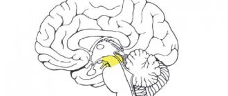
Pineal gland
The pineal gland looks like a very small spruce cone, so the second name for the pineal gland is the pineal gland (glandula pinealis). The pineal gland is located in the area of the thalamic cushion; it is, as it were, suspended on leashes. The quadrigeminal plate of the midbrain is also a good landmark, because the epiphysis literally hangs over it, and many people mistake it for the “fifth colliculus.” In our illustration, the pineal gland corresponds to the number 11:
Here I highlighted the leash triangles in green, and the pineal gland in lilac:
What is the pineal gland for? This small gland controls our body's circadian rhythms through the synthesis of the hormone melatonin. Melatonin is what helps us fall asleep when we close our eyes. Melatonin inhibits the production of stress hormones, lowers body temperature, and also increases the activity of cellular immunity.
By the way, some fish, amphibians and reptiles literally have a third eye - an unpaired light-sensitive organ that is located just above the forehead. Histologically, this organ, also called the parietal eye, is a greatly simplified traditional eye, with receptors capable of detecting only the difference between light and dark.
The parietal eye is directly or indirectly connected via conductive pathways to the pineal gland - this is how the synthesis of melatonin is triggered in response to the onset of darkness, so that the daytime animal falls asleep. In humans, the function of the parietal eye is performed by the traditional organs of vision. This is why physiologists advise turning off the lights a few minutes before bedtime - this creates optimal conditions for the production of melatonin and falling asleep.
Metathalamus
The metathalamus is the part of the thalamic brain that connects the diencephalon and midbrain. The metathalamus includes the medial and lateral geniculate bodies, as well as the superior and inferior arms. It's actually very simple.
Top and bottom handles
As you remember, the dorsal surface of the midbrain is formed by the quadrigeminal plate, in which the subcortical centers of vision (superior colliculi) and hearing (inferior colliculi) are localized. From each hillock towards the thalamus, bundles of pathways extend, which on the outside look like small cylindrical thickenings. These are the upper and lower arms (brachia colliculi superior et inferior).
There is a certain circumstance - in some sources the upper and lower arms of the quadrigeminal region are attributed to the midbrain, and in others - to the metathalamus. Maybe the whole point is that having a midbrain and arms and legs would sound too funny?
In my guide, I attributed the handles to the metathalamus (this is exactly what happened in Professor Izranov’s lecture), so let’s add them to our drawing as numbers 12 (upper) and 13 (lower):
geniculate bodies
The geniculate bodies are another subcortical centers of hearing and vision. Externally, the geniculate bodies appear as four small protuberances on the posterolateral part of the thalamic cushion. Unlike the colliculi and the manubrium, the geniculate bodies are divided into lateral and medial.
The lateral geniculate bodies (corpora geniculata lateralia) are the subcortical centers of vision, which are approached by the upper arms, starting from the superior colliculi. Accordingly, the medial geniculate bodies (corpora geniculata medialia) are responsible for subcortical sound processing and are approached by the lower arms, starting from the lower colliculi. The geniculate bodies, unlike the handles, are indicated in absolutely all sources as part of the thalamic brain. Let us also mark them - number 14 for the lateral geniculate bodies, and number 15 for the medial ones.
I found this illustration on the Internet, where we see the dorsal side of the brain stem. Here I have marked the upper arms and lateral geniculate bodies in yellow, and the lower arms and medal geniculate bodies in blue:
Without any secretions, you will also be able to identify these anatomical formations. And, I hope, you can easily find in this illustration the quadrigeminal plate, the pineal gland, the massive middle cerebellar peduncles, the rhomboid fossa and the medulla oblongata:
Embryonic stages of development
At the end of the first month of intrauterine development, the fetus develops three brain vesicles - rhomboid, anterior and middle.
The diencephalon is formed from the anterior bladder, which becomes the wall of the third cerebral ventricle. The anterior vesicle is divided into two parts, which serve as the basis for the development of the diencephalon and telencephalon. The most fleshy are the lateral walls, from which the geniculate bodies and the thalamus and hypothalamus are later formed. The walls of the vesicles consist of three layers - marginal (includes a small number of cells), interstitial and germinal (in the latter, the cells are poorly differentiated and have not formed into full-fledged tissue). The nuclei of brain structures develop from the interstitial layer of the ventral walls. The lateral protrusions of the diencephalon develop into optic cups, which in the later stages of intrauterine development become the optic nerves.
Where the diencephalon vesicle fuses with the adjacent one, the epiphysis and its leashes with triangles are formed from the protrusion of the upper wall. From the dorsal wall, the thinnest of all, the rudiment of the epiphysis buds, and the wall itself fuses with the choroid, forming the roof of the third cerebral ventricle. From a single protrusion of the posterior wall of the diencephalon, the posterior lobe of the pituitary gland and the gray tubercle are formed. Protrusions of the lower wall become prototypes of the gray tubercle, mastoid formations, intermastoid and mastoid recesses.