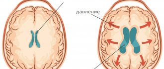
The present time is referred to as the age of brain research. One of the most interesting topics in scientific research on this organ has been the ability of the brain to change its structural and functional properties in response to a person's experiences throughout life. For most of history, neuroscientists believed that the basic structure of the brain was predetermined before birth, and the only changes that could occur were degenerative, the result of disease, injury (concussions, TBI). Modern scientists have directed research towards brain restoration. What conclusions did they come to? Is the brain recovering or not?
Research results
Two major discoveries were made by scientists involved in neural networks and human brain research. A study published in Cell Stem Cell reports that Japanese doctors have begun culturing human brains. The journal Science presented material on how chemical destruction was prevented by stimulating the regeneration (renewal) of the brain and spinal neural network.
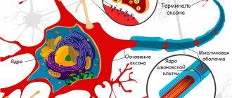
A neuron is a structural unit of nervous tissue that, under a microscope, resembles a body with tentacles. The task of a neuron is to receive and process information.
The Japanese started from brain cells, which, through appropriate cultivation, were multiplied tenfold and enriched in accordance with the structure of the brain of a human embryo. It was also discovered that in the resulting particles of brain matter, the size of which is 1-2 mm, neural activity, measured in electromagnetic impulses, spontaneously arises. Scientists from the city of Kobe believe that in the future it will be possible to create structures of brain tissue that can be implanted in place of parts damaged by disease (ischemic stroke, multiple sclerosis, etc.) or injury.
Brain neurons are not able to regenerate like their counterparts in nerve endings. Another way to save damaged parts of the brain or spinal cord (damage often leads to serious consequences, including paralysis, coma) is to activate the possibility of regeneration in both main organs of the nervous system. In experiments on mice, a team led by Dr. Che Qiang at Harvard Medical School in Boston was able to answer the question of whether brain cells repair themselves by chemically influencing the process. In mice, scientists used genetic engineering to stimulate the release of a substance called mTOR, which responds to neuronal regeneration. It is present in the newborn, but is destroyed in the adult, especially after injury. Thanks to this process, scientists were able to restore almost half of the damaged optic nerve in a short time (2 weeks). Even the formation of new axons has been recorded.
Che Qian concluded: “We knew that after development ends, networks stop growing due to genetic mechanisms. We believe that one of these mechanisms may also restore regeneration and stop death after injury."
Advances in emergency medicine have ensured that more brain-injured patients survive. Today it is known that the adult brain is capable of rearranging its functional connections, creating new ones, and changing physiological parameters. This phenomenon is called neuroplasticity; it has become the basis for the treatment of central nervous system diseases of various origins.
Fewer cells die and more form in autistic people. It can be said that autism, paradoxically, is a disorder that has beneficial effects on the brain.
Hippocampus and brain recovery
According to recent data, the human brain contains about 85 billion nerve cells (neurons). It is known that throughout life there is a gradual loss of these cells (they begin to die around the age of 30).
One of the first studies to generate interest in brain plasticity among lay people was conducted by Eleanor Maguire of University College London. She found that London taxi drivers have much more developed hippocampi than bus drivers. The hippocampus is the part of the brain responsible, among other things, for the perception of space. Given the fact that taxi drivers must remember many street names, their placement and connections, it has been suggested that this change is caused by training in spatial orientation, which bus drivers lack.
The problem with this study is that it does not distinguish between innate and acquired function. In this context, interesting results were provided by studies of violinists, which established that these musicians have a much larger surface area of the motor cortex related to the fingers of the left hand. This corresponds to the fact that when playing the violin, each finger of the left hand must make an independent movement. In this case, on the right hand, all fingers work together. The objection to the possibility of a genetic predisposition is countered by the fact that the difference between the organization of the left and right hemispheres is directly proportional to the age at which musicians began to play the violin.
Reorganization of the cerebral cortex has also been observed in people with congenital visual or auditory defects. According to the “use it or loose it” principle, the unused cerebral cortex can be used by another function. Areas originally dedicated to processing visual or auditory stimuli are deprived of them, and their space is used for other functions, such as tactile. Reorganization is the result of the growth of long processes of neurons, axons. After a head injury that damages the brain, neural connections can be restored or replaced with new connections that compensate for the lost function in another part of the brain.
One of the great surprises of recent times is the discovery that the adult brain can, in some areas, create entirely new neurons from stem cells, a process influenced by human experience.
Other effective exercises
One of the simplest and most effective exercises for training the brain is changing the pace when performing habitual actions. If you are used to having breakfast quickly, trying to do it in half an hour, try to slow down the process of eating.
Conversely, if you like long meals, reduce your lunch.
It is equally useful to change your usual routes - to school, university, store or work.
Advertising:
Choose a different road - a longer or shorter one, take a walk or take public transport. This exercise has a complex effect. It not only helps to activate the work of unused areas of the brain, but also has a positive effect on the muscles, helping to burn extra calories.
A good brain workout is moving around the house or apartment with your eyes closed. Due to the fact that the eyes are closed, other senses become more acute and become maximally sensitive to external factors and irritants. This improves, activates and sharpens their work. You need to start training by covering short distances, after which you can gradually increase them.
Neurogenesis
Information not known to the general public is that the brain creates new cells throughout life. This phenomenon is called neurogenesis.
The human brain is made up of many parts (but not all of them undergo cellular renewal). Neurogenesis occurs in the area responsible for the sense of smell and in the hippocampus, which plays an important role in memory quality.
Experts have also discovered that damaged brains also produce new cells. Evidence of higher neurogenesis during illness was presented by New Zealand's University of Auckland, which studied people with Huntington's disease, in which a person's mental abilities decrease and uncoordinated movements appear. The creation of new neurons was most intense in the most affected tissues. Unfortunately, this is not enough to suppress the disease. Identifying the conditions under which this process occurs and stimulating it could lead to treatments for Huntington's or Parkinson's disease by transplanting stem cells into the affected areas of the brain.
Medical science is taking its first steps in studying brain neuroplasticity. The next step is a precise description of the conditions under which its changes occur, determining the specific impact on individual functions in human life. Understanding and using knowledge of neuroplasticity also requires analysis of genes associated with the growth of axons or neurons from stem cells.
Neuroprotective therapy for chronic cerebral ischemia
New demographic trends today are of particular relevance in geriatric practice. In the second half of the 20th century, the age composition of the population changed significantly. Improvements in socio-economic conditions and the quality of medical care in industrialized countries have led to a significant increase in life expectancy. The result of this was a significant increase in the number of elderly and senile people. According to the 2010 All-Russian Population Census, about 47 million elderly people live in our country, 71.8% are people over working age [1]. The fastest growing age group in Europe is older people over 80 years of age.
The main group of patients visiting the clinic in our country are elderly people. Thus, for 1 patient aged 50 years and older there are from 1.7 to 3.6 diseases (while for people 70 years and older there are 5–7 diseases). The most common complaints with which elderly patients consult a doctor are associated with manifestations of cerebrovascular disease (insomnia, depression, cognitive disorders), exacerbation of chronic somatic diseases, and side effects from frequent and sometimes uncontrolled use of medications [1].
Cerebrovascular diseases (CVD) are one of the main causes of mortality and permanent disability in patients. According to the classification of cerebrovascular disorders adopted in our country in 1958, dyscirculatory encephalopathy (DEP) is the main clinical form of chronic cerebrovascular insufficiency [2–4]. Despite the fact that in the International Statistical Classification of Diseases and Related Health Problems, the IX and X revisions, DEP as a nosology is absent and it was replaced by the term “cerebral ischemia (chronic) (CHI)” - ICD-10, class IX “Diseases of the circulatory system” "(I00-I99), I67.8 - "Other specified cerebral vascular lesions: 1) acute cerebrovascular insufficiency of 5-bromo-2'-deoxyuridine (NOS) and 2) cerebral ischemia (chronic)", the term is still widely used "encephalopathy".
Definition. CCI is a special type of vascular cerebral pathology, caused by a slowly progressive diffuse disorder of the blood supply to the brain with gradually increasing various defects in its functioning [5]. DEP is a chronic progressive form of cerebrovascular pathology, characterized by the development of multifocal or diffuse ischemic brain damage [3, 4, 6, 7]. DEP, like stroke, is a cerebrovascular syndrome [2] - a syndrome of chronic progressive brain damage of vascular etiology, which develops as a result of repeated acute cerebrovascular accidents and/or chronic insufficiency of blood supply to the brain [8].
The clinical picture of DEP is characterized by [4]: 1) a progressive increase in cognitive impairment (decreased memory, attention, intelligence), reaching the level of dementia in the final stages, which is manifested by a combination of pronounced impairment of cognitive functions, personality changes with significant difficulty in normal social activity and the inability to continue work; 2) a gradual increase in emotional impoverishment, loss of interest in life; 3) gradual increase in coordination and walking disorders, destabilization of the tempo and rhythm of movements, tendency to falls; in severe cases, walking becomes impossible, despite the absence of paresis; 4) subcortical syndrome: oligobradykinesia, hypomimia, acheirokinesis, increased muscle tone of the extrapyramidal type (like parkinsonism syndrome); 5) pseudobulbar syndrome of varying severity: dysarthria, dysphagia, forced laughter and crying, symptoms of oral automatism; 6) decreased strength in the limbs, mild paresis with severe brain damage; 7) gradual appearance of disturbances in control of the function of the pelvic organs.
There are three stages of DEP: I - mild or moderate (compensation), II - severe (subcompensation), III - pronounced (decompensation) [9, 10]. For DEP Art. I typical [4]: complaints of increased fatigue, frequent headaches, irritability, moderate memory impairment (primarily operational), moderate decrease in performance, sleep disturbances, which are accompanied by fairly persistent objective disorders in the form of anisoreflexia, discoordination phenomena, mild oculomotor disorders, symptoms oral automatism. There are diffuse neurological symptoms, and the identified disorders are subsyndromal in nature [9]. The early stages of DEP are most characterized by vascular depression and emotional lability, and, as is known, emotional disorders can have an adverse effect on the cognitive sphere [8]. The gradual progression of the disease and worsening symptoms are a powerful factor in social maladjustment [1].
With inadequate treatment, DEP progresses and turns into stage II DEP, in which the noted diffuse neurological symptoms develop into a separate dominant syndrome, which most significantly reduces the patient’s professional and social adaptation [4, 9]. Anxious and depressive reactions appear, cognitive disorders worsen, mental production and volitional activity decrease, professional memory deteriorates, there is a viscosity of thinking, a narrowing of the range of interests, a decrease in criticism and a change in personality. Circadian rhythms are disrupted in the form of daytime sleepiness with poor night sleep. For DEP II Art. characteristic: deepening memory impairment and decreased attention function, increasing intellectual and emotional disorders, and a significant decrease in performance. Somewhat less frequently, there are complaints of chronic fatigue, headache and other manifestations of asthenic syndrome. In some patients, mild subcortical disturbances and changes in gait are detected (it becomes shuffling, mincing). Focal symptoms are more clearly manifested in the form of revitalization of reflexes of oral automatism, central insufficiency of the facial and hypoglossal nerves, coordination and oculomotor disorders, pyramidal insufficiency, and amyostatic syndrome [4].
With DEP stage III. the number of complaints decreases, which is due to a decrease in patients’ criticism of their condition, the severity of intellectual-mnestic and neurological disorders increases, unmotivated actions and inadequate reactions are observed, emotional disorders are characterized by a dysthymic-dysphoric mood background with irritability, dissatisfaction with others and weakness. This stage is characterized by: clearly defined discoordination, amyostatic, psychoorganic, pseudobulbar, pyramidal syndromes. Paroxysmal conditions are more often observed - falls, fainting, epileptic seizures. The main difference between DEP Art. III. from DEP II Art. is that with DEP stage III. In the clinic, several fairly pronounced syndromes are observed, while with DEP stage II. one of them clearly predominates [4, 9].
The relationship between the proportion of complaints and neurological symptoms at different stages of DEP is shown schematically in Fig.
Diagnosis of DEP is possible if the following criteria are met [4]: 1) objectively identified neuropsychological or neurological symptoms; 2) signs of CVD, including risk factors (arterial hypertension, hyperlipidemia, diabetes mellitus, heart rhythm disturbances, etc.), and/or anamnestic signs, and/or instrumentally confirmed signs of damage to cerebral vessels or brain matter; 3) evidence of a causal relationship between (1) and (2); 4) correspondence of the dynamics of neuropsychological and neurological deficits to the characteristics of the course of CVD (a tendency to progression with alternating periods of sharp deterioration, partial regression and relative stabilization); 5) correspondence of changes in the brain substance of vascular origin detected by computed tomography/magnetic resonance imaging with the leading clinical manifestations; 6) exclusion of other diseases that can explain the clinical picture.
Diagnostic criteria for DEP [11]: 1) presence of signs (clinical, anamnestic, instrumental) of brain damage; 2) the presence of signs of acute or chronic dyscirculation (clinical, anamnestic, instrumental); 3) the presence of a cause-and-effect relationship between disorders and the development of clinical, neuropsychological, and psychiatric symptoms; 4) clinical and paraclinical progression of cerebrovascular insufficiency.
Course of DEP and prognosis. There are stable, slowly progressive (with paroxysms and transient cerebrovascular accidents and without vascular episodes), paroxysmal, rapidly progressive course. Stable and slowly progressive are more typical for stage I DEP, which can last for 7–12 years. With a rapidly progressive variant of stage II or III DEP. develop in less than 5 years of illness. Clinical prognosis of stage III DEP. unfavorable, at this stage of the disease patients often need care, and sometimes are completely helpless in everyday life.
Cognitive impairment is a key manifestation of DEP, which largely determines the severity of the patient’s condition. They can serve as the most important diagnostic criterion for DEP and are perhaps the best guide for assessing the dynamics of the disease [2]. At the early stage of DEP, moderate neurodynamic disturbances predominate in the form of slowness, aspontaneity, decreased performance, exhaustion, and weakened concentration. However, such patients generally perform well on timed tests, consistent with mild cognitive impairment [2]. Further progression of the cognitive defect in DEP is associated with the development of dementia, in which cognitive deficit (regardless of motor and other symptoms!) leads to limitation of daily activity and at least partial loss of household independence [2].
Treatment of patients with DEP is a complex medical and social problem and, in fact, is limited to the therapeutic effect on the manifestations of DEP stages I and II. The main directions of management of these patients are reversing the developed decompensation of the pathological process, preventing the progression of the disease, including stroke, reducing the severity of cognitive disorders and neurological deficits [10, 11]. The most effective measure to prevent further progression of the disease is to influence vascular risk factors, primarily proper antihypertensive therapy, correction of carbohydrate and lipid metabolism, changes in vascular tone, increased cerebral perfusion, and improvement of brain tissue metabolism [2].
The basis of therapy aimed at improving the well-being of a patient with DEP and improving his quality of life are drugs that affect cerebral circulation at the microcirculatory level (vasoactive drugs) and drugs that improve metabolic processes in the brain (nootropic drugs). Vasoactive drugs improve blood supply to the brain by dilating the microvasculature. These include: phosphodiesterase blockers (such as pentoxfilline), including those of plant origin (Ginkgo biloba); calcium blockers, the effect of which is most pronounced if the blood flow is impaired in the vertebral arteries supplying blood to the brain stem; α-blockers acting on receptors of the vascular wall. Nootropic drugs can increase neuronal plasticity, thus increasing the adaptive capabilities of nerve cells and reducing their susceptibility to damaging factors by improving metabolic processes in neurons. Nootropic drugs have a positive effect on the higher integrative functions of the brain, facilitate learning processes and memory consolidation.
Today, neurotrophicity, neuroprotection, neuroplasticity and neurogenesis are considered fundamental neurobiological processes involved in the implementation of endogenous protective activity, as well as in attempts to counteract pathophysiological damaging mechanisms and stimulate endogenous repair. The classical concept of neuroprotection involves suppression of a specific pathophysiological mechanism using an appropriate drug [12]. The action of various drugs is aimed at enhancing the oxidation of glucose in mitochondria (Actovegin), inhibiting the oxidation of long-chain fatty acids, which increases the synthesis of adenosine triphosphoric acid (ATP) and neutralization of oxygen radicals, the production of which increases under ischemic conditions [13].
Today, there are several pharmacological groups of drugs with neurometabolic action. They can be roughly divided into classic ones, used for several decades to treat patients with cognitive impairment, and drugs that have relatively recently become widespread in the practice of rehabilitation of patients with vascular diseases of the brain, and were originally proposed for the treatment of Alzheimer's disease. The first group includes Actovegin, piracetam, pyriditol and Cerebrolysin [6]. One of the drugs that has a wide range of pharmacological effects (antihypoxic, antioxidant) is Actovegin, a drug that has a neurotrophic effect. Antioxidant and anti-apoptotic mechanisms of action underlie the neuroprotective properties of Actovegin. It not only improves glucose transport and oxygen absorption, but also stimulates their utilization, which improves oxygen metabolism even under hypoxic conditions.
Impaired cognitive functions (lack of attention, concentration, impaired ability to quickly orientate in a changing environment; decreased memory, especially for current events; slowness of thinking, rapid exhaustion during intense mental work, narrowing of the range of interests) is one of the most common manifestations of CVD. Treatment of cognitive impairment in DEP should, first of all, include measures to prevent further damage to cerebral vessels and brain matter, improve and long-term stabilization of cognitive functions, and correct other clinical manifestations of the disease [2]. On the 1st and 2nd stages. DEP cognitive impairment is present in 88% of cases. A wide range of nootropic and neuroprotective drugs are used to improve cognitive functions [8].
A double-blind, placebo-controlled study was conducted to evaluate the therapeutic effect of the drug Actovegin (tablet form containing 200 mg of the active substance) on mnestic-intellectual abilities in patients with CVD [14]. The study included 120 patients with CVD; the average age of the patients was about 67 years (60–72 years), and the average duration of the disease was 2.5 years. All patients were randomized into 3 groups: group 1 (n = 40), taking Actovegin 3 tablets 3 times a day; group 2 (n = 40) - placebo, of which: 20 patients took 2 tablets 3 times a day, 20 patients took 3 tablets 3 times a day; group 3 (n = 40) - Actovegin 2 tablets 3 times a day. The duration of therapy was 12 weeks, with assessment of the dynamics of the condition before the start of therapy, after 4, 8 and 12 weeks of treatment. To assess the clinical effectiveness of Actovegin, tests were used that assessed mnestic-intellectual functions (memory, synthetic and analytical abilities, concentration, comprehension). The study showed high effectiveness (92%) of 12-week therapy with Actovegin forte on mnestic-intellectual abilities in patients with CVD. Thus, the tablet form of Actovegin can be recommended for long-term outpatient treatment of elderly patients with DEP.
A multicenter study was conducted to evaluate the effectiveness of Actovegin in 1549 elderly patients with impaired cerebral functions [15]. The average age of the patients was 74.1 years. Patients included in the study received treatment according to a standard regimen: 2 weeks of intravenous injections of 10 ml of Actovegin solution, and then 4 weeks of oral administration of Actovegin film-coated tablets (2 tablets 3 times a day). Patients were examined before the start of therapy, two weeks after IV therapy and after 4 weeks of oral therapy. All baseline values were compared with results after 2 and 6 weeks of treatment, and results obtained after 2 weeks were also compared with results after 6 weeks of treatment. After 4 weeks from the start of Actovegin therapy, in 80% of cases, an improvement in the general condition was noted: a decrease in the severity (or cessation) of headaches, dizziness, anxiety and feelings of fear, improvement in memory and concentration (according to psychological testing). In 10.9%, undesirable effects of the drug were noted (feelings of heat and nausea), which were observed in the first, parenteral phase of therapy. The study shows the practical significance of combination therapy: start therapy with iv 10 ml of Actovegin to achieve a quick and good response, followed by continued oral administration of Actovegin forte tablets). The proposed treatment regimen with Actovegin can be recommended for long-term therapy of elderly patients with organic syndrome.
A number of clinical studies have shown that the use of the drug Actovegin has a positive effect on cognitive functions, improves psychological and behavioral reactions, and is also most effective for mild and moderate cognitive impairment. Currently, therapeutic regimens for the treatment of CVD have been developed and are widely used with the prescription of Actovegin: 10 ml (400 mg) per 200 ml of saline solution intravenously for 7–10 days, then 1–2 tablets (200–400 mg) 3 times /day orally for 1-2 months, in the presence of mnestic-intellectual disorders in the elderly - up to 12 weeks, 2-3 tablets 3 times a day. Repeated courses after 6–8 months [16].
In the prevention and treatment of moderate cognitive disorders of vascular origin, neurometabolic drugs have been shown to be highly effective, including the drug Ceraxon (citicoline) [17–19]. The drug has a targeted effect on the key links in the processes of death of nerve cells of vascular, traumatic, toxic and other etiologies. Since Ceraxon is a natural metabolite of biochemical processes in the body and combines neurotransmitter and neurometabolic effects in its spectrum of action. The most important of them is the activation of the biosynthesis of membrane phospholipids of brain neurons, primarily phosphatidylcholine. Citicoline can correct cognitive impairments already at the initial stages of their manifestations in patients with DEP [18]. Its use promotes regression of cognitive impairments and reduces concomitant emotional, affective and behavioral disorders. Cerakson is able to potentiate the effect of other drugs in the treatment of acute cerebrovascular pathology, including thrombolytics, antiplatelet agents and neurotrophics [17]. Prescription regimen for the drug Ceraxon: 1000 mg for 10 days IM or IV once a day, then Ceraxon solution 2 ml orally 3 times a day for 3 months [18] or according to the scheme [20]: daily at a dose of 3 ml 2 times a day for 6 months. The duration of neuroprotection can last up to 12 months.
Conclusion
Moderate and severe cognitive impairment of a cerebrovascular nature can serve as an equivalent to DEP and is detected in 3–16 people over 60 years of age [2]. The main thing in the treatment of a patient with stage I or II DEP. in an outpatient setting is to relieve the developed decompensation of the pathological process, prevent the progression of the disease, including stroke, reduce the severity of cognitive disorders and neurological deficits [21].
A number of studies have shown the high clinical effectiveness of administering the drugs Actovegin and Cerakson as long-term neuroprotection in patients with dyscirculatory encephalopathy.
Literature
- Shavlovskaya O. A. Medical and social aspects of the elderly / Articles of the All-Russian Scientific and Practical Conference with International Participation “Society and Health: Current State and Development Trends”. 2013, vol. 2, p. 365–371.
- Levin O. S. Use of Ginkgo biloba extract (EGb 761) for the treatment of cognitive impairment in dyscirculatory encephalopathy // Breast Cancer. 2009, vol. 17, no. 20, p. 1356–1361.
- Maksudov G. A. Dyscirculatory encephalopathy. In the book: Vascular diseases of the nervous system. Ed. E. V. Schmidt. M.: Medicine, 1975, pp. 501–512.
- Temnikova E. A. The use of the drug Omaron in the practice of a general practitioner when working with elderly patients // RMZh. 2009, vol. 17, no. 20, 1345–1356.
- Putilina M.V. Modern ideas about the treatment of anxiety and depressive disorders in chronic cerebral ischemia // RMJ. 2011, no. 9, p. 569–573.
- Polyakov I. A., Malozemov I. V., Stepanova N. S. Antioxidant therapy in the complex treatment of dyscirculatory encephalopathy // Terra Medica Nova. 2009, no. 4–5, p. 22–24.
- Shtulman D. R., Levin O. S. Neurology. Handbook of a practicing physician. M.: MEDpress-inform, 2008, 1025 p.
- Zakharov V.V., Lokshina A.B. Cognitive impairment in discirculatory encephalopathy // Breast Cancer. 2009, vol. 17, no. 20, p. 1325–1329.
- Dadasheva M.N., Podrezova L.A., Shuchalin O.G. et al. Algorithm for the treatment of dyscirculatory encephalopathy in patients with arterial hypertension in general medical practice // RMZh. 2009, vol. 17, no. 20, p. 1320–1324.
- Kadykov A. S., Shakhparonova N. V. Vascular diseases of the brain. Miklos, 2006, 192 p.
- Damulin I.V., Parfenov V.A., Skoromets A.A., Yakhno N.N. Circulatory disorders in the brain and spinal cord. In the book: Diseases of the nervous system. Guide for doctors. Ed. N. N. Yakhno, D. R. Shtulman. M., 2003. pp. 231–302.
- Shakhparonova N.V., Kadykov A.S. Antioxidant therapy for cerebrovascular diseases // Breast Cancer. 2010, vol. 18, no. 26, p. 1570–1572.
- Astashkin E.I. The influence of Actovegin on the energy metabolism of cells during ischemia. Proceedings of MMA im. I. M. Sechenov. M., 2009: 1–4.
- Jansen V., Bruckner G.V. Treatment of chronic cerebrovascular insufficiency using Actovegin forte tablets (double-blind, placebo-controlled study) // RMZh. 2002. T. 10. No. 12–13. pp. 543–546.
- Letzel H., Schicktiger U. Use of Actovegin in elderly patients with organic syndromes. Multicenter study of 1549 patients // Breast Cancer. 2003. T. 11. No. 25. P. 1428–1431.
- Shavlovskaya O. A. Use of Actovegin in neuroprotective therapy of patients with cerebrovascular diseases // Journal of Neurology and Psychiatry named after. S. S. Korsakova. 2013, v. 113, no. 6, p. 74–76.
- Boyko A. N., Kabanov A. A. Cytocoline: new possibilities of neuroprotection and pharmacotherapy for diseases of the nervous system // Farmateka. 2007, no. 15, pp. 42–48.
- Putilina M.V. Features of combined neuroprotective therapy for acute cerebrovascular accidents // Breast Cancer. 2009, vol. 17, no. 4, p. 261–267.
- Putilina M.V., Shabalina N.I. Possibilities of early correction of mild and moderate cognitive disorders in patients with dyscirculatory encephalopathy // Treating Doctor. 2010, no. 9, p. 100–103.
- Shavlovskaya O. A. Neuroprotective therapy of neurological deficit in cerebrovascular pathology // Practicing doctor today. 2012, no. 3, p. 39–44.
- Shavlovskaya O. A. Efficacy of antioxidant drugs in the treatment of mild and moderate cognitive disorders // Breast Cancer. 2013, vol. 21, no. 10, p. 476–480.
O. A. Shavlovskaya, Doctor of Medical Sciences
GBOU VPO First Moscow State Medical University named after. I. M. Sechenova Ministry of Health of the Russian Federation, Moscow
Contact Information
The importance of neurogenesis
Recent estimates indicate that the hippocampus produces about 700 new brain cells every day. At first glance, this number does not seem large, but the creation of each new neuron is very important, especially for the psychological state of a person. If the formation of new cells stops, depression and psychosis begin to appear. Restoration of brain neurons is important for learning, memory, intelligence (study of certain places, orientation in space, quality of memories).
Recent scientific research has shown that it is possible to improve the production of new brain cells on your own, i.e. at home. What types of activities have a positive effect on the formation of neurons?
Neuron production increases:
- education;
- sex;
- training of cognitive functions;
- mnemonics;
- physical activity (significant help);
- nutrition (regular meals, longer pauses between meals)
- vitamin P (flavonoids);
- omega-3 (also a good antidepressant).
Neuron production decreases:
- stress;
- depression;
- lack of sleep;
- diets rich in saturated fats;
- anesthesia used during surgery;
- alcohol;
- drugs (especially Amphetamine);
- smoking;
- age (with age, neurogenesis continues, but slows down).
Neurons can die in a number of diseases:
- epilepsy – cell death occurs during an attack;
- cervical osteochondrosis – neurons die due to poor circulation;
- hydrocephalus;
- encephalopathy;
- multiple sclerosis;
- Parkinson's disease is a disease characterized by impaired mobility of the legs, arms, and cerebellar signs (due to damage to the amygdala);
- Alzheimer's disease is a disease that leads to dementia, a disorder of speech functions (due to damage to speech receptors).
Neurons may temporarily stop renewing when you take certain cancer drugs. Therefore, after treating cancer with pharmaceutical drugs, people suffer from depression. After restoration of neurogenesis, depression disappears.
It is safe to say that the formation of new brain cells occurs naturally in healthy people. However, whether the process will speed up or slow down largely depends on the person himself.
The importance of the rehabilitation period
For a complete and final victory over the disease, a full-fledged rehabilitation course is important, which is best completed in a specialized center. Proven programs, a friendly microclimate and medical support will help restore the brain after alcoholism. An individual rehabilitation plan is developed for each patient, which takes into account the dominant brain disorders.
The restoration method is based on the following principles:
- Strict adherence to the daily routine. At the same time, optimal ratios of sleep patterns, medical procedures, rest and feasible work are adhered to.
- Physiotherapeutic activities. Massages, contrast showers, magnetic therapy, physical therapy, ultrasound, etc. help restore the normal functioning of the central and peripheral nervous system.
- Neuropsychological adjustment. To get rid of disorders of memory, intelligence, speech functions and attention, special sessions with a psychologist are prescribed. Certain physical exercises and activities can help improve fine motor skills and regain previous skills.
Recovery from brain damage caused by alcohol does not occur within a few weeks. Everything will depend on the severity of the damage and the duration of the person’s exposure to alcohol. This period can last for months, or even years. The main thing is not to return to drinking alcohol and follow all the doctor’s recommendations .
What supports the creation of new neurons?
In addition to the ability to renew itself, the brain is constantly changing, adapting to the external environment, optimizing its activity in accordance with human living conditions. In case of injury, severe intoxication with poisons, medications, or micro-stroke, circulatory disturbance occurs (blood flow to the brain decreases), hypoxia (oxygen starvation) develops, and functions from the affected areas can be transferred to undamaged segments, from one hemisphere to the other. This way a person is able to learn new things and create new habits at any age.
The brain is affected by everyday life, ways of acting, and constant habits. To maximize the manifestation of his wonderful abilities, activity is required, stimulating brain activity in all possible ways.
Electrical stimulation
Targeted electrical stimulation supports the cooperation of neurons in a specific center. This is a non-invasive, drug-free therapy performed by passing a low current through electrodes placed on the scalp. Electrical stimulation can restore brain activity and restore neurons, selectively activating protective mechanisms in the brain, causing an increased release of endorphins and serotonin.
Physical activity
Physical activity and the process of neurogenesis are closely related. As heart rate and vascular blood flow increase during exercise, the levels of factors that stimulate neurogenesis increase. Physical activity also helps release endorphins, reducing stress hormones (especially cortisol). At the same time, testosterone levels increase, which also promotes neurogenesis.
To prevent the negative effects of aging on both the body and brain, physical activity is an excellent choice. It combines both of these goals. You don't have to lift dumbbells or do exercises at the fitness center. Regular vigorous walking, swimming, dancing, cycling is enough. These actions strengthen weakened muscles, improve blood circulation and mental abilities.
Any action aimed at reducing tension and stress promotes neurogenesis. Choose an activity that suits your preferences.
Freshness of mind
There are many ways to regenerate neurons while maintaining a fresh, sharp mind. Various actions can help with this:
- reading - read every day; reading makes you think, seek connections, supports the imagination, arouses interest in everything, including other possible types of mental activity;
- learning or developing knowledge of a foreign language;
- playing a musical instrument, listening to music, singing;
- critical perception of reality, study and search for truth;
- openness to everything new, sensitivity to the environment, communication with people, travel, discovery of nature and the world, new interests and hobbies.
An underrated yet effective method of supporting brain activity is handwriting. It supports memory, develops imagination, activates brain centers, coordinating the movement of muscles involved in the writing process (up to 500). Another advantage of handwriting is maintaining elasticity, mobility of joints, hand muscles, and coordination of fine motor skills.
Nutrition
In connection with the topic at hand, it must be said that the human brain is 70% fat. Fat is a part of every cell in the body, incl. brain tissue, where in the form of myelin it is the insulation surrounding nerve endings. Brain cells create it from sugar, i.e. do not wait for fat to come from food. But it is important to eat healthy fats that do not contribute to the occurrence and development of inflammation. Health benefits come primarily from fats containing omega-3.
Many people, hearing the word “fat,” involuntarily shudder. In an attempt to maintain a slim waist, they buy low-fat products. These foods are unhealthy, often even harmful, because fat is replaced with sugar or other ingredients.
Eliminating fat from your diet is a mistake. Its restriction must be strictly selective. Hydrogenated fats found in margarines and industrially processed foods are harmful to the body. Unsaturated fatty acids, on the contrary, are beneficial. Without fat, the body is unable to absorb vitamins A, D, E, K. They are soluble only in fat, which are of great importance for brain activity. But you also need saturated fats found in animal sources (eggs, butter, cheese).
Low-calorie nutrition is good, but it should be varied and balanced. It is known that the brain consumes a lot of energy. Provide it in the morning. Oatmeal with yogurt and a spoon of honey is an ideal breakfast option.
How to restore the brain with the help of products and folk remedies:
- Turmeric. Curcumin affects neurogenesis and increases the expression of the neuropathic factor, which is necessary for a number of neurological functions.
- Blueberry. The flavonoids contained in blueberries stimulate the growth of new neurons and improve the recognition functions of the brain.
- Green tea. This drink contains EGCG (epigallocatechin gallate), which promotes the growth of new brain neurons.
- Brahmi. Clinical studies examining the effects of the brahmi plant (bacopa monnieri) on brain function showed that after 12 weeks of use, volunteers significantly improved verbal learning, memory, and the speed of processing received information.
Three C's:
- Sun. Healthy exposure to sunlight on the body is 10-15 minutes a day. This promotes the formation of vitamin D, affects the secretion of serotonin, the growth of brain factors that directly affect neurogenesis.
- Dream. Its abundance or deficiency significantly affects brain activity. Lack of sleep causes inhibition of neurogenesis in the hippocampus, disrupts the balance of hormones, and reduces the degree of mental activity.
- Sex. Sexual activity increases the secretion of happiness hormones, endorphins, reduces anxiety, tension, stress, and promotes neurogenesis.
Meditation
The positive effects of meditation on the human brain and overall health have been scientifically documented. Regular meditation has been shown time and again to increase gray matter growth in several areas of the brain, including the hippocampus.
- Meditation stimulates the development of certain cognitive abilities, especially attention, memory, and concentration.
- Meditation improves understanding of reality, focus on the present, and prevents the mind from burdening the mind with fears of the past or future.
- During meditation, the brain works in a different rhythm. In the first phases, increased activity occurs, which is manifested by a higher amplitude of α-waves. During the process of meditation (during the following phases), δ-waves arise, associated with the regeneration of the body and rehabilitation after illness.
- Meditation done in the evening stimulates the brain, increasing the production of melatonin, which is part of the process of neurogenesis. The body relaxes.
Monoatomic gold
Ormus, monoatomic (monatomic) gold is often associated with increased intelligence and overall brain health. David Hudson, who discovered ormus and began analyzing it, said that the substance is capable of restoring the body at the genetic level. Ormus professionals also claim that monoatomic gold can correct DNA errors and even activate dormant DNA.
Methods for restoring the brain after alcoholism
The main prerequisites for normalizing the functional capabilities of the brain are a complete abstinence from alcoholic beverages, timely initiation of treatment and a full rehabilitation course.
The most effective is considered comprehensive medical care, which consists of the following stages:
- counseling and examination;
- detoxification;
- relief of withdrawal symptoms;
- hardware research;
- drug treatment;
- psychotherapeutic support;
- rehabilitation;
- adaptation.
The most effective way is to undergo a treatment program in an inpatient setting. In addition to the fact that the patient is under close supervision of medical staff, he does not have contact with his former environment and does not have the opportunity to return to alcoholism again. The doctor can adjust the medical plan at any time, prescribe additional tests or consultations with other specialists.
Detoxification promotes the removal of toxic substances from the body, which promotes the active division of neurons and the formation of additional neural connections. The course of drips and intravenous injections is selected individually for each patient. This normalizes the water-salt and acid-base balance and accelerates the cleansing of the intercellular fluid.
In drug treatment, pharmacological agents are used that activate restorative brain processes, accelerate tissue metabolism, and protect neural connections from toxic effects.
For example, to restore the brain after alcoholism, drugs from the following groups are used:
- nootropics help restore memory, intellectual capabilities, attention;
- antioxidants support the functions of cell membranes, stimulate cell reproduction;
- Antihypoxants help overcome oxygen starvation of organs and systems and increase oxygen absorption.
Note! Medicines should be taken only as prescribed by a doctor. There is no need to rely on the advice of friends, acquaintances or commercials. Only a qualified specialist can correctly select the necessary list of medications in accordance with the patient’s indicators and calculate the dosage. Then your recovery will be quick and without complications.
During treatment, professional psychological support is very important. It is aimed at mobilizing the patient’s internal strength, developing persistent motivation for full recovery, and creating the prerequisites for the resumption of social and family ties. For this purpose, individual and group psychotherapeutic sessions, hypnotic sessions, art therapy and other psychocorrection techniques are used.
What not to do?
Mental health (according to experts) is more important than the physical condition itself. So how do you support brain function? First of all, you need to know what harms him.
Contaminated air
The brain consumes a significant amount of oxygen, which is necessary for it to function properly. But modern people are constantly exposed to polluted air (vehicle exhaust, dust from industrial production). People from larger cities experience frequent headaches and short-term memory disorders. Longer exposure to polluted air causes permanent changes in the brain.
Alcohol and cigarettes
In addition to causing cancer, heart disease and a range of other health problems, alcohol and nicotine can impair brain function, new research shows.
Unlike alcohol, nicotine compounds do not damage brain cells directly, but lead to other neurological disorders, including. to multiple sclerosis. Long-term consumption of alcohol, prolonged binges, in addition to delirium tremens, cause a chemical imbalance leading to structural disorders. Alcoholics have been shown to have decreased cranial volume.
Lack of sleep
The body, including the brain, recovers most during sleep. Long-term sleep deprivation can cause damage to a vital organ. The body does not have time to create new neurons, and old ones lose the ability to interact with nerve cells. For insomnia caused by overexertion, it is better to take a sleeping pill.
Relaxation for neurons
There are several points on the head that stimulate the overstressed nervous system. Place the fingers of both hands just above your ears and gently massage the skin, creating light pressure. Do the same on the top of the head. Finally, massage your temples and masseter muscles on your cheeks.
Don't cover your head
And one interesting thing. The fact that the brain needs sufficient oxygen is explained above. But did you know that children can have problems with this? They like to hide under the covers, often falling asleep this way. During sleep, the amount of carbon dioxide exhaled increases. This reduces oxygen levels, which interferes with the proper functioning of the brain.
This also applies to adults. Provide sufficient fresh air while sleeping.
Change your brain
The scientists' findings are significant for everyone. Research shows that people of any age can learn new things and form new habits. What we learn in life, who we surround ourselves with, what and how we decide to do, how we think, determines who we are, what our vision of the world is. The more a person is open to new stimuli and knowledge, the more he develops his brain.
Thanks to a proactive approach, habitual but disadvantageous stereotypes can be eliminated. Using various psychological methods, it is possible to replace “trampled” pathways in the brain with new ones. You can transform anxious thought patterns into realistic ones, and replace a negative attitude towards the world with a positive one. It all depends on the restoration of the brain and on the person himself.
Some interesting features of our brain
Before we jump into tips, check out some interesting facts about our brains:
- The brain weighs approximately 2-3% of the body's weight, but consumes about 20-25% of the body's total energy.
- The brain does not distinguish reality from fantasy and thoughts. Everything you think, feel, imagine, see affects the structure of the brain and leaves its imprint.
- The brain never rests. It processes information even when you sleep.
- The more fears, anxieties, and stress there are in a person’s life, the more the area responsible for memory is affected.
- It is believed that we use only about 10% of our brain capacity. In fact, its entire structure is constantly involved, by 10% we mean consciousness. About 90% of processes occur in the subconscious, which is inaccessible to us in our normal state. Therefore, it would be more correct to say that we are aware of only about 10% of the work of our brain.
- All our habits, beliefs, ways of emotional and psychological response are simply stable neural connections in the brain. They can be destroyed and new ones built.
- The destruction of the old neural connection occurs in 40 days. This is how many days religious fasts last - mental cleansing occurs. New neural connections are built in 21 days.