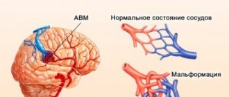
They are a dense tangle of dilated vessels, in which the veins are in contact with the arteries without the participation of the capillary network due to its absence. As a result of the deviation, arterial blood is shunted into the system of deep and superficial veins without releasing nutrients and removing carbon dioxide. The size of vascular malformations can be both small and huge.
The causes of the disorder are not fully understood, but it is believed that it is not related to heredity. Symptoms can be vague and nonspecific, as a result of which the AVM is detected during a CT/MRI of the brain or spinal cord for an unrelated reason (for example, a stroke or injury). The mechanisms and consequences of the influence of AVM on human life are unpredictable, so the disease requires adequate treatment.
Causes of vascular malformations
Vascular malformation is a congenital defect in the structure of the blood vessels of the central nervous system. It has a correlation with disorders of intrauterine development of the fetus and birth injuries, but the specific provoking factors for this phenomenon have not been clarified.
Local anomalies in the formation of arteries and veins of the brain occur in the embryo no later than 1-2 months of pregnancy.
To date, no direct relationship has been found between the presence of this anomaly and the age or gender of the patient.
Symptoms and clinical picture
An AVM may not make itself felt for many years, so patients often learn about their diagnosis by chance, while undergoing diagnostic tests for related reasons. Malformations that increase in volume can put excess pressure on the brain, and only in this case do neurological (focal) symptoms appear.
The clinical picture of the disorder is nonspecific and therefore requires in-depth differential diagnosis.
In everyday life, AVMs can manifest themselves in the following signs:
• Intracranial hypertension with a characteristic pain syndrome of a pressing or pulsating nature;
• Lethargy, apathy, decreased performance;
• Movement coordination disorder;
• Visible decrease in intellectual abilities;
• Motor aphasia and other speech disorders;
• Suppression of innervation of certain parts of the body;
• Unsteadiness of gait and sudden falls (often on the back or side);
• Muscular hypotonia and convulsive attacks;
• Visual impairment (squint, loss of visual fields, partial or complete blindness);
• Epileptic seizures of varying duration and intensity;
• Paresis of the limbs.
Symptoms of the disease are completely dependent on the location of the malformation (frontal or temporal lobe, cerebellum, base of the brain, spinal cord).
The rupture of pathologically altered vascular tangles also entails certain acute manifestations. This complication leads to internal bleeding into the brain or spinal cord, associated with a sudden disruption of their blood supply. With subarachnoid hemorrhage and the formation of a hematoma in the brain, the manifestations of the pathology become distinguishable.
The patient may complain of the following symptoms:
• A sharp and sudden attack of severe headache;
• Nausea and intense vomiting without objective reasons (the eruption of gastric contents does not bring relief);
• Syncope;
• Photophobia (pain in the eyeballs when looking at a light source or being in a lit room);
• Immobility of the eyes and visual impairment up to its complete loss;
• Violations of expressive and impressive speech (the patient pronounces inarticulate sounds and fragments of phrases, does not perceive information from outsiders);
• Convulsive seizures;
• Paralysis of limbs.
If you or your loved ones experience the following clinical manifestations of the pathology, seek professional help as a matter of urgency.
results
Of 223 patients with AVM of the cerebral hemispheres, malformations within one lobe were localized in 151 children (damage to the frontal and temporal lobes predominated), lesions of adjacent lobes were detected in 72 patients. Type I AVMs according to SM were observed in 52 (24%) patients, type II - in 83 (39%), type III - in 63 (29%), type IV - in 9 (4%), type V - in 7 ( 3.35%). 9 children had fistula type AVMs.
In the group of patients with AVMs of deep brain structures (77 children), malformations were most often localized in the corpus callosum region - 23 children, in the subcortical ganglia - 22, in the optic thalamus - 19, AVMs of the choroid plexus were in 13 children. Among them, small AVMs (up to 2.5 cm) were noted in 46 (60.5%) children, medium (2.5-5 cm) - in 22 (29%) and large (5 cm or more) - in 8 ( 10.5%) patients. In 1 child, a fistula-type AVM was detected in the area of the subcortical ganglia.
Damage to the right hemisphere was noted in 49.3% of cases, to the left - in 44.2%.
No relationship was found between the size of the malformation, the features of angioarchitecture and the age of the children.
Clinical manifestations
When comparing two groups of patients with AVM - hemispheric (group 1) and deep localization (group 2), it was noted that in children of group 2 intracranial hemorrhages were observed more often (83.0%) than in patients of group 1 groups (66.4%). In deep-lying AVMs, intraventricular hemorrhages were observed in 20%, often with hemotamponade of the ventricular system. Intraventricular hemorrhages in hemispheric AVMs were observed in 6% of cases. Repeated hemorrhages in deep-seated AVMs occurred 2 times more often than in patients of group 1 - 10.4 and 4.6%, respectively, and were more severe. The duration of recurrent hemorrhages in both groups varied from several hours to several years. The general trend was characterized by a more frequent threat of hemorrhage in small AVMs in both groups (Table 1).
In most cases, intracerebral hemorrhage occurred suddenly. Provoking factors were physical or emotional stress, mild traumatic brain injury. Intracranial hemorrhages with AVMs of deep localization were characterized by the occurrence of a sudden headache with nausea and vomiting, decreased level of consciousness, severe meningeal symptoms, and hyperthermia. In children of the younger age group, in most cases, the hemorrhage was erased, periods of short-term headache alternated with periods of psychomotor agitation, repeated vomiting, which was more reminiscent of the symptoms of an acute intestinal infection.
The combination of subarachnoid-parenchymal hemorrhage with intraventricular hemorrhage significantly worsened the prognosis of the disease. However, even with massive intraventricular hemorrhage, the child’s condition could remain stable and of moderate severity, and only with repeated hemorrhages did it progressively worsen.
Some patients had epileptic seizures [24]. The frequency of epileptiform syndrome was 21% of cases. With AVMs of deep localization, this syndrome was observed almost 3 times less often. Convulsive syndrome, as a manifestation of the disease, was more typical for children of the younger age group (up to 7 years). Of these, 2 patients had epileptic seizures from the first days of life. Most of the malformations in these children were large (for AVMs of the cerebral hemispheres SM III-V and more than 5 cm for AVMs of deep structures) and in most cases drained into the superficial veins of the brain. The attacks in most children occurred with loss of consciousness and generalized convulsions, tended to become more frequent, and were difficult to correct with medication.
The combination of intracranial hemorrhage with epileptic seizures was detected in 4.6% of AVMs of the cerebral hemispheres, and in 1.3% of cases with AVMs of deep brain structures.
Cephalgic syndrome was characterized by constant headache, aggravated by physical or mental stress, or paroxysmal pain. In most cases, the pain was poorly controlled with painkillers. Cephalgic syndrome was more typical for AVMs of the cerebral hemispheres (5.5%), especially when the malformation was localized in the occipital region.
Treatment methods
Microsurgical excision of AVM.
AVM excision using the microsurgical method was performed in 84 patients aged 4 to 18 years. 73 children had AVMs of the cerebral hemispheres, which corresponded to variants according to SM I-III. In 70 (94%) patients with AVM of this localization, the disease manifested itself as SIC, in 2 (4%) children - drug-resistant epileptic seizures, and in 1 (1%) case, cephalgic syndrome was noted; 11 children had malformations of deep brain structures measuring up to 2.5 cm, in 100% of cases SIC appeared.
Upon admission, the children had the following neurological symptoms: pyramidal disorders - in 18 (21%) children; speech disorders of the sensorimotor aphasia type - in 5 (6%) patients; damage to the cranial nerves (most often insufficiency of the III and VII pairs) - in 4 (5%) and 7 (8%) children, respectively. In 15 (18%) patients with AVM of the occipital region and posterior parts of the thalamus, visual field impairments of the homonymous hemianopia type were detected.
The indications for this treatment method were: 1) the possibility of total excision of the AVM; 2) localization of AVM outside functionally important areas of the brain.
87 operations were performed. In 82 cases (according to the surgical protocols), total excision of the malformation was achieved, partial excision in 5 cases. In the last group, in 3 patients this was confirmed by postoperative angiographic control. 2 children had repeated intracranial hemorrhages: in 1 observation, 5 months after surgery, in the other, 1 year later.
In the postoperative period, the following dynamics of neurological symptoms could be noted: by the time of discharge from the clinic, children with motor disorders showed improvements in the functions of paretic limbs in 7 (39%) cases, mainly in children of preschool and primary school age. In 4 (22%) observations, an increase in movement disorders was noted, which was typical for patients over 15 years of age. In 6 (33%) children no dynamics were observed. In 3 patients, speech impairments regressed. Changes in visual fields and damage to cranial nerves were persistent.
In the postoperative period, 12 (29%) children admitted without neurological deficits experienced an increase in focal symptoms in the form of hemiparesis in 4 children and visual field impairment in 8.
An example of total excision of an AVM is shown in Fig. 3.
Figure 3. Patient Sh., 10 years old. AVM of the left parietal region. Angiograms: A and B - before surgery; C and D — after surgery (AVM excision).
Endovascular embolization.
85 endovascular operations were performed on 64 children aged 3 months to 18 years. There were 55 children with AVMs of the cerebral hemispheres, and 9 with AVMs of deep structures. Most of the AVMs of the cerebral hemispheres corresponded to SM I-III. Among AVMs of deep structures, malformations up to 2.5 cm predominated.
In this group of patients, children who underwent SIC also predominated - 34 (53%) patients. It should be noted that in this group, compared with the group of patients who underwent AVM excision, children with drug-resistant epileptic seizures predominated - 25 (40%) patients. There were 5 (8%) patients with cephalgic syndrome.
Clinical symptoms were represented by pyramidal disorders in 3 (5%) children, visual field disorders of the homonymous hemianopia type in 3 (5%) children, and dysfunction of the VI pair of cranial nerves in 2 patients.
Indications for endovascular operations were: 1) palliative method for large AVMs manifested by pharmacoresistant epileptic seizures; 2) fistula type AVM; 3) AVMs located in functionally significant areas of the brain, manifested by SIC; 4) the presence of afferent vessels accessible for catheterization.
In 1 stage, embolization was performed in 34 patients, in 2 stages - 24, in 3 stages - 1. Angiographically confirmed total obliteration of AVM was achieved in 27 (32%) children, subtotal - in 12 (14%), partial - in 15 (18). %) of patients. An example of total embolization is shown in Fig. 4.
Figure 4. Patient S., 14 years old. AVM of the posterior parts of the left temporal region. Angiograms: A and B - before surgery; C and D — after surgery (total embolization with histoacryl).
In the postoperative period, there were no positive or negative dynamics of the symptoms present before the operation. In 6 children who had previously had no neurological symptoms, a transient deficit in the form of motor aphasia was observed in 2 children, and persistent impairment in the form of homonymous hemianopsia was observed in 4 patients.
The dynamics of the epileptiform syndrome attracted attention. After endovascular operations and with adequate anticonvulsant therapy, 12 (48%) children showed a decrease in the number of seizures to 1-2 per month; in 6 (24%) children a change in the nature of seizures was detected; in 4 (16%) observations, the disappearance of attacks was noted. In 1 (4%) child there was a sharp increase in the frequency of epileptic seizures, and in 2 (8%) children there was no dynamics of epileptiform syndrome.
A special group consisted of children with fistula AVMs - 10 patients. All patients underwent endovascular treatment. In 3 patients, total exclusion of the AVM from the bloodstream was achieved (Fig. 5); in 7 cases, subtotal exclusion was achieved.
Figure 5. Patient B., 15 years old. Fistula AVM of the right temporal region. Angiograms: A and B - before surgery; C and D — after surgery (embolization with histoacryl).
Radiosurgical treatment.
Stereotactic radiosurgery (SRS) was performed on 77 children. The age of patients is from 4 to 18 years. In this group of patients there were age restrictions - treatment was indicated only for children over 3 years of age. There were 37 children with AVMs of the cerebral hemispheres, and 40 with AVMs of deep structures. The dimensions of the AVMs did not exceed 3 cm. For malformations larger than 3 cm, radiotherapy treatment was used. The patients were in clinically stable condition.
In the majority of children, the disease manifested itself as SIC—32 (86%) patients with AVMs of the cerebral hemispheres and 38 (95%) patients with AVMs of deep structures. There were significantly fewer children with drug-resistant epileptic seizures. It should be noted that this group included patients with previously inoperable AVMs, i.e. those patients for whom it was impossible to undergo excision or embolization of the malformation.
Indications for radiosurgery were: 1) deeply located malformations; 2) if the risk of endovascular surgery or direct surgical intervention exceeded the risk of recurrent hemorrhage.
An analysis of the results of radiosurgical treatment of 14 children in periods ranging from 6 months to 4 years was carried out. According to the control MRI data after 2-2.4 years, 7 children had total obliteration of the AVM, but according to the control angiographic study this was confirmed only in 4 children, in 1 patient a decrease in the AVM was detected and in 2 patients the AVM was fully functioning. In 7 children, MRI revealed the presence of a functioning AVM.
Combined treatment.
Combined treatment was carried out on 42 children aged 7 to 18 years. There were 34 children with AVMs of the cerebral hemispheres, and 8 with AVMs of deep structures. Most malformations were classified as type II-III SM. In 29 (80%) children in this group, the disease began with SIC, and 7 (20%) had drug-resistant epileptic seizures. Among the deep-seated AVMs there were malformations from 2.5 to 5 cm, all children had SIC.
During the examination, pyramidal symptoms were revealed in only 2 children; the remaining children had no neurological deficit.
The indication for the combined treatment method was the impossibility of total embolization or total excision of the AVM.
In 36 children, combined treatment was carried out in the form of embolization followed by radiosurgery (Fig. 6); 4 children underwent embolization followed by AVM excision, and 2 patients underwent AVM excision at the first stage, and radiosurgery at the second stage.
Figure 6. Patient K., 15 years old. AVM of the right occipital region. Angiograms: A and B - before surgery; C and D — after partial embolization with histoacryl (before radiosurgery); D and F - 3 years after radiosurgery (total obliteration of AVM).
In the postoperative period, 1 child developed persistent hemiparesis after the first stage of treatment (AVM embolization).
2 patients died. A 7-year-old child with an AVM of the left occipital lobe died from repeated intracranial hemorrhage during the second stage of endovascular surgery. An 8-year-old girl died as a result of intraoperative hemorrhage from an AVM in the right occipital region. Overall postoperative mortality was 0.7%.
Long-term results of surgical treatment
Long-term treatment results were assessed in 73 (27%) children with cerebral AVMs after using various methods of AVM treatment: with AVMs of the cerebral hemispheres - 57 children, with AVMs of deep brain structures - 16. The observation period ranged from 3 months to 15 years .
An analysis of the long-term results of treatment of 22 patients after endovascular operations was carried out. According to RS, excellent results (grade 0) were found in 5 children, good results (grade 1-2) in 10 patients, satisfactory (grade 3-4) in 5, poor (grade 5) - in 2 children.
Repeated intracranial SICs were observed in 2 children after 7 years. However, they proceeded without significant neurological deficit.
The follow-up of 25 children after AVM excision was studied. In this group of children, excellent and good results were observed in all patients (grade 0-3 according to RS). It is important to note that 4 patients created families, 2 of them had healthy children.
Long-term results after radiosurgery were analyzed in 14 patients. It can be noted that an excellent result (0th degree according to RS) was detected in 4 children, good (1-2nd degree according to RS) - in 6 patients, satisfactory (3-4th degree according to RS) - in 4 patients .
The results of combined treatment of AVM in 12 patients were analyzed. In all children, the first stage was subtotal or partial AVM embolization; In 8 children, the second stage was radiosurgery; in 4 cases, excision of the malformation was performed. Between 2.6 and 3.5 years, 4 children underwent cerebral angiography, which confirmed total obliteration of the AVM. In the remaining patients, it was possible to talk about a functioning malformation only based on MRI data. In this group of patients, excellent and good results were noted in the long-term period (grade 0-3 according to RS).
In the long-term period, 3 (1%) children died: 2 patients from repeated intracranial hemorrhages, and in 1 observation (a 7-month-old child with multiple AVMs), the cause of death was complications after an acute respiratory viral infection (Table 2).
Group of non-operated patients
Operations were not performed on 30 children aged 1 to 18 years. In this group, there were 23 patients with AVMs of the cerebral hemispheres. It should be emphasized that in these children malformations of large and giant sizes predominated - SM IV-V. Deeply located AVMs were present in 7 patients. Giant AVMs—more than 5 cm—predominated in this group.
In 15 patients (without SIC), surgical intervention was not required, since clinical symptoms were either absent or minimal, and AVMs were diagnosed after MRI as an incidental finding. In 10 patients, AVMs were considered inoperable due to their large size and location in functionally important areas. In 5 observations, the children's parents refused the proposed operations.
Catamnesis was followed in 7 non-operated patients. The follow-up period ranged from 6 months to 15 years. In 2 patients, an increase in epileptic seizures was noted, but after correction with anticonvulsants, the seizures stopped. In 5 patients there is currently no increase in neurological symptoms; children are adapted socially and at home. Only 1 child with concomitant pathology of cerebral vessels (AVM SM V, hypoplasia of the vertebral arteries) retained epileptiform syndrome, resistant to anticonvulsant therapy, with severe delay in psychomotor development. It is important to note that patients in this group did not have intracranial hemorrhages during the observation period.
Differential diagnosis of AVM
The best specialists in the vascular surgery department in our center organize optimal diagnostic measures to recognize the disease in any form. Differential diagnosis in our department is carried out in accordance with the highest medical standards.
Vascular malformations are determined using the following studies:
1. Assessment of the patient’s current condition, analysis of his complaints, in-depth study of the medical history;
2. Primary neurological examination, during which signs of a specific location of the AVM are identified in accordance with the clinical picture of damage to certain areas of the brain or spinal cord;
3. Computed tomography (CT) and magnetic resonance imaging (MRI), which make it possible to study the structure of the brain layer by layer, visually see the location of the AVM and its volume, and assess the general condition of the organ being studied;
4. Doppler ultrasound, which makes it possible to observe the spatial position of blood vessels in the affected area in real time, as well as differentiate pathology from other modifications of their anatomical structure;
5. Angiography, performed using a contrast agent that is injected into the vascular bed; helps evaluate the condition of veins and arteries using x-rays.
If it is difficult to make a diagnosis, you may need additional consultation with a neurologist and neurosurgeon. Typically, vascular malformation (if the patient has one) is diagnosed immediately.
Ischemic brain lesions
The main cause of ischemic stroke is a decrease in cerebral blood flow due to stenotic and occlusive lesions of the great vessels of the neck and arteries of the brain.
In the development of ischemic stroke (cerebral infarction), an important role is played by the blockage of cerebral vessels by microthrombi, which are formed as a result of atherosclerotic plaques breaking off and moving through the vessels.
The main etiological factors leading to damage to both extracranial and intracranial vessels include atherosclerosis, hypertension, and rheumatism. Much less often, the cause of obstruction of the patency of the carotid arteries is cicatricial traumatic and inflammatory extravascular lesions, fibromuscular dysplasia resulting from radiation therapy, as well as pathological bends of blood vessels.
The clinical picture of cerebral infarction depends on the location of the lesion, and the severity and persistence of symptoms depends on the size and condition of the collateral systems. Thrombosis of the internal carotid artery is characterized by decreased visual acuity or blindness on the affected side and hemiparesis or hemiplegia on the opposite side. With arterial stenosis, these disorders can be temporary,
Methods of treating the disease
Complete therapy of patients with vascular malformations requires the ability to carry out the main treatment options - embolization, radiosurgery and surgery. Tactics are selected by an experienced specialist. At the same time, the potential risk of complications from the chosen method of therapy is assessed in relation to the risk of spontaneous progression and course of the disease.
Treatment is aimed strictly at obliterating the malformation to eliminate the threat of hemorrhage. The specialist decides how this goal will be achieved, based on the diagnostic results, individual characteristics and age category of the patient.
Surgical treatment is a rather complex procedure during which the AVM is removed from the brain through a burr hole (mechanical opening of the cranial cavity).
Radical extirpation is used in cases of large volume of AVM tangles.
Radiosurgical treatment (where the malformation is excised with a gamma knife using specific radiation) is suitable only in the case of a small vascular tangle.
The endovascular method is not so aggressive and involves blocking the lumen of the AVM through the vessel.
If treatment is ignored, vascular malformation can cause severe complications :
• Hemorrhage in the brain or spinal cord due to rupture of the AVM focus;
• Persistent movement disorders (paresis of limbs and other parts of the body);
• Severe neurological impairment.
If treatment is impossible due to the large size of the AVM, prevention of its rupture should be undertaken.
Bibliography
1. Astakhova L.S. Clinic and diagnosis of intracranial arteriovenous malformations and aneurysms in children and adolescents: Abstract of thesis. dis. cand. honey. Sci. St. Petersburg, 1995. 2. Belousova O.B. , Filatov Yu.M., Sazonova O.B. and others. Third Congress of Neurosurgeons of Russia: Materials of the Congress. St. Petersburg, 2002. P. 303-3-4. 3. Voznesenskaya N.N., Svistov D.V., Maksimets V.A. and others. III Congress of Neurosurgeons of Russia: Materials of the Congress of St. Petersburg, 2002. P. 310-311. 4. Dzyak L.A. Arterial aneurysms and arteriovenous malformations of the brain. 2003. 5. Zagorodniy V., Shcheglov V. Features of arteriovenous malformations of the brain in children. Materials of the 4th Congress of Neurosurgeons of Russia. M., 2006. P.261. 6. Zubkov Yu.N., Khilko V.A., Yankin V.F. Intravascular neurosurgery. M.: Medicine, 1982. P. 200. 7. Zlotnik E.I., Nabezhko I.I. Issues of neurosurgery. 1977. No. 2. P. 11 – 13. 8. Nikitin Yu. M., Trukhanov A. I. Ultrasound Doppler diagnosis of vascular diseases. – M., 1998. 9. Konovalov A.N. Surgical treatment of arterial cerebral aneurysms. M.: Medicine, 1973. P. 326. 10. Krylov V.V., Tkachev V.V., Dobrovolsky G.F. Microsurgery of aneurysms of the Willis polygon. M.: Medicine, 2004. P. 159. 11. Lebedeva B.V. Handbook of childhood neurology. M. Medicine. 1995 pp. 22-23. 12. Lebedev V.V. Krylov V.V. Shchelkovsky V.N. Clinic, diagnosis and treatment of intracranial arterial aneurysms in the acute period of hemorrhage. M.: Medicine, 1996. P.189. 13. Lugovsky A.G., Shaversky A.V., Tsimeyko O.A., Verbova L.N. Giant saccular aneurysm of the bifurcation of the basilar artery. Questions of neurosurgery. 2001. No. 1. pp.27–28. 14. Medvedev Yu.A., Zabrodskaya Yu.M. Corrections to the classification of cerebral aneurysms. 1991 New classification. Neurosurgery. 2001. No. 3. P.20–26. 15. Oleshkevich F.V., Skorokhod A.A. Successful treatment of a patient with a giant aneurysm of the ophthalmic segment of the internal carotid artery. Neurosurgery. 2005. No. 2. pp.42–44. 16. Orlov M.Yu. Arteriovenous malformations of the brain in children. Ukrainian Journal of Neurosurgery, No. 1, 2007. 17. Rameshvili T.E., Svistov D.V., Trufanov G.E., Fokin V.A. Radiation diagnostics of vascular malformations and arterial aneurysms of the brain. Second edition. 2008. 18. Smeyanovich V.A., Smeyanovich A.F. Arteriovenous malformations of the posterior cranial fossa (diagnosis, surgical treatment). State Research Institute of Neurology, Neurosurgery and Physiotherapy of the Ministry of Health of the Republic of Belarus, Republic of Belarus State Institution LP SKU UD President of the Republic of Belarus, Minsk. Conference proceedings 2002, volume 2: pp. 179-182 19. Filatov Yu.M., Eliava M.M. Question neurosurgery. 1985. No. 6. pp. 19 – 27. 20. Batjer., Samson DJ Neurosurg. 1986. Vol. 64 P. 849 – 856. 21. Giller CA Acta neurochir. (Wien). – 1991. – Vol.108, N1. – P. 7-14. 22. Krivoshapkin A., Golub A., Jacobson M. Proceedings Intern. Cong. On Advances in Brain Revascularization, Eilat. 1993. P.47 23. Hansen PA, Knudsen F., Jacobsen M., Haase J., Bartholdy NJ Neurosurg. Anesthesiol. 1995. Vol. 7, No. 2. R. 117-20. 24. Morgan MK, Day MJ, Little N, Grinnell V, Sorby WJ Neurosurg. 1995. Vol. 82, No. 2.R. 296-9. 25. Kader A., Young W. L. Neurosurg. Clin. N.Am.1996. Vol. 7, No. 4. R. 767-81. 26. Miyasaka Y., Yada K., Ohwada T., Kitahara T., Endon M., Saito M., Kurata A., Ohtaka H. J. Neurosurgery. 1990. Vol. 72. P. 540-545. 27. Hademenos GJ, Massoud TF, Stroke. 1996. Vol. 27. P. 1072-83. 28. Sekhon LHS, Morgan MK, Spence IJ Neurosurg. 1997. Vol. 86, N 3. P. 519-524.
Team of authors: Kudakova A.M., Litvinov N.I., Kalenova I.E., Kazantseva I.V., Zubanov A.G.