MRI of neck vessels Magnetic resonance imaging is one of the most modern and informative methods for diagnosing diseases of internal organs and soft tissues, including in the neck area. The procedure is actively used to assess the condition of the walls, the diameter of the lumens of blood vessels and their patency. One of the most important areas is the diagnosis of pathologies of the veins and arteries of the neck. The vessels of this area provide the functionality of the brain, upper spine, arms, and are also directly connected to the heart. Human life depends on their normal state. MRI of neck vessels can detect diseases in the early stages of its development. Using this technique, the doctor makes the correct diagnosis and prescribes treatment.
Neck vascular diseases
The largest arteries (left and right carotid), as well as their branches (internal and external), are located in this area. They provide 70% of the blood supply to the brain. The remaining 30% comes from the vertebral arteries, which rush deep into the skull through bony canals. The latter are formed by the transverse processes of the cervical vertebrae.
The large jugular veins take blood from the brain and face and carry it to the heart. The preservation of the functions of all of these vessels ensures continuous trophism of the head structures. Any damage to the veins and arteries is fraught with paralysis, loss of the ability to see or hear, impaired cognitive function and even death.
MRI of neck vessels allows you to diagnose:
- anomalies in the structure and location of veins or arteries;
- stenosis (narrowing of individual areas);
- pathological expansion (aneurysm);
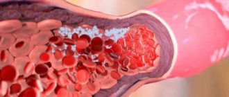
- atherosclerosis (formation of cholesterol plaques on the walls) of arteries;
- inflammatory lesions (vasculitis);
- narrowing due to external pressure - a growing tumor, etc.;
- thrombosis and thromboembolism (blockage of the lumen with a blood clot);
- proliferation of scar tissue;
- dissection of the walls (dissection).
MR angiography (MRA) is used to assess the long-term consequences of injuries and to determine the causes of chronic and acute cerebral circulatory disorders. Often, the diagnosis of extracranial vessels is combined with a brain scan, which gives a more detailed picture of pathological changes.
Neck vessels - MRA and digital angiography
MRI of the neck artery + digital angiogram of the neck vessels.
1) Brachiocephalic trunk 2) Right subclavian artery. 3) Right vertebral artery 4) Right common carotid artery 5) Right internal carotid artery 6) Left vertebral artery 7) Left internal carotid artery Left external carotid artery 9) Left common carotid artery 10) Left subclavian artery. 11) Aorta
1. Brachiocephalic trunk (or innominate artery) 2. Right subclavian artery 3. Right vertebral artery 4. Right common carotid artery 5. Right internal carotid artery 6. Left vertebral artery 7. Left internal carotid artery 8. Left external carotid artery 9. Left common carotid artery 10. Left subclavian artery 11. Aorta
Digital angiography.
Other photographs show the x-ray anatomy of the neck vessels. A – digital angiogram of the right common carotid artery and its branches. Lateral projection.
B – digital angiogram of the vertebral and subclavian arteries. Direct projection
1 Pars prevertebralis arteriae vertebralis - Prevertebral part of vertebral artery
2 Pars transversaria arteriae vertebralis Transverse process part of the vertebral artery Cervical part of vertebral artery
3 Pars atlantica arteriae vertebralis Atlantic part of vertebral artery
4 Pars intracranialis arteriae vertebralis Intracranial part of vertebral artery
5 Atreria subclavia Subclavian artery
6 Truncus thyrocervicalis Thyrocervical trunk
7 Arteria cervicalis ascendens Ascending cervical artery
8 Arteria suprascapularis Suprascapular artery
9 Arteria thyroidea inferior Inferior thyroid artery
10 Arteria thoracica interna Internal thoracic artery Internal thoracic artery
11 Arteria carotis communis Common carotid artery
12 Arteria carotis externa External carotid artery
13 Arteria carotis interna Internal carotid artery Internal carotid artery
14 Sinus caroticus Carotid sinus
15 Bifurcatio carotidis Bifurcation of the carotid artery Carotid bifurcation
16 Arteria thyroidea superior Superior thyroid artery Superior thyroid artery
17 Arteria lingualis Lingual artery
18 Arteria facialis Facial artery Facial artery
19 Arteria pharyngea ascendens Ascending pharyngeal artery
20 Arteria auricularis posterior Posterior auricular artery
21 Arteria occipitalis Occipital artery Occipital artery
22 Arteria maxillaris Maxillary artery
23 Arteria temporalis superficialis Superficial temporal artery
Title: Anatomy according to Pirogov. Volume 2. Head. Neck Author: Shilkin V.V., Filimonov V.I. Year of publication: 2013
How to check the vessels of the neck?
Imaging methods are extremely important for assessing the condition of veins and arteries. For this purpose the following are used:
1. Computed tomography (CT). The method is used to detect acute circulatory disorders and is used for additional diagnostics in case of low information content of MRI. Its advantages are high accuracy and speed, but its disadvantages are radiation exposure and the need to use an iodine-based contrast agent, which creates a number of contraindications to CT.
CT scan of neck and head vessels in 3D modification
2. Digital subtraction angiography. Invasive radiographic procedure. It is not prescribed as a screening because there are faster and safer research methods. Angiography involves the injection of a radiopaque contrast agent into the vascular bed for subsequent assessment of the functions of the arteries and veins in real time. Disadvantages include the need for special training, risk of allergies, radiation exposure, and invasiveness. Advantages: highest accuracy, possibility of simultaneous angioplasty. Angiography should only be performed in the operating room.
3. Duplex scanning. This is a variant of ultrasound examination of blood vessels, which is based on measuring the speed of blood flow. The method is used for emergency diagnosis of obstruction or other pathologies of extracranial (brachiocephalic) vessels and dynamic observation. The advantages are information content, safety, painlessness, lack of preparation and high speed of obtaining results. The disadvantage is the relative accuracy. The method is not very informative at the initial stages of disease development.
4. MR angiography. It does not require special preparation and can be performed with or without contrast. For more accurate visualization of blood vessels, preparations based on a rare earth metal (gadolinium) are used. These compounds are bioinert and safer than iodine-containing products. The advantages of the method are the minimum number of contraindications. MR angiography is safe and painless. The results of the study are quite informative. Native diagnostics is allowed for pregnant women according to indications, starting from the second trimester.
The need for contrast during MRA is determined by the attending physician. Depending on what the MRI of the neck vessels shows and how clear the images are, the radiologist may also order a repeat examination with contrast.
MR angiography is a highly informative procedure that allows you to study not only blood vessels, but also soft tissues of the neck, nerves, and lymphatic structures. The scanning results are layer-by-layer images of the area under study (slices) in three mutually perpendicular projections, with the help of which the localization of pathological changes (tumors, aneurysms, etc.) can be accurately determined.
MRI of head vessels and brain matter
Tomography is based on the phenomenon of nuclear magnetic resonance. The method does not carry radiation exposure and has no restrictions on the frequency of application. The magnetic field is safe for tissues, organs and systems. MR angiography can be used to monitor the dynamics of disease development and evaluate the effectiveness of prescribed treatment.
The disadvantage of MRI is its cost and procedure time. Studying one area in native mode takes 15-20 minutes, and with contrast - 30-35.
Ultrasound examination of the vessels of the head and neck
Ultrasound examination of the vessels of the head and neck Synonyms for the name of the technique:
- duplex scanning
- triplex scanning
- ultrasound examination with color mapping of blood flow
- Doppler ultrasound
Ultrasound examination of the vessels of the head and neck includes 2 separate techniques:
- First of all, the patency of the vessels of the neck (brachiocephalic arteries, main arteries of the head in the neck) is assessed, since pathological changes that can lead to a stroke are most often detected in them.
- If indicated, the doctor may additionally prescribe a study of the cerebral vessels (intracranial arteries) - transcranial duplex (triplex) scanning. In 10-20% of people, especially the elderly, the study cannot be performed due to poor permeability of the skull bones for ultrasound, therefore, this study is performed first, and then payment is made.
Indications
- Prevention of stroke in people with risk factors (smoking, high cholesterol, diabetes, obesity, arterial hypertension, age over 40 years)
- Suspected stroke: weakness and numbness in an arm or leg
- "facial distortion"
- speech disorder
- Lost vision in one eye
- dizziness or unsteadiness when walking
What is being studied?
- In the neck are the carotid, vertebral and subclavian arteries.
- In the head - middle, anterior and posterior cerebral arteries, basilar artery, vertebral arteries (intracranial segment).
What is visible?
- Thickening of the vascular wall
- Atherosclerotic plaques
- Blood clots
- Dissection (separation) of the wall
- Tortuosity
- Aneurysms and arteriovenous malformations
Accurate assessment of the degree of carotid artery stenosis to determine indications for surgery. There are several ways to measure the degree of stenosis. Only one of them is the reference for determining indications for surgery. In our laboratory, the assessment of the degree of stenosis is performed in accordance with domestic and international recommendations. If there are indications for surgery, a consultation with a vascular surgeon is provided on the same day.
Preparation No special training is required.
Cost Duplex (triplex) scanning of the brachiocephalic arteries (main arteries of the head) - 3,000 rubles. Transcranial duplex (triplex) scanning - RUB 3,000.
Indications for MRI of neck vessels
Pathologies of extracranial arteries and veins lead to circulatory disorders in the brain and deterioration of the trophism of its tissues. Such conditions have various manifestations. Indications for MRI of neck vessels are:
- dizziness;
- nausea, morning vomiting;
- fainting;
- sudden attacks of weakness;
- blood pressure surges;
- headache;
- discomfort in the neck;
- speech and swallowing disorders;
- hearing loss;
- blurred vision;
- memory disorders;
- sleep disorders;
- noise in ears;
- poor coordination of movements;
- impaired sensitivity of the skin of the hands, etc.
The method is used in planning operations, as well as for assessing brachiocephalic circulation a certain time after injury. It is necessary to monitor the condition of important vessels in some systemic diseases, for which MRI of the arteries and veins of the neck is prescribed, namely:
- multiple sclerosis;
- diabetes mellitus;
- atherosclerosis;
- vegetative-vascular dystonia;
- hypertension.
Most often, MR angiography is prescribed to clarify the diagnosis when duplex scanning has low information content.
Anatomy of the Human Cerebral Vessels - information:
The arteries of the cerebrum originate from the branches of a. carotis interna and a. basilaris, forming the circulus arteriosus cerebri at the base of the brain. The anterior, middle and posterior cerebral arteries branch on the surface of each hemisphere.
A. cerebri anterior supplies blood to the medial surface of the hemisphere up to the sulcus parietooccipitalis, on its outer surface to the superior frontal gyrus and the upper edge of the parietal lobe, and on the lower surface of the hemisphere to the gyrus rectus of the frontal lobe.
A. cerebri media supplies blood to the insula, both central gyri, the inferior frontal gyrus and most of the middle frontal gyrus, the parietal lobe and the superior and middle temporal gyri.
A. cerebri posterior branches on the medial, inferior and lateral surfaces of the temporal and occipital lobes, with the exception of the superior and middle temporal gyri.
The listed arteries, with their branches in the pia mater, form an arterial network, from which they penetrate vertically into the thickness of the medulla:
- cortical arteries - small branches that branch only in the cerebral cortex, and
- medullary arteries, which, after passing through the cortex, go into the white matter.
The central arteries enter from the base of the brain. Cortical, medullary and central arteries anastomose with each other, forming a single vascular network. The cerebellum receives blood from three arteries on each side.
Two a. cerebelli inferior anterior (branch of a. basilaris) and a. cerebelli inferior posterior (branch of a. vertebralis), branch on the lower surface of the cerebellum, the third branch, a. cerebelli superior (branch of a. basilaris), goes to its upper surface. From a. cerebelli superior is also supplied to the lower colliculi of the roof of the midbrain, and the superior colliculi receive their branches from a. cerebri posterior. The arteries of the remaining parts of the brain, related to the pons and medulla oblongata, originate from a. vertebralis, a. basilaris and their branches. In addition to the described arterial vessels, there are also special arteries of the choroid plexus, four in number on each side.
The cerebral veins are divided into superficial and deep. The superficial veins mostly collect blood from the cerebral cortex and flow partly into the sinus sagittalis superior (superior veins), partly (lower veins) into the sinus transversus and the sinuses of the base of the skull. The veins lack valves and are distinguished by their numerous anastomoses. The deep veins collect blood from the central gray nuclei and ventricles of the brain and merge into one large v. cerebri magna, flowing into the sinus rectus. The veins of the cerebellum form groups: the upper ones pour blood into the sinus rectus and v. cerebri magna, lower - in sinus transversus, sigmoideus, petrosus inferior.
How is an MRI of the cervical spine done?
At the Magnit diagnostic clinic, scanning is carried out by appointment. During its registration, the patient must inform the medical staff about the presence of any metal structures in the body, namely:
- pins;
- knitting needles;
- endoprostheses;
- middle ear implants, etc.
The presence of products and metals with ferromagnetic properties in the body can affect the quality of the images. Clinic staff must be informed of the specific material the structure is made of. You can obtain this information from the medical institution where the operation was performed. You need to ask for an extract from there and take it with you to the procedure to show the radiologist.
MRA of head and neck vessels
You need to arrive for the MRI 5-10 minutes earlier than the appointed time so that you can fill out the documents without rushing. The study is preceded by a consultation at which the patient is asked about contraindications to the procedure, which are:
- first trimester of pregnancy;
- presence of cardiac pacemakers (pacemakers, defibrillators);
- implantation of an insulin pump;
- the presence of metal stents or hemostatic clips in arteries and veins;
- other ferromagnetic implants.
During the interview and introduction, the X-ray technician explains how an MRI of neck vessels is performed and the rules of behavior in the diagnostic room. Patients with claustrophobia are offered to undergo the procedure on an open tomograph. In case of increased anxiety, it is possible to use sedatives, but only as prescribed by a doctor. For patients with acute pain and neuropsychiatric diseases, MRI is performed under sedation or anesthesia in a hospital setting.
Preparing for the examination involves removing all metal objects. The patient must remove jewelry, glasses, hairpins, and items of clothing with such accessories. Electronic devices (phone, watch, hearing aid) remain outside the tomography room. The procedure for performing MRI of neck vessels is as follows:
- The patient is taken to a special room and placed on the tomograph platform. The conveyor moves so that the neck area is in the center of the device frame.
- The laboratory assistant fixes the position of the person’s head, places cushions for convenience, hands the emergency button and leaves the office.
- An x-ray technician watches the procedure from the next room through glass. He checks the functionality of the speakerphone, reminds the patient to remain still until the end of the study, and turns on the device.
During scanning, the tomograph makes sounds (clicking, humming) that may cause discomfort. At the Magnit diagnostic clinic, patients are offered headphones to listen to pleasant music during the examination. You can get regular earplugs instead. Preparation takes 5-10 minutes, scanning lasts 15-20.
Vertebral artery syndrome with neck osteochondrosis
According to statistics, the majority of disorders (more than 70%) are degenerative-dystrophic changes in the body or osteochondrosis. The rest consists of congenital anomalies of the structure of the vertebrae or vertebral arteries, postpartum injuries, scars, tumors, herniated intervertebral discs and overgrown osteophytes, vertebral instability, etc.
Vertebral artery syndrome is a symptom complex of cerebral, vascular, and autonomic abnormalities that occurs as a result of compression of the vertebral arteries, deformation of their walls, or narrowing of the lumen of the blood canal.
Anatomy
The vertebral artery is a paired formation that arises from the subclavian arteries and runs in the canal formed by the openings of the transverse processes of the cervical vertebrae. After passing through the atlas and occipital foramen of the skull, the vertebral arteries merge into a single basilar artery, which supplies blood to the posterior parts of the brain. Any circulatory disorders immediately affect the nutrition of brain and nerve cells, causing severe headaches and dysfunction of the organs of vision and hearing.
On the one hand, the cervical spine has a rather delicate structure, a thin muscular corset and ligamentous apparatus, and does not have such reliable protection as other segments. On the other hand, he constantly experiences colossal loads (supports a fairly heavy skull, turns and tilts his head, etc.). Due to the natural deflection, the neck does not rest even during sleep.
In a normal healthy state under normal physiological conditions, the vertebral arteries are also compressed, limiting blood flow, but it is practically not impaired due to sufficient compensating conditions. The condition of the vessels can change due to anatomical narrowing of the lumen of the channels, atherosclerotic stenosis. Soft tissues, for example, the scalene or oblique muscles of the neck, can also have a compressive effect on the arteries, which intensifies after sharp turns or tilts of the head.
Degenerative-dystrophic changes in the body lead to disruption of metabolic processes in the cartilage of the intervertebral discs. Over time, the disc shell dries out, cracks, and flattens, pinching the vertebral arteries. Constant mechanical compression of blood channels entails chronic irritation of periarterial nerve clusters and the development of angiospastic syndrome (constant vasospasm).
Signs of vertebral artery syndrome
Functional symptoms of compression of the vertebral arteries:
- Headaches in combination with autonomic abnormalities. There may be pulsating, burning, aching, constant. As a rule, they originate in the back of the head, then spread to the temples, parietal and frontal parts. They intensify paroxysmally, especially during physical exertion (sudden movements of the head, after a long stay in a forced position, fast walking, running, with strong shaking in transport).
- Cochleovestibular disorders, manifested in the form of paroxysmal systemic or non-systemic dizziness. It manifests itself as a feeling of instability of the soil, rotation of surrounding objects or the body in space, instability of gait, and inability to maintain balance. Sometimes accompanied by slight hearing loss, inability to perceive a certain frequency of sounds.
- Visual disturbances are expressed in the flickering of butterflies or diverging circles before the eyes, pain, a feeling of sand, flashes. During an attack of severe headache, photophobia is noted. An ophthalmological examination shows a slight change in the tone of the vascular system of the fundus.
Organic symptoms accompanied by persistent impairment of cerebral circulation manifest themselves in the form of sudden dizziness, nausea, vomiting, difficult or slow articulation (the ability to pronounce sounds). Drop attacks (sudden falls with preservation of consciousness) lasting up to several minutes, loss of consciousness lasting up to 10 minutes may occur.
The attack usually subsides after the patient assumes a horizontal position. After the attack, there is a feeling of general weakness, sweating, tinnitus, irritation from bright lights, increased heart rate, blood pressure may increase, etc.
Neurological disorders include neuralgia of the occipital nerves, inflammation of the cervicobrachial plexuses, loss of sensitivity or reflex reactions of the lower part of the head, neck, shoulder girdle, and upper extremities. Torticollis may form - a forced tilt of the head, usually in the direction where the artery is pinched, and pain develops. The development of the disease against the background of existing cardiovascular pathologies is dangerous due to the occurrence of angina pectoris, coronary artery disease, etc.
Diagnosis and treatment
Diagnosing vertebral artery syndrome is quite difficult due to the variety of clinical complaints and symptoms. X-ray images do not always give a clear picture (changes are noticeable only in the bone structures of the spine), so an additional CT or MRI examination is required to identify the location and degree of vascular compression.
Magnetic resonance imaging can visualize in more detail intervertebral discs damaged by osteochondrosis, the presence of protrusions or hernias, subluxations of articular processes, vertebral hypermobility and other pathologies.
It is possible to identify compression of the vertebral arteries, their congenital anomalies of structure, and determine the asymmetry of the linear velocity of blood flow and the intensity of blood circulation in them using duplex scanning or vertebral Dopplerography. Tests are carried out both in a calm state (relaxed) and with functional loads (with flexion and extension of the neck, lateral bending, rotation).
From hardware diagnostics, electrocardiography and electromyography (determining the passage of nerve impulses) are mandatory. If necessary, a consultation with a cardiologist and neurologist is carried out.
Treatment of patients with vertebral artery syndrome requires an integrated approach. At first, it consists of complete rest, elimination of the compressive effect on blood vessels, reduction of neurogenic inflammation and traditional drug therapy:
- To relieve pain and inflammation - analgesics and non-steroidal anti-inflammatory drugs. In this case, anti-inflammatory drugs are best taken in the form of intravenous injections. Then they have a more gentle effect on the gastrointestinal tract, are more effective and the speed of onset of relief is much higher.
- Muscle relaxants are substances that relieve muscle spasms (relax muscles).
- Vasoactive drugs are prescribed to eliminate vascular inflammation and changes in the arterial bed, as well as to expand the lumen of the canals.
- Vitamins (a high content of B vitamins is especially useful) and anabolic agents - to activate trophic processes and improve the general condition of the body.
- Antidepressants and tranquilizers - for the correction of psycho-vegetative pathologies, improving emotional and psychological mood.
As the pain syndrome subsides, physiotherapeutic procedures (UHF, electrophoresis, ultrasound, etc.), massages, reflexology, and exercise therapy are added to the treatment.
If necessary, wearing a support collar (for example, Shants) and posture correction using traction (spinal traction) may be prescribed. The use of manual techniques (acupuncture, plantar and Thai massages, acupuncture, etc.), swimming, classical and passive yoga has a good effect. Author: K.M.N., Academician of the Russian Academy of Medical Sciences M.A. Bobyr
MRI of neck vessels with contrast
Sometimes native MR angiography turns out to be uninformative. Vessels (even of small diameter) can be visualized more clearly by contrast-enhanced examination. It involves the injection into a vein of a drug that increases the vibrations of hydrogen atoms in water molecules in tissues and makes the latter more visible in photographs.
MR angiography of neck vessels
Contrast is required if abnormal vascular development or tumor changes are suspected. With its help, you can assess the condition of the veins and arteries and the speed of blood flow in them. The procedure for MR angiography with contrast is as follows:
- After standard patient preparation, native scanning begins.
- Once the shooting is complete, a contrast agent is injected into a vein in the forearm.
- Repeat the study.
The procedure takes 30-35 minutes. Contrast MR angiography is contraindicated in pregnant women. During lactation, you need to make a supply of breast milk for the next 2 feedings, which you will have to skip.
Interpretation of photos of MRI images of neck vessels
The scanning results are layer-by-layer monochrome images of the area under study. They are deciphered by a radiologist. The specialist sees and records any deviations in tissue structure from the norm. All pathological changes will be recorded in the conclusion, and the images will be recorded on digital media.
Preparing results takes from 15 to 60 minutes. During this time, the patient can walk or rest in the waiting area. Study protocols are sent by email if you specify it when filling out the documents. You can pick up the original on any other day.
The radiologist describes what MRI shows in the arteries and veins of the neck, he gives explanations when presenting the results and recommendations on which doctor to contact. The specialist does not diagnose, make prognosis or prescribe treatment. If results are received by email, the patient will not be able to consult with a radiologist.