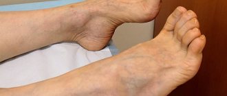
Toxic polyneuropathy is a syndrome that occurs due to damage to peripheral nerves by external or internal toxins. It develops as a result of the direct toxic effect of poisons on various structures of the central and peripheral nervous system (exogenous toxicosis) or as a result of damage to parenchymal organs and body systems responsible for detoxification (endogenous toxicosis).
Neurologists at the Yusupov Hospital provide complex therapy for toxic polyneuropathies, which includes:
- removing toxins from the body using infusions of blood substitutes that have a detoxifying effect;
- impact on the mechanisms of development of damage to the nervous system;
- reduction and reversal of symptoms of the disease.
To determine the cause of peripheral nerve damage, toxicology tests are performed. Changes in the patient’s body are determined using modern laboratory tests. Neurophysiologists perform electroneuromyography, which makes it possible to determine the location and extent of damage to nerve fibers.
Candidates and doctors of medical sciences, doctors of the highest category have an individual approach to the choice of treatment method for the patient. Leading neurologists and toxicologists use modern medications to treat patients with polyneuropathy. At the Yusupov Hospital, patients suffering from toxic polyneuropathy of the lower extremities undergo plasmapheresis.
Causes and mechanisms of development of toxic polyneuropathy
The developing toxic process is based on damage to any structural element of the nervous system by modifying energy and plastic metabolism, disrupting the generation and conduction of nerve impulses along excitable membranes, and signal transmission at synapses. All known poisons have a neurotoxic effect. Any acute intoxication is accompanied by dysfunction of peripheral nerves.
The nervous system is affected by many narcotic substances and industrial poisons:
- metallic mercury;
- arsenic compounds;
- manganese;
- tetralead;
- carbon disulphide.
Nerve fibers are involved in the pathological process during intoxication with many chemicals: lead, benzene and its homologues, fluorides, acrylates, carbon monoxide, chromium. The nature of damage to the nervous system depends on the chemical structure of the substance, the total dose received by the body, and the methods of entry of these substances into the body.
Toxins can selectively affect individual structural elements of the nervous system:
- mercury compounds, manganese, aluminum, glutamate, cyanide, thallium damage neurons and dendrites (branched processes of neurons);
- tetrodotoxin, saxitoxin, carbon disulfide, colchicine have a toxic effect on axons (long cylindrical processes of a nerve cell);
- nicotine, organophosphorus compounds, carbamates, bicyclophosphates, norborian, picrotoxicin, cannabinol diethylamide disrupt synapses (points of contact between two neurons or between a neuron and a cell receiving a signal;
- hexachlorophenol, triethyltin, tellurium destroy the myelin sheath and myelinating cells.
Specific points of application of most toxic substances have not been identified. The selectivity of the toxic effect is relative. As the dose of poisons increases, the damage becomes less selective.
Diffuse damage to neurons of the central nervous system is caused by organophosphorus substances, organic solvents, manganese, and thallium. Some poisons preferentially affect the basal ganglia (striatum, globus pallidus, and cerumenus). Arsenic, mercury, thallium, and carbon disulfide have a toxic effect on sensitive nerve fibers of peripheral nerves. Lead, thallium, arsenic, and mercury disrupt the functions of motor nerve fibers of peripheral nerves. The autonomic ganglia and dorsal roots of the spinal cord are affected by methylmercury.
Toxic encephalopathy of the brain: what kind of disease, main causes
The toxicogenic effect of most neurotropic poisons is based on their negative impact directly on neuronal membranes and the mechanisms of nerve impulse transmission. Violations of energy and plastic metabolism play an important role in the pathogenesis of the disease. In addition, some toxins inhibit the hematopoietic and circulatory systems, breathing, which also causes toxic hypoxic encephalopathy. Due to the peculiarities of the anatomical structure, cells localized in the cerebral cortex, cerebellum and hippocampus are especially sensitive to the influence of pathological factors, which causes the clinical manifestations of the disease.
Major neurotoxins
The main causes of toxic encephalopathy include:
alcohol and volatile solvents: destroy cell membranes, receptors, cause changes (irreversible with long-term exposure) in energy metabolism;- psychoactive substances (opiates, cannabinoids, amphetamines, cocaine, ephedrine, hallucinogens, some drugs, etc.): pathologically affect the reuptake system and the production of norepinephrine, dopamine and serotonin
- heavy metals (mercury, lead, manganese, arsenic, thallium, etc.): have a direct destructive effect on the structures of the central nervous system and also affect the functioning of the peripheral nervous system.
Accordingly, the risk of toxic brain encephalopathy is especially high among people working in hazardous industries. These are, for example, chemical, construction, metallurgy, and agriculture, where insecticides and pesticides are used. The disease is often diagnosed in alcoholism and drug addiction. The pathology is less common among artists; those who use neurotoxic paint solvents are at risk.
Symptoms of toxic polyneuropathy
Depending on the conditions of exposure, the structure of the toxic substance, and its neurotoxic potential, pathological processes in polyneuropathy occur acutely, subacutely or chronically. A manifestation of an acute neurotoxic effect is a violation of the conduction of nerve impulses along motor and autonomic fibers and blockade or distortion of incoming sensory information.
In polyneuropathies - clinical syndromes characterized by diffuse damage to peripheral nerve fibers, the unit of damage is the fibers that are part of various nerves. The likelihood of damage depends on their length, caliber, and metabolic rate. Clinical manifestations of polyneuropathies - widespread, symmetrical, usually distal and progressive lesions - vary widely. They differ in the rate of progression, severity of symptoms, the ratio of sensory and motor disorders, and the presence of irritation symptoms.
Toxic polyneuropathy of the lower extremities begins with paresthesia (a crawling sensation) and pain in the feet, then in the hands. The intensity of the pain quickly becomes unbearable. In some patients, the tip of the nose and ears are involved in the process. They are worried about pain in the teeth and itching of the gums in the upper jaw, and short-term nasal congestion.
The process of polyneuropathy spreads from bottom to top: on the lower extremities - to the knee joints, on the upper extremities - to the shoulder girdle. First, damage to the lower extremities is detected. Neurologists diagnose the extinction of the Achilles reflex and sensory disturbances in the form of “socks”. Then flaccid paralysis develops with gradual extinction of reflexes. Knee and abdominal reflexes are preserved, sometimes strengthened, and symmetrical. Patients develop a fear of pain from touching objects, which leads to a forced position of their legs and arms. The following symptoms are noted:
- pain upon palpation of the skin of the extremities and upon pressing on the exit points of the second branch of the trigeminal nerve;
- hyperesthesia (increased sensitivity of the skin) like high socks and stockings;
- impairment of proprioceptive sensitivity (sense of the position of parts of one’s own body relative to each other and in space);
- decreased muscle tone and strength;
- hypotrophy (loss of mass) of the muscles of the limbs.
As the disease progresses, swelling of the feet and lower third of the legs and hands appears. Some patients with toxic polyneuropathy develop trophic ulcers and skin irritation due to the fact that, in an attempt to relieve pain, they take cold water baths for the extremities. Other patients experience atrophic changes in the skin of the hands. Sometimes the epidermis of the hands and feet exfoliates, hyperkeratosis (excessive growth of the stratum corneum of the epidermis) and mosaic pigmentation. Appetite decreases, sleep is disturbed, and severe neurosis develops. There is pallor of the skin of the face and moderate tachycardia (increased heart rate).
Peripheral neuropathy is sometimes caused by drugs:
- chloramphenicol;
- colistin;
- phenelzine;
- ergotamine.
Mixed sensory-motor polyneuropathy develops as a result of uncontrolled intake of ethambutol, isoniazid, metronidazole, streptomycin, chlorpropamide. Motor polyneuropathy is mainly caused by amphotericin B, sulfonamides, amitriptyline, cimetidine, and tetanus toxoid. Neurotoxicity is one of the side effects of drugs for the treatment of malignant neoplasms and immunosuppressants. Polyneuropathies often occur in acute and chronic intoxication with metals and salts of arsenic, tin, thallium, and zinc vapor.
Neurologists at the Yusupov Hospital make a diagnosis of toxic polyneuropathy based on a characteristic medical history, clarification of the cause of chronic intoxication, and neurological examination data. An electroencephalogram, ECG, Doppler study of cerebral vessels are performed and the diagnosis is confirmed by the results of electroneuromyography.
Neuropathies caused by somatic diseases
Somatic diseases often cause damage to the nervous system, despite the protective function of the blood-brain barrier. Peripheral nerve fibers are most sensitive to dysmetabolic disorders caused by somatic pathology. Simultaneous damage to more or less peripheral nerves leads to the formation of polyneuropathic syndrome.
Table 1. Spectrum of somatic diseases accompanied by polyneuropathy
Table 2. Clinical characteristics of neuropathic pain
Rice. 1. Classification of alcoholic neuropathy
Table 3. Tactics of using ALA for the treatment of polyneuropathies
Often, damage to the peripheral nervous system causes more harm to the patient than the physical disease that caused it, since it leads to limited ability to work and disability. The range of somatic diseases that most often lead to damage to the peripheral nervous system is presented in Table. 1.
Classification and diagnostic approaches
Symptoms of neuropathy include widespread sensory and motor deficits, loss of tendon reflexes and, subsequently, muscle atrophy. In general, polyneuropathic syndrome is a complex of sensory, motor and autonomic disorders. However, symptoms associated with damage to a certain type of nerve fiber usually predominate. Depending on this, all neuropathies are divided into somatic and autonomic (autonomic) neuropathies. In turn, somatic neuropathies are divided into polyneuropathies and multiple or isolated mononeuropathies. Autonomic neuropathies can affect the cardiovascular, gastrointestinal, urogenital and other systems. A correct descriptive syndromic diagnosis is important in the diagnosis of neuropathy. It is advisable to analyze the clinical picture of neuropathy based on the following criteria:
- predominant clinical signs;
- lesion distribution;
- rate of development of symptoms.
The peripheral nerve consists of thin and thick fibers. All motor nerves are thick myelinated fibers. The conduction of impulses of proprioceptive (deep) and vibration sensitivity is also provided by thick fibers. The fibers that transmit impulses for pain and temperature sensitivity are unmyelinated and thin myelinated. Both thin and thick fibers take part in the transmission of tactile sensations. Autonomic fibers are thin, unmyelinated. Damage to thin fibers can lead to selective loss of pain or temperature sensitivity, paresthesia, spontaneous pain in the absence of paresis, and even with normal reflexes. Pain sensations during neuropathy (Table 2) differ in their characteristics from ordinary mechanical pain. When diagnosing neuropathic pain, it is necessary to take into account both the nature of the pain and positive and negative symptoms.
Thick fiber neuropathy is accompanied by muscle weakness, areflexia, and sensory ataxia. In addition, unusual symptoms such as tremor (indicating the activity of a pathological process affecting the peripheral nervous system) and cramps (paroxysms of painful spasms of groups of muscle fibers) may be observed. Damage to autonomic fibers leads to the appearance of somatic symptoms. Autonomic neuropathy is considered as potentially the most dangerous complication of somatic diseases. Unfortunately, autonomic failure often goes unrecognized. Depending on the leading syndrome, cardiac, urogenital, gastrointestinal (neuropathic gastroparesis) and trophic forms are distinguished. The most common symptom is sphincter dysfunction, manifested by sphincter insufficiency or bladder atony, attacks of diarrhea, especially at night, and impotence. Other symptoms of peripheral autonomic failure are tachycardia, orthostatic hypotension, swelling of the feet and joints, dry skin. The dominance of these symptoms is characteristic of the most dangerous cardiac form of autonomic neuropathy. As a result of impaired sympathetic and parasympathetic control of the heart, in patients with autonomic neuropathy the pulse is fixed and is associated with resting tachycardia. A more serious symptom is a violation of orthostatic blood pressure, as a result of which the patient experiences orthostatic hypotension and tachycardia when standing. Clinicians often ignore this symptom, although it is orthostatic hypotension that is the cause of complaints of dizziness in this category of patients. The main complications of the cardiac form of neuropathy are a denervated heart, silent or asymptomatic myocardial infarction, and arrhythmias leading to sudden death syndrome. 25–50% of patients die 5–10 years after diagnosis of cardiac autonomic neuropathy.
Damage to all fibers leads to mixed sensorimotor and autonomic polyneuropathy. In addition, it is diagnostically important to determine the substrate of the lesion: axonopathy and/or myelinopathy. The most accurate determination method is electroneuronomyography. Most somatically caused neuropathies are axonopathies, although the participation of segmental demyelination as an additional factor of damage has also been described. Based on the nature of the distribution of the lesion, distal/proximal and symmetrical/asymmetrical lesions of the limbs are distinguished. In most cases, somatically caused polyneuropathies manifest as distal symmetrical sensory or motor disorders of the limbs. Demyelinating neuropathies are characterized by symmetrical, predominantly proximal lesions of the extremities. In contrast, multiple mononeuropathy is characterized by asymmetric proximal lesions. There are also polyneuropathy with predominant involvement of the upper extremities and polyneuropathy with predominant involvement of the lower extremities. The latter option significantly prevails in frequency of occurrence among neuropathies associated with somatic diseases. In general, all neuropathies can be divided into diffuse symmetrical, caused mainly by metabolic disorders, and focal asymmetrical (mainly ischemic damage to the nerve trunk). According to the nature of the course, an acute form is distinguished (symptoms develop over several days to four weeks); subacute form (symptoms develop over several weeks); chronic form (several months or years). Recurrent polyneuropathies are chronic forms. An acute onset is characteristic of toxic, vascular or immune etiology of polyneuropathy. Most toxic and systemic diseases develop subacutely over several weeks or months. Finally, some neuropathies of metabolic origin can develop extremely slowly (over several years). Thus, the syndromic diagnosis of polyneuropathy should include the nature of the course, distribution of the lesion and the predominant clinical symptoms (example: subacute symmetrical distal sensory neuropathy).
The diagnostic algorithm for polyneuropathic syndrome includes electrophysiological studies and biochemical studies of cerebrospinal fluid, blood and urine. The most informative is stimulation electroneuromyography. To determine the nature (axonopathy or myelinopathy) and level of peripheral nerve damage, it is important to study the speed of excitation along the motor and sensory fibers of the peripheral nerves.
Pathogenetic mechanisms of development of neuropathies associated with somatic diseases
The main factors of damage to nerve fibers are vascular, metabolic, neurotrophic and immunological disorders. Of course, the degree of their participation in the development of neuropathy depends on the type of somatic disease. However, even the same etiological factor can trigger different mechanisms of nerve fiber damage. For example, in diffuse chronic diabetic neuropathies, the main damaging factor is metabolic disorders (hyperglycemia). On the contrary, in acute and subacute focal and multifocal diabetic neuropathies, the main damaging factor is ischemia and, possibly, immunological disorders. One of the universal mechanisms that damage nerve fiber is oxidative stress, which always accompanies metabolic disorders. The participation of oxidative stress in disruption of nerve fiber functioning explains the lack of direct correlation between the severity of somatic disease and the development of neuropathy. Oxidative stress is defined as an imbalance between the formation of free radical oxidation (FRO) and lipid peroxidation (LPO) products and their neutralization and removal from the body. Oxidative stress may be based on both an increase in the production of FRO and LPO derivatives and the depletion of antioxidant defense systems, but more often both of these links are involved in the pathological process. Normal cell activity and processes occurring in the intercellular space lead to the formation of free radicals (FR). CP are extremely unstable substances and are capable of spontaneous decomposition. However, the formation of FRO and LPO products increases significantly with any metabolic disorders. The balance between the production and elimination of derivatives of oxidative processes depends on the effectiveness of various cellular and tissue specific antioxidant mechanisms, the violation of which leads to the development of OS. Insufficient activity of antioxidant enzymes in various somatic diseases is primarily determined by genetic factors, which is confirmed by the study of polymorphism of genes of the body's antioxidant system [1]. Dysfunction of antioxidant systems under different conditions ultimately causes the same consequences.
The most common somatic neuropathies
Diabetic neuropathies
are the most common type of somatic neuropathies. Damage to the peripheral nervous system occurs in 20–40% of patients with diabetes. As a rule, clinical symptoms of neuropathy develop 5–10 years after the onset of the underlying disease. But in at least 10% of patients, the diagnosis of diabetes is verified only after the onset of neurological deficit. The individual combination of clinical signs and symptoms of diabetic neuropathy varies widely. However, symptoms can be grouped into characteristic syndromes to more accurately describe the clinical picture. The most common clinical syndromes are:
- distal symmetrical sensorimotor diabetic polyneuropathy;
- proximal motor diabetic neuropathy (diabetic amyotrophy);
- mononeuropathy in diabetes;
- neuropathy of cranial nerves in diabetes;
- damage to the autonomic nervous system in diabetes.
According to the algorithm presented above, all syndromes can be divided into diffuse or symmetric polyneuropathies (sensory, motor and autonomic) and focal neuropathies (mononeuropathies, multiple mononeuropathies, plexopathies, radiculopathies and cranial neuropathies). The need for such a classification is due to the difference in pathogenetic mechanisms and therapeutic approaches to the treatment of different types of neuropathy. Distal symmetric sensorimotor neuropathy is the most common type of damage to the peripheral nervous system in diabetes, which develops slowly (chronically), usually several years after the onset of the underlying disease, the first symptoms appear in the lower extremities, sometimes unilaterally. Distal symmetric sensorimotor neuropathy often causes the development of chronic neuropathic painful pain syndrome. Severe forms of polyneuropathy occur in patients with early onset diabetes (juvenile forms) and poorly controlled diabetes. In the most severe forms of polyneuropathy, loss of proprioceptive sensitivity can lead to sensory ataxia (pseudotabetic form). In contrast to diffuse forms of neuropathy, focal forms develop acutely or subacutely; the main damaging factor in these forms is ischemia. Among the cranial nerves, the third and sixth oculomotor nerves are most often affected. Proximal asymmetric diabetic neuropathy is less common than distal forms. The clinical picture is characterized by an acute onset and dominance of pain symptoms, which often worsen at night. Typically, pain is localized proximally and affects the lower extremities to a greater extent than the upper extremities. At the same time, muscle weakness occurs followed by atrophy (a characteristic symptom is difficulty in climbing the stairs). Proximal asymmetric diabetic neuropathy predominantly affects older people with type 2 diabetes, with a peak incidence at age 65 years. The average time from diagnosis of diabetes to the onset of neuropathy is approximately four years. Autonomic disorders in patients with diabetes are usually associated with other neurological deficits, but can also occur as an isolated pathology. The most common symptom, as mentioned above, is sphincter dysfunction.
Uremic polyneuropathy
occurs in chronic renal failure. Characterized by predominantly sensory, symmetrical distal disturbances. The disease may begin with cramps and restless legs syndrome. Then dysesthesia, burning and numbness of the feet occur. Uremic polyneuropathy is sometimes called hot leg neuropathy. There is a positive effect of hemodialysis on the course of neuropathy. At the same time, 25% of patients on dialysis have symptoms of neuropathy. Dialysis-associated arteriovenous fistula can lead to focal ischemic neuropathy of the median nerve.
Neuropathies in systemic connective tissue diseases are primarily caused by vasculitis. The most common neuropathies are periarthritis nodosa (in 25% of patients), rheumatoid arthritis (in 10% of patients). Neuropathies associated with systemic vasculitis are usually sensory mononeuropathies (with a pronounced pain component and/or spontaneous pain) or asymmetric polyneuropathies with acute or subacute onset [2]. Symmetric sensory or sensorimotor polyneuropathies are less common.
Polyneuropathies caused by exogenous causes
These disorders account for about 25% of all types of neuropathy. Among the exogenous causes are the effects of stimulants, medications, industrial poisons and other substances. The scope of this work does not allow us to consider the entire spectrum of exogenous neuropathies, so we will focus only on individual types. Alcoholic polyneuropathy
(AP) ranks second in prevalence after diabetic neuropathy. Clinical manifestations of damage to the peripheral nervous system in patients suffering from alcoholism (alcoholic polyneuropathy) occur, according to various authors, in 12.5–29.6% of cases [3]. Previously, it was believed that the development of AP is associated primarily with a nutritional deficiency of vitamin B1 (thiamine), caused by a monotonous, unbalanced, predominantly carbohydrate diet. However, the toxic effects of alcohol are more diverse. Currently, a whole range of different reactions of the body in response to the direct and indirect effects of alcohol is described. The leading role in alcohol damage is given to the excessive formation of free oxygen radicals (FRRs) [4]. With chronic alcohol consumption, the production of TFR increases, and the activity of antioxidants decreases, which leads to the development of oxidative stress. Free radicals disrupt the activity of cellular structures, primarily the endothelium, causing endoneurial hypoxia and leading to the development of neuropathy. Phenomenologically, alcoholic polyneuropathy most often represents a symmetrical distal sensorimotor neuropathy, which is based on axonal degeneration. However, the spectrum of nerve fiber damage may include different patterns (Fig. 1).
Drug-induced neuropathy.
Many medications have neurotoxic side effects, the most common of which is damage to the peripheral nervous system (polyneuropathy). The classic variant of drug-induced neuropathy is isoniazid polyneuropathy. This sensorimotor neuropathy occurs due to vitamin B6 deficiency caused by isoniazid in individuals with a genetically determined disorder in the metabolism of this vitamin. Prescribing pyridoxine together with isoniazid made it possible to practically relieve tuberculosis patients from this type of neuropathy. Neuropathy can be a dose-limited side effect of most drugs used in the treatment of life-threatening conditions such as cancer and HIV infection. Epidemiological studies confirm earlier reports that cytotoxic drugs cause axonal sensorimotor neuropathy or, less commonly, small fiber lesions in some patients [5]. The prognosis of drug-induced neuropathies is unfavorable, since discontinuation of drugs does not lead to improvement in neuropathy symptoms. Treatment with antiviral drugs may lead to sensory neuropathy. A whole spectrum of damage to the peripheral nervous system is characteristic of HIV infection; it is caused by the immunodeficiency virus itself, metabolic disorders and the neurotoxic effect of antiviral therapy. Neuropathies associated with HIV infection include distal symmetric sensorimotor polyneuropathy, toxic (drug-induced) symmetric sensory neuropathy, inflammatory demyelinating polyneuropathy (proximal symmetric sensorimotor polyneuropathy), multifocal mononeuropathy, and progressive polyradiculopathy [6].
Therapy of somatogenically caused neuropathies
The mainstay of therapy is treatment of the underlying disease that led to the development of neuropathy, for example, optimal control of diabetes mellitus. Sometimes compensation for the underlying disease leads to spontaneous regression of neuropathy. However, quite often clinicians are faced with refractory cases of neuropathy. The absence of a direct correlation between the severity of the underlying disease and concomitant neuropathy, as well as the influence of additional factors (endogenous and exogenous) on the development and course of neuropathy, was discussed above. Therefore, another strategic approach to treatment is to influence the known links of pathogenesis and additional factors influencing the course of neuropathy. These measures include vitamin therapy (priority is given to B vitamins), taking vasoactive drugs, and antioxidant therapy. Symptomatic treatment, aimed mainly at correcting pain, deserves special attention. Approaches to the treatment of neuropathic pain are currently quite well developed. As a rule, anticonvulsants and antidepressants are used sequentially or together. Among the anticonvulsants, the most successfully used are: pregabalin, gabapentin, oxcarbazepine, carbamazepine, and valproic acid preparations. Among antidepressants, tricyclic antidepressants (amitriptyline) and dual-action antidepressants (duloxetine, venlafaxone) have high analgesic activity.
Let's take a closer look at antioxidant therapy, which can affect both neurological deficits and the intensity of pain. Antioxidant therapy is considered as one of the possible ways to level toxic-dysmetabolic effects on the nervous system. One of the first places among antioxidants today is alpha-lipoic (thioctic) acid (ALA). ALA is formed naturally in the body and its chemical structure is 1,2-dithiolane-3-pentanoic acid (C8H14O2S2). In humans, ALA is synthesized in the liver and other tissues. ALA was originally known as an essential biochemical cofactor for mitochondrial enzymes. However, in the last decade it has been discovered that ALA and its metabolic intermediate, dihydrolipoic acid, are powerful antioxidants. Due to the fact that ALA is both a fat- and water-soluble substrate, it has the ability to influence oxidative stress in cell membranes and in the cytosol. ALA leads to a decrease in lipid peroxidation processes in the brain and peripheral nerves (shown on sciatic nerve fibers) [7].
Alpha-lipoic acid was first used in 1966 by German clinicians for the treatment of diabetic polyneuropathy and liver cirrhosis, as evidence of low ALA levels in this category of patients appeared [8]. Over decades of use of ALA, numerous evidence has accumulated regarding the effectiveness of this drug against the symptoms of diabetic polyneuropathy. The drug is well tolerated by patients and has a high safety profile. One of the latest meta-analyses, based on four randomized placebo-controlled studies (ALADIN I, ALADIN III, SYDNEY, NATHAN II), which included a total of more than 1000 patients, convincingly showed that after 3 weeks of IV administration of ALA at a dose of 600 mg, a positive the effect is observed in more than 50% of patients [9]. However, improvement was observed both in terms of positive neuropathic symptoms and in terms of neurological deficits. Russian neurologists and endocrinologists have also accumulated extensive clinical experience in the use of ALA for diabetic neuropathy.
The ability of ALA to improve the function of autonomic fibers is important for the treatment of many somatogenic neuropathies. In the Deutsche Kardiale Autonome Neuropathie (DEKAN) study, 73 patients with type 2 diabetes mellitus with impaired heart rate variability were treated with ALA tablets (200 mg 4 times daily, total dose 800 mg) or placebo for 4 months. An analysis was made of the dynamics of heart rate variability at rest, during breathing, and during 24-hour ECG monitoring (high and low frequency power spectrum) during treatment. A significant increase in heart rate variability was found in the ALC group compared to the placebo group (p
A study of the mechanism of action of ALA in patients with diabetic neuropathy showed the complex effect of ALA on the normalization of endoneurial blood flow [11], reduction of oxidative stress [12] and improvement of vascular dysfunction [13]. The complex mechanism of action of ALA suggests its effectiveness against all somatically caused neuropathies, the pathogenesis of which is associated with a toxic-dysmetabolic and vascular damaging component. Indeed, the successful use of ALA in a number of somatogenic neuropathies has been described. For example, ALA has been successfully used to treat uremic neuropathy [14]. ALC has been shown to be effective in 70% of patients with alcoholic neuropathy: it affects sensory and motor symptoms, reduces pain and paresthetic manifestations of neuropathy that are painful for patients [15]. When analyzing the effectiveness and tolerability of ALA in comparison with thiamine, it was found that ALA was significantly more effective than vitamin B1 in clinical and electrophysiological parameters. ALA is recommended for widespread use in the treatment of alcoholic neuropathy [16]. Recently, observations have emerged about the beneficial effect of ALA on neuropathies induced by cytostatics [17]. During the next course, patients were prescribed ALC at a dose of 600 mg IV for 3–5 weeks, and then 1800 mg in tablet form until the symptoms of neuropathy resolved, for a maximum of 6 months. The average duration of treatment was two months. Improvement in neuropathy symptoms was observed after an average of 4 weeks of treatment. The authors conclude that the preventive use of ALA in combination with cytostatics is useful for the treatment and prevention of damage to the peripheral nervous system. Table 3 outlines various tactics for using ALC depending on the etiological factors and the severity of neuropathy.
Russian clinicians have extensive experience in the use of ALA, in particular the drug Berlition® (Berlin-Chemie AG/Menarini Group, Germany). The effectiveness of the drug in reducing motor, sensory and autonomic symptoms of somatic neuropathies has been convincingly shown in many Russian studies, including in conditions close to free clinical practice. Treatment with Berlition® at a dose of 600 mg/day for 3 months led to a decrease in the severity of clinical (sensory, motor) and neurophysiological changes in the peripheral nerves of the lower extremities in patients with diabetes mellitus [18]. Patients noted a decrease in the intensity of the pain syndrome, and in 46% the pain syndrome completely resolved during treatment. The positive effect of Berlition® on autonomic diabetic neuropathy has been confirmed at the level of various tissues and systems [19, 20], including erectile dysfunction (one of the common complications of diabetes mellitus). Many research works and clinical experience indicate that Berlition® has high compliance and practically does not cause side effects. As a universal stabilizer of cell membranes, Berlition® can be used for various neuropathies associated with somatic diseases.
Thus, ALA is indicated for the treatment of polyneuropathies, in the genesis of which oxidative stress plays a significant role (diabetic, alcoholic, drug-induced, etc.).
Treatment of toxic polyneuropathy
Treatment of toxic polyneuropathy of the lower extremities involves eliminating the toxic effects of the causative factor. If we are talking about endogenous poisoning, then endocrinologists prescribe drugs for the treatment of diabetes mellitus, diseases of the thyroid gland, liver and kidneys.
Doctors at the Neurology Clinic provide complex therapy for toxic polyneuropathy of the lower extremities in several areas:
- etiotropic – aimed at removing toxic substances from the body and treating the disease that caused chronic intoxication;
- pathogenetic – influencing the mechanisms of damage to peripheral nerves;
- symptomatic – relief of pain and other signs of the disease;
- restorative - restoring the myelin sheath of the nerves of the lower extremities and conducting impulses through them.
For acute toxic polyneuropathy, plasmapheresis is performed at the Yusupov Hospital. Vitamins have a direct neurotropic effect. They are necessary for the normal functioning of nervous tissue. Thiamine (vitamin B1) maintains the protein, carbohydrate and amino acid balance of the nerve cell and is a modulator of neuromuscular transmission. Pyridoxine (vitamin B6) affects the structure and function of nerve fibers, regulates amino acid metabolism and prevents the accumulation of neurotropic poisons. Vitamin B12 (cyanocobalamin) promotes regeneration (restoration) of nervous tissue. The optimal dosage form of B vitamins for toxic polyneuropathies is Neurovitan. It contains high dosages of all three B vitamins.
Alpha lipoic acid normalizes metabolic processes, helps compensate for polyneuropathy, and normalizes the functions of antioxidant protection. Berlition®, due to its optimal concentration of alpha-lipoic acid, improves endoneurial blood flow, trophism and function of neurons, and helps eliminate symptoms of polyneuropathy such as burning, pain, and numbness of the lower extremities. The versatility of the drug allows neurologists to prescribe it for the treatment of toxic polyneuropathy of the lower extremities.
To maintain muscle tone, rehabilitation therapists conduct courses of massage and physical therapy, electrical stimulation of the spinal cord and magnetic therapy. You can undergo a course of complex therapy for toxic polyneuropathy at the neurology clinic by first making an appointment by calling the Yusupov Hospital, where they use modern methods for removing toxic substances from the body. Neurologists use modern medications that are highly effective and have minimal side effects.


