September 24, 2019
59307
0
3.8 out of 5
The presence of occipital neuralgia is indicated when, as a result of irritation by certain factors of two large or two small occipital nerves, severe headaches occur. Attacks can reach such a frequency that they completely deprive a person of his ability to work.
But you can cope with the disease if you consult a neurosurgeon. The doctor will be able to accurately determine the causes of neuralgia and select the most appropriate treatment tactics in a particular case, including surgery. SL Clinic specialists have extensive experience and knowledge in the field of neurological disorders and are able, using modern treatment tactics, to significantly improve the patient’s quality of life or achieve complete elimination of signs of neuralgia. The price of a specialist consultation is available to everyone and is given in the price list.
Anatomy of the occipital nerve
There are 2 major and 2 minor occipital nerves. They are symmetrically located in pairs on both sides of the back of the head. The large ones are responsible for the innervation of the skin of the back of the head and are formed from the posterior branches of the second cervical spinal nerve. They pass under the lower edges of the oblique muscle of the head, in the thickness of the semispinalis, along the trapezius tendon and branch into branches of different sizes in the skin of the occipital and part of the parietal zone. The greater occipital nerves are not responsible for innervating muscle fibers.
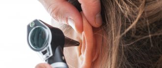
The minor occipital nerves are responsible for the sensitivity of the lateral surfaces of the head, the area behind the ears and partially the auricles. They are formed from the anterior branches of the 2nd and 3rd spinal nerves and pass under the sternosubclavian muscle.
Knowledge of the location of nerve fibers allows a neurologist to correctly diagnose the cause of pain through manual manipulation of special points. With neuralgia, this provokes a sudden attack of pain.
Prevention
To prevent occipital neuralgia, you need to:
- Avoid injury and hypothermia.
- Don't ignore exercise.
- Provide adequate nutrition with the required amount of vitamins.
- Always maintain correct neck position.
- Do not delay treatment for influenza and similar diseases.
- Do not miss scheduled physical therapy sessions.
- Physical activity should be evenly distributed.
In addition, it is necessary to deal with respiratory diseases in a timely manner, as the occipital nerves may become inflamed..
Causes of occipital neuralgia
Neuralgia becomes a consequence of irritation of nerve fibers at any part of their passage. This could be a consequence:
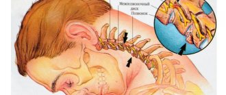
- degenerative-dystrophic processes of the cervical spine, provoking the formation of osteophytes, thinning of cartilage tissue, deformation of the intervertebral discs, which leads to pinched nerves (osteochondrosis, herniated discs, spondylosis, spondyloarthrosis);
- receiving neck injuries;
- formation of tumors of various nature in the area of passage of the occipital nerves;
- severe spasm of the neck muscles resulting from prolonged stay in a static forced position;
- hypothermia;
- anomalies of the craniovertebral junction;
- diabetes mellitus, spinal tuberculosis, gout, rheumatoid arthritis, etc.
In these cases, it can be considered as a symptom of an existing disease, since its elimination leads to the elimination of headaches. Due to the nature of their professional activities, office workers and drivers belong to the risk group.
There is also spontaneously occurring neuralgia of the occipital nerve, called Arnold's neuralgia. Its presence is indicated in the absence of other factors that can lead to compression or irritation of the nerves, i.e., the idiopathic nature of the development.
Causes of the disease
As soon as he hears a diagnosis from a specialist, the patient begins to be interested in the reasons that prompted the problem. There are many of these:
- diseases of the cervical vertebrae;
- sore throat, flu, other infectious diseases;
- spondyloarthrosis;
- spinal injuries;
- vertebral artery aneurysm;
- arthritis or osteochondrosis of the cervical spine;
- gout;
- oncological diseases of the spinal cord and brain;
- lymphadenitis of the glands of the neck.
Symptoms of the disease
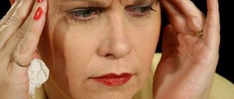
The main manifestation of the disorder is attacks of pain. They can occur with different frequencies and be of different nature. But the pain always initially appears in the back of the head and radiates to the neck and ear area. More often it is unilateral, in some cases there is bilateral damage.
The pain can be lumpy, electric-like, or pulsating. It is always acute and can reach painful intensity, intensifying with head movements and coughing. A patient can experience from one attack to hundreds per day, which completely deprives him of the joy of life. The duration of each varies from a few seconds to a couple of minutes.
Attacks occur spontaneously, more often after sudden movements, sneezing or coughing. A distinctive feature is that patients try to take a forced position with their head slightly pulled back and to the side. This further increases tension in the neck and can make attacks more frequent.
Additionally there may be:
- nausea and vomiting;
- lacrimation;
- feeling cold;
- photophobia;
- intolerance to loud sounds;
- hair loss on the back of the head;
- redness or paleness of the skin in the affected area.
Between attacks, usually nothing bothers the person. Although in some cases it is possible to retain a dull, aching pain in the occipital region, a sensation of goosebumps, burning, and tingling.
A decrease in the sensitivity of the skin of the occipital region is often observed. The patient does not feel touching it, and when injected he only has a slight sensation of touch. But this is only true if you don’t touch the so-called trigger points.
Treatment with folk remedies
You can try to treat this disease using traditional medicine recipes.
These recipes will help alleviate the condition:
- Sleep-herb tincture . The raw materials are poured with boiling water and kept for 24 hours. I use the product once a day. The infusion is also very effective for neurasthenia and insomnia.
- Lilac ointment . Boil lilac buds and add pork fat. The resulting ointment is rubbed into areas of the neck and head that are inflamed.
- Willow bark decoction . Pour boiling water over dry bark. Then simmer over low heat for 20 minutes. The product is taken one spoon 5 times a day.
- Horseradish compress.
It is necessary to take into account that all folk remedies are used only after consultation with the attending physician.
Diagnostics
Diagnosing occipital neuralgia is not difficult. This can usually be done at the first consultation with a neurosurgeon. Pain in the skin of the back of the head or numbness are clear specific symptoms of the disease. The doctor can finally confirm its presence by influencing special trigger points located along the greater or lesser occipital nerve on the affected side. This:
- Impact on a point located on a conventional line passing between the mastoid process and the occipital protuberance. If you divide it into 3 parts and press on the point located on the border of the upper and middle third, the patient will feel severe pain due to neuralgia of the greater occipital nerve.
- Pressure on the site at the junction of the sternocleidomastoid muscle with the mastoid process when the lesser occipital nerve is damaged causes an attack of pain.
But the main goal of diagnosis is to search for factors and diseases that caused its development. Otherwise, any treatment will be ineffective, and painful attacks will become more frequent, longer and more frequent. To detect the causes of the development of occipital neuralgia, a vertebrologist may prescribe to the patient:
- radiography;
- MRI;
- CT;
- UAC;
- blood chemistry;
- blood sugar test.
If during the studies no pathological abnormalities are found, neuralgia is recognized as primary, which determines further tactics for treating the disease.
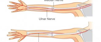
Shooting pain and numbness in the arm/leg/hand
Our clinic provides treatment for tunnel syndromes (carpal tunnel, cubital tunnel), treatment of piriformis muscle syndrome under ultrasound guidance. Along with radicular pain syndromes , shooting (radiating) pain and/or numbness give rise to nerve compression syndromes throughout. The most common examples of such syndromes are:
1) Pinching of the sciatic nerve by a spasmodic piriformis muscle. In this case, aching, “brainy” pain occurs in the gluteal-sacral region with shooting (irradiation) along the back and/or lateral surface of the thigh, lower leg and foot.
2) Compression of the median nerve in the carpal tunnel begins with numbness of the thumb, index and middle fingers after sleep. As the process progresses, a burning pain in these fingers and involvement of the ring finger in the process occurs. In advanced cases, it becomes difficult to hold objects due to muscle atrophy.
3) Compression of the ulnar nerve in the cubital canal is also clearly manifested after sleep by numbness of the little finger and half of the ring finger. Also characteristic is a shooting “electric” pain when tapping the elbow in the area of the cubital canal (Tinnel’s sign).
In advanced cases, muscle atrophy also occurs. For nerve blocks in these tunnel syndromes, ultrasound control is especially important! The likelihood of needle damage to an already suffering nerve with a “blind” technique is extremely high.
Treatment of occipital neuralgia
Without influencing the cause of the disorder, it is impossible to completely eliminate its manifestations. The only case where therapy is purely symptomatic is idiopathic occipital neuralgia.
Until research results are obtained, all patients are prescribed symptomatic therapy. Once the root cause is known, treatment is supplemented with remedies aimed at eliminating it.
In most situations, neuralgia can be dealt with directly using conservative therapy. It assumes:
- drug treatment;
- massage;
- exercise therapy;
- physiotherapy.
If left untreated, occipital neuralgia can lead to destruction of nerve tissue and the development of complications, including hair loss and depressive disorders.
Drug therapy
Depending on the nature of the clinical manifestations and causes of nerve irritation, patients may be prescribed:
- muscle relaxants - a group of drugs that help eliminate muscle spasms;
- NSAIDs are drugs that have anti-inflammatory and analgesic properties;
- corticosteroids – have powerful anti-inflammatory properties, prescribed in short courses when NSAIDs are insufficiently effective;
- anticonvulsants – help reduce muscle tone;
- antidepressants – necessary to normalize the psycho-emotional state of patients;
- B vitamins - necessary to improve the condition of nervous tissue and impulse conduction.
In case of a severe attack of pain, a blockade can be performed, which involves pinpoint injections of anesthetics at certain points along the affected nerve.
Physiotherapy
To increase the effectiveness of drug therapy, patients are recommended to undergo courses of physiotherapeutic procedures:
- UVR method involves exposure to mid-wave ultraviolet rays. This leads to an intensification of the release of specific neurotransmitters, which has a beneficial effect on the transmission of nerve impulses. Typically, the course of treatment includes 10 procedures.
- UHF - the procedure is based on the action of high frequency currents, which provoke an improvement in the quality of blood circulation and restoration of the sodium-potassium membranes of nerve cells. Treatment duration is from 15 to 20 sessions of 15 minutes each.
- Electrophoresis is a procedure during which painkillers or other drugs are injected directly into the affected area using an electric current. Traditionally, 10 procedures are prescribed, performed every other day.
- inductometry;
- Laser therapy is a method based on the positive effect of the thermal energy of laser beams on nerve fibers. This effect reduces their sensitivity, which reduces the degree of their irritation and the frequency of attacks. Patients are recommended 10 procedures of 4 minutes each.
- Diadynamic currents - the procedure involves fixing electrodes on trigger points and passing Bernard current through them. This is an ultra-high frequency current, the impact of which provides a rapid decrease in the pain threshold and inhibition of the transmission of nerve impulses. The duration of the treatment course is 5 sessions, performed every other day.
- Spinal traction is a procedure aimed at increasing the size of intervertebral discs and normalizing the position of the vertebrae. It can be performed using dry or underwater methods. Its duration ranges from several minutes to an hour, and the course of treatment includes about 15 sessions. With the dry method, traction is carried out under the influence of its own weight in a vertical or horizontal position. When underwater, the procedure is carried out after the patient is immersed in water. The second method has a more gentle effect on the spine.
Additionally, patients may be recommended reflexology sessions. It has long been noted that targeted effects on biologically active points provoke increased blood circulation, improved lymph flow and a decrease in the severity of pain.
Massage
A properly performed massage helps eliminate pressure on nerve endings, improve blood circulation in the head, and eliminate muscle spasms. The mastoid process and the area under it are exposed to manual intervention. It is important that the procedure is carried out by a qualified specialist. Otherwise, you may accidentally hit trigger points, which will provoke a new attack of pain.
Traditionally, a course consisting of 12–14 procedures is prescribed. The duration of each of them is about a quarter of an hour.
Therapeutic exercise (physical therapy)
Regular exercise therapy classes according to individually designed programs will help normalize the functioning of the cervical spine. They are especially important for osteochondrosis, as they allow you to accelerate the processes of natural restoration of cartilage tissue and eliminate the prerequisites for compression of nerves.
For each patient, a set of exercises is selected separately, taking into account the characteristics of existing diseases. They should be performed daily at least once a day. On average, one physical therapy session takes about 20–30 minutes.
All exercises are performed carefully, without sudden movements. If a new attack of neuralgic pain or discomfort in the cervical spine occurs, you should immediately stop exercising and consult a doctor as soon as possible to understand the causes of pain and change the nature of the exercises to a more gentle one.
Treatment
Treatment consists of following the general recommendations of the doctor, treatment of concomitant diseases or injuries, medication, physiotherapy, reflexology and surgical treatment.
General recommendations include maintaining bed rest, normalizing sleep, and eliminating psycho-emotional stress.
Drug treatment
Anti-inflammatory drugs
Drugs in this group stop the inflammatory process both in the nerve itself and in the surrounding tissues. Of the drugs in this group, drugs from the diclofenac group are prescribed: diclofenac, naklofen, voltaren, diclobrew, diclac; drugs from the coxib group: rofnix, denebol; drugs of the meloxicam group: Movalis, Movalgin. The side effects of these drugs include abdominal pain, nausea, vomiting, stomach ulcers, headaches, and dizziness.
Hormonal drugs
Hormonal drugs are prescribed to enhance the effect of anti-inflammatory drugs. The most commonly prescribed medications are Kenalog, hydrocortisone, and dexamethasone. Side effects of drugs in this group are: abdominal pain, diarrhea, stomach ulcers, metabolic disorders in the body.
Muscle relaxants
Muscle relaxants are used to relieve spasms in the muscles that surround the nerve. Mydocalm, midostad, tolperil, sirdalud are prescribed. Side effects of drugs in this group may include: abdominal discomfort, nausea, vomiting, muscle weakness, headache.
Painkillers
To relieve severe pain, dexalgin, baralgin, and catadolone are prescribed. Side effects of drugs in this group include abdominal discomfort, nausea, dizziness, headaches, and drowsiness.
Anticonvulsants
Anticonvulsants are prescribed to relieve severe pain in the absence of effect from painkillers. Medicines from the gabaleptin group are used: gabalept, pregabalin, tebantin. Side effects of drugs in this group include blood disorders, drowsiness, anxiety, increased blood pressure, palpitations, and allergic reactions.
Dibenzazepine group drugs are also prescribed: carbamazepine, finlepsin. The drugs can cause abdominal pain, nausea, dizziness, headache, and blood and liver disorders.
Novocaine blockades
Novocaine blockades are performed to potentiate the effect of painkillers and in case of their low effectiveness. The nerve exit site is injected with novocaine. A side effect of this type of treatment may be an allergic reaction.
Antidepressants
Antidepressants enhance the effect of pain therapy. Amitriptyline, doxepin, zalox and other drugs are prescribed. Side effects of antidepressants include nausea, heartburn, palpitations, dizziness, tinnitus, and blurred vision.
Physiotherapeutic treatment
Physiotherapeutic treatment is carried out after the acute signs of the disease disappear. Appointed:
- laser therapy;
- ultrasound therapy;
- electrophoresis;
- magnetotherapy.
Reflexology
Reflexology includes:
- acupuncture;
- centrotherapy.
Surgical treatment of occipital neuralgia
For idiopathic neuralgia, as well as when conservative treatment is ineffective, patients may be offered surgical treatment. It may consist of:
- microvascular decompression;
- radiofrequency ablation;
- neurostimulation.
Patients may also be recommended surgeries aimed at eliminating the cause of neuralgia. Most often, they lie in disturbances in the structure of the cervical spine, which is why surgical interventions are performed in this same area with high frequency.
Microvascular decompression
Microvascular decompression allows complete preservation of the nerve. It can be used when a nerve fiber is compressed by a nearby passing blood vessel, from which blood pulsations are transmitted to it, which causes pain.
The operation is performed openly under general anesthesia. Its essence is to separate the vessel from the nerve at its junction and install a Teflon gasket between them. This is a reliable means of eliminating pain and leads to complete recovery, provided that neuralgia was a consequence of irritation of the occipital nerve by pulsation of a blood vessel.
But this method of surgical treatment requires great caution from the operating neurosurgeon, since it can easily damage the nerve fiber. This will result in loss of sensation in the entire area it innervates. Therefore, you can trust its implementation only to a highly qualified neurosurgeon.
Radiofrequency ablation
The procedure is characterized by minimal trauma and a high level of safety. Its essence consists in introducing a thin needle through the skin under the control of an image intensifier. It is brought directly to the nerve, after which the active or damaging electrode is immersed through the cavity in it. It is connected to a generator, due to which, after turning on the device, current is supplied to the non-insulated end of the electrode. By acting on the nerve fiber, it is destroyed, which leads to the elimination of pain.
For neuralgia of the occipital nerve, the use of current with a frequency of 100 Hz is indicated. This leads to heating of the tissue around the bare end of the electrode to 42°C. The ablation itself lasts about 2 minutes, and the extremely low temperature effect eliminates the risk of thermal damage to surrounding tissues.
Because the procedure does not require significant tissue damage, the patient can move and return to daily activities almost immediately afterward. No hospitalization required.
Radiofrequency ablation is performed under local anesthesia, so choosing it completely avoids the risks associated with general anesthesia. The duration of the procedure is about half an hour. Almost always it leads to complete recovery and immediate pain relief.
The method is also used if the cause of pain is spondyloarthrosis, which is not amenable to conservative treatment. In this case, the essence of the procedure is the same, but the damaging electrode is immersed under the control of the image intensifier into the soft tissue in the immediate vicinity of the affected joint.
Neurostimulation
The essence of the procedure is to implant electrodes under the skin that generate electrical impulses, which helps reduce pain. A total of one or two 8-pin electrodes are installed. The quantity depends on whether the process is one-sided or two-way.
Electrodes are installed in the area of the greater and lesser occipital nerves. They are connected to a generator with the ability to recharge and are equipped with a remote control. Patients can independently turn on the supply of electrical impulses when an attack occurs, which allows them to completely get rid of the painful sensations.
Operations aimed at eliminating pathologies of the cervical spine
Since one of the causes of the development of occipital neuralgia is degenerative-dystrophic diseases of the cervical spine, patients may be prescribed surgical interventions aimed at eliminating them. The type of operation depends on the nature of the detected deviations. It can be:
- Microsurgical methods for the treatment of intervertebral disc protrusions (cold plasma, radio wave, laser nucleoplasty) - make it possible to reduce the size of the disc protrusion by sclerosing part of the nucleus pulposus with cold plasma, thermal laser energy or high-frequency current. They are not performed under local anesthesia, allow you to immediately return home and do not leave scars on the skin, since all the necessary instruments are inserted into the patient’s body through a thin cannula.
- Microdiscectomy is an operation to remove a herniated intervertebral disc, performed under general anesthesia through an incision, usually on the side of the neck, up to 3 cm in size. It requires a short hospitalization and compliance with a number of restrictions during the recovery period, but has a significantly lower risk of relapse. At the same time, with the help of microdiscectomy, hernias of almost any size can be removed.
- Endoscopic surgery is performed for herniated intervertebral discs, but is carried out through a pinpoint puncture of soft tissue up to 1 cm in size. It has a more gentle effect on the body, which facilitates the rehabilitation period. It is also indicated for severe spondylosis. In such cases, excess bone tissue is removed, the intervertebral disc is moved or removed, and, if necessary, implants are installed.
Back pain
Our clinic provides the following types of ultrasound-guided treatment for back pain: paravertebral blockades, facet joint blockades, trigger point blockades for myofascial syndrome, caudal blockade. Back pain is quite varied and directly depends on the severity of the pathological process of the musculoskeletal structures of the spine.
This may be local pain in one of the parts of the spine (cervical, thoracic, lumbosacral), accompanied by reflex muscle spasm and limitation of movements. As the pathological process worsens, so-called “radicular” syndromes may occur (with herniated intervertebral discs) - pain and/or numbness in the area of innervation of the affected spinal nerve.
Such pain can spread to the arms, legs, torso (girdle), back of the head, and anogenital area. All manipulations are performed under ultrasound control - if the level of damage is correctly determined, the effect of the procedure is immediate. In the spine area (especially in the thoracic and cervical regions), ultrasound control is especially necessary! In the absence of this, the effect of the procedure will be minimal, and the risk of complications (including life-threatening ones) will be maximum.
Treatment of occipital neuralgia at SL Clinic
At SL Clinic you can receive qualified treatment for occipital neuralgia. Our vertebrologists will be able to thoroughly understand the causes of pain and purposefully influence them. This approach ensures maximum effectiveness of the therapy and increases the chances of a full recovery.
The cost of radiofrequency treatment of neuralgia of the greater occipital nerve is 68,000 rubles and depends on: - The cost of needles for radiofrequency ablation; — Clinics and class wards. The price includes: — Stay at the clinic before and after surgery; — Operation; — Cost of needles for radiofrequency ablation; — Observation and consultation during the rehabilitation period. All clinic services and costs are listed in the price list.
In most cases, conservative therapy, correctly constructed by our doctors, in combination with radiofrequency treatment, gives good results and eliminates the manifestations of neuralgia. Help from vertebrologists can be provided at the SL Clinic at the highest level. Modern operating rooms, the best equipment and highly qualified neurosurgeons who master advanced techniques are the key to the success of surgical treatment and its safety. All this can be provided to you by SL Clinic.
With us you can quickly complete all the necessary research at a time convenient for you. If necessary, our specialists will carry out a blockade or radiofrequency treatment to stop a severe attack and normalize the patient’s condition. Our prices make advanced European-level surgery affordable.
Don’t tolerate pain, sign up for a consultation at SL Clinic.
How to treat inflammation?
There are several ways to treat occipital neuralgia. Let's look at them below.
Conservative method
Use of medications:
- NSAIDs (Naproxen, Meloxicam, Diclofenac, Ibuprofen).
- Drugs that relax muscles and relieve spasms (Mydocalm, Sirdalud).
- Anticonvulsants (Pregabalin, Carbamazepine).
- Antidepressants (Amitriptyline).
Injections:
- Hormonal substances (Hydrocortisone, Dexamethasone, Diprospan, Kenalog).
- Anesthetic drugs (Novocaine, Lidocaine).
Ointments for neuralgia in the neck:
- Voltaren. It works better when taken with tablets from the NSAID group.
- Ketonal.
- Apisatron. After applying the medicinal composition, redness and burning appear, but this is normal.
- Finalgon.
- Menovazin.
- Turpentine ointment.
- Miaton.
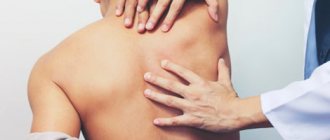
Massage and manual therapy
It is carried out daily for 8 - 10 minutes during the period of pain subsiding. The main attention is paid to the upper back, neck, head, especially those places where the occipital nerves exit.
The patient should be in a sitting position, with the head positioned vertically. This pose helps relax the muscles of the neck and head.
Massage stages:
- Stroking the back, kneading the long dorsal muscle with the edge of the palm, the pads of four fingers and fingers bent into a fist.
- Stroking the scalp and sides of the neck. Then squeezes are performed with the edge of the palm from the top of the head down along the spine. After this, the trapezius muscle is stroked and kneaded with the pads of four fingers.
- This ends the preparatory stage and begins a deeper massage:
- rubbing the back of the head;
- rubbing the side of the mastoid neck;
- rubbing the edge of the skull from ear to ear;
- rubbing the exit site of the occipital nerve.
Spinal traction and manual therapy:
- Traction traction of the spine (quickly and safely eliminates compression of the radicular nerve).
- Osteopathy (improves the flow of blood and lymph, starts the process of restoration of damaged nerve tissue).
- Therapeutic exercises and kinesiotherapy (strengthens the muscles of the neck and back of the head. This prevents recurrence of attacks of pain).
- Acupuncture (provokes restoration of the physiological state of nerve fibers).
Exercise therapy
- Neck stretch: pressing the chin to the neck. At this moment it should be smooth.
- Strengthening the neck muscles that hold the head in the correct position: lying on your back, press the back of your head onto the pillow for 10 - 15 seconds. The neck should be tense, breathing should be even. The same thing when lying on your stomach - pressing the pillow with your forehead.
- While standing on your feet, rotate your head in a circular motion to the maximum amplitude. First clockwise, then vice versa.
- While standing, turn your head left and right at an average pace. Try to turn your head as much as possible to look over your shoulder.
- Make circular movements with your shoulders forward, then back. 5 times on each side.
In addition to the above methods, physiotherapy is also used.
Surgical
It is resorted to only when conservative treatment is useless. Methods:
- Microvascular decompression . It is used for pinched nerve fibers, using special microsurgical equipment that prevents compression of the nerve endings.
- Neurostimulation . Wires are inserted into the affected nerve fibers and transmit electrical impulses. The neurostimulator is implanted in the neck area, its operation is adjusted depending on the patient’s condition.
- Pulsed radiofrequency ablation of the occipital nerves is a safe and effective treatment method. Provides long-term relief from pain by interrupting the transmission of nerve signals from the affected organ to the central nervous system.
Traditional recipes for use at home
Often decoctions and infusions for compresses are used against the disease at home:
- Mix chopped potatoes, onion and pickled cucumber. Pour wine vinegar over all this and leave. This compress is placed on the forehead and back of the head twice a day for one hour.
- Willow bark decoction. Boil a tablespoon of bark and take orally. This should be done three times a day and apply grated horseradish to the affected area.
- Herbal baths will help you relax. You can use an infusion of oregano, thyme and mint.
More information about traditional methods of treating neuralgia here.
Joint pain
In our clinic, we treat joint pain using ultrasound-guided blockade techniques, and we also perform intra-articular administration of hyaluronic acid preparations. Joint pain is so typical that there is no point in describing it. We perform drug blockade of large joints (knees, shoulders, elbows) under ultrasound control using local anesthetic and anti-inflammatory drugs.
Intra-articular injection of hyaluronic acid preparations is a kind of medical joint replacement. For a number of reasons, many people experience a decrease in the amount of synovial fluid (the natural “lubricant” of the joint), which leads to rapid wear and tear of the joint. The introduction of hyaluronic acid preparations allows you to restore the shock-absorbing properties of the joint and, in many cases, avoid surgical treatment.
Ultrasound control during this manipulation allows you to be sure that you are not pouring an expensive hyaluronic acid preparation “into nowhere”!